Journal of Craniovertebral
Junction and Spine
J J C V S Editor-in-Chief : Atul Goel (INDIA) Open Access HTML Format
For entire Editorial Board visit : http://www.jcvjs.com/editorialboard.asp
Practitioner Section
C1-2 posterior arthrodesis technique with a left
segmental and right transarticular fixation. A hybrid
novel (Kotil) technique
Kadir Kotil, Murat Muslumanoglu
Department of Neurosurgery, T.C. Istanbul Arel University, Istanbul, Tepekent/Turkey
Corresponding author: Prof. Kadir Kotil, Department of Neurosurgery, T.C. Istanbul Arel University, Istanbul, Tepekent/Turkey. E-mail: [email protected]
Journal of Craniovertebral Junction and Spine 2014, 5:23
Abstract
Introduction: The most commonly used techniques for C1-C2 posterior arthrodesis are Goel and Magerl
fixation techniques. Due to the anatomical variations of the region, the prior determination of the surgical technique might be hard. Right side Magerl, left side Goel’s C1-C2 posterior arthrodesis case is presented as a
new surgical combination technique used due to anatomical difficulties. MaterialsandMethods: Posterior
C1-C2 arthrodesis operation was indicated for a 56-year-old female patient for the treatment of atlanto-axial subluxation caused by os odontoideum. First it was fixed from the nondominant arterial side (right vertebral artery) with Magerl (transarticular) technique. The left side was not suitable for the anatomical transarticular fixation, and the contralateral Goel fixation technique (segmental) was performed. Eventually, right side transarticular left side segmental fixation techniques were combined in one patient for the first
time and C1-C2 fusion combination technique was presented. Results: Both Goel and Magerl techniques
of C1-C2 posterior fusion techniques were successfully used simultaneously. The operation was initiated with Magerl technique with one screw on the nondominant side. The contralateral side was not suitable for Magerl technique therefore we changed to Goel’s technique. Although, fluoroscopy was used 3 times as much during the introduction of the Drill with Magerl technique, twice as much operative time was spent during hemostasis and bleeding, preparation of the C1 entry point, and the reconstruction of polyaxial screws
for Goel technique. No neurovascular complications were occurred during both procedures. Discussion:
Combination of two C1-C2 posterior fusion techniques, Goel and Magerl, in suitable cases caused by anatomical or other reasons appears to be an alternative surgical procedure that protects the patient from
complications. For a collection of better data, other studies that include large numbers of patientswith high
evidential value should be conducted.
Key words: C1-C2 posterior arthrodesis, novel technique, segmental fixation, transarticular fixation, Kotil technique
INTRODUCTION
Posterior fusion could be done with various techniques for
C1-C2 atlanto-axial dislocations.[1,2] Now-a-days, the most
popular techniques are Goel, which is performed with C1-C2 segmental fixation, and Magerl, transarticular fixation techniques.
Segmental fixation was first defined by Goel and Laheri.[3,4]
Transarticular fixation was first defined by Magerl and Seeman in 1970.[5] There is no clear view in the literature assessing which
technique causes complications.[6] In fact, the determination of
Access this article online
Quick Response Code:
Website:
www.jcvjs.com
DOI:
103 Journal of Craniovertebral Junction and Spine 2014, 5:23 Kotil and Muslumanoglu: C1-C2 posterior arthrodesis with hybrid novel technique
the surgical technique depends on the radiological investigations and the experience of the surgeon. Intraoperative difficulties might cause problems during the operation and increase the risk of complications. We tried to present this case as a new surgical technique we performed in an operation that we had some problems with the application of the intraoperative transarticular fixation and switched to another technique.
CASE REPORT
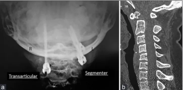
Old type-2 fracture or os odontoideum view was present on direct cervical X-ray of the 56-year-old female patient complaining of occipital nevralgia that has been present for years and has been increasing gradually. In addition, the patient had a ponticulus posticus anomaly of C1 [Figure 1a]. Os odontoideum was seen on cervical computed tomography (CT) and magnetic resonance imaging (MRI). A hyperintense signal was detected from spinal cord on T2 sagittal MRI sections [Figure 1b and c]. Neurological exam was normal except for the increase of deep tendon reflexes and Hoffman sign. C1-C2 fusion surgery was performed on a patient on whom it was decided to perform posterior C1-C2 transarticular arthrodesis caused by ponticulus posticus anomaly of C1. Transarticular fixation was done first, on the right side which is the nondominant vertebral artery (VA) side. Same application was tried on the left side, but when we saw that we would fail in the reduction unilateral fixation with C2 pedicle, C1 mass screw technique was performed to avoid neurovascular complications. In this manner, surgical technique combining Goel on one side and Magerl on the other side was performed on the patient. The patient was discharged from the hospital on day 2 postoperative following a control X-ray [Figure 2a and b]. The patient is still on follow-up with no problems.
Surgical technique
A midline incision between C0 and C4 was performed while patient was fixed in the prone position with her head flexed.
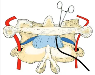
Laminae of C1 and C2 were exposed after the paravertebral muscles were scraped. At first lateral fluoroscopy view was taken for the last views. To try and reduce the atlantodental distance thick spiral silk sutures [Figure 3] passing under laminae of C1 and C2 before fixation process was used. Transarticular fixation with a single screw was performed on the right side because the nondominant side of the patient was the right. The same procedure was attempted on the left side afterwards, but the transarticular trial was unsuccessful because we failed on reduction, and C1-C2 was fixed with segmental fixation. The surgical technique and details are the same as the segmental and transarticular technique applications, no different application was performed. Thus, C1-C2 complex fixation was done with the transarticular fixation on the right side and segmental fixation on the left side. Afterwards, an autograft harvested from the iliac crest was tightened with sutures of number 0 silk thread over spiral sutures passing under laminae between laminae of decorticated C1 and C2 and posterior arthrodesis was performed [Figure 4].
RESULTS
Posterior arthrodesis using the Goel and Magerl techniques was performed on the patient. Magerl technique, which is the first technique ever performed for posterior arthrodesis, was performed without complication on the right side, which is the nondominant side with the guidance of fluoroscopy. Then, we switched to Goel’s technique because Magerl technique was not suitable for the contralateral side. Fluoroscopy was used 3 times longer while introducing the drill in Magerl technique when compared to the other side. Operative duration was 3 times longer for the Goel technique because of the bleeding and hemostasis, preparation of the entry point of C1 and reconstruction of polyaxial screws. Goel technique appears as a technique which might prevent neurovascular complications, while on the other hand Magerl technique shows some disadvantages in the case of anatomical unsuitability or when reduction is failed.
DISCUSSION
Magerl and Goel are the most commonly used techniques for
C1-C2 posterior arthrodesis.[3-5] Comparative biomechanical
Figure 1: (a) The direct X-ray lateral graphy imaging of the cervical spine showing a ponticulus posticus (arrow) anomaly of C1. (b) Sagittal computed tomography was showing an os odontoiedum. (c) T2-weighted sagittal magnetic resonance imaging of the cervical spine showing hyperintense signal
a b
c
Figure 2: Postoperative direct X-ray posterior-anterior (a) and coronal computed tomography (b) imaging of the upper cervical spine showing both segmental and transarticular screws
Journal of Craniovertebral Junction and Spine 2014, 5:23
and long-term studies of the surgical fixation techniques performed in this region are present in the literature.[2-12] Lately,
translaminar fixation technique is becoming more popular.
[13] The selection of the fixation technique for C1-C2 posterior
arthrodesis surgery depends on a lot of factors such as the anatomy of the upper cervical vertebrae (C1 and C2), anomalies, deformities, VA variations, nonreductable fractures and most importantly the experience of the surgeon.[6-8,14] The
location of VA and presence of anomalies on preoperative detailed anatomical investigations are the decisive factor for the technique of the operation, because osteovascular variations are mostly present in this region than the other regions.[8,15] But the
most trustable way to determinate the most suitable operation preoperatively is the experience of the surgeon. We planned to perform transarticular technique on both sides preoperatively because our team has a lot of experience on transarticular technique, but perioperatively we had to change our decision and perform two different techniques on each side.
When performed with the posterior wiring transarticular fixation, which is known as the Magerl technique, provides the strongest fixation and fusion (Magerl and Seeman, 1970). When observed from the point of VA damage (1-8.2%), it might be high risk when performed on cases with high riding VA, wide transverse foramen; hypo-plastic pars interarticularis because it requires experience.[8,15] Vergara et al. compared Magerl and
Goel techniques on 122 cases retrospectively, and reported that intraoperative complications are 10 times, VA injuries are 6 times, and broken screws were 3 times more with the Magerl technique.[6] Despite that, it is stated in the latest publications
that the complication rates with Goel technique might just be as high as in Magerl technique.[11] Finn et al. performed Magerl
technique on 269 cases, indicated 15% complication rate of which one case was fatal, and reported that this technique has many disadvantages.[16]
Yoshida et al. examined their 62 consecutive cases with 3D
CT and stated that there was no difference between the two
techniques related to the VA damage.[8] The latest update on the
subject was by Elliott et al. who conducted a class 3 metanalysis
study in 2014, stating that segmental fixation technique is superiorly related to fusion and VA injury.[11]
Technically Goel technique is more advantageous, but it is reported to have some disadvantages caused by the risk of
venous bleedings and C2 nerve root neuropathy.[9] To avoid
these problems we have chosen the entry point of the C1 screw just above the intersection of massa lateralis and lamina, so bleedings are less, and C2 nerve root contact is minimum. Fluoroscopy was used 6 times with Magerl technique while on the contralateral side it was used only twice with Goel technique. However, other than this, more bleeding and application of more implants on the side we performed Goel technique are the disadvantages. Another important point, while performing
Goel technique is that VA damage[16,17] should be taken into
consideration during the application of a screw on the lateral mass of C1 because of the ponticulus posticus anomaly which is present in 15.5% of the cases.[17] Even though we planned to
perform Magerl on both sides, we switched to Harms technique because the ponticulus posticus anomaly was present in our case as well and we could not penetrate the lateral mass of C1 with a transarticular screw on the contralateral side. Operation duration is longer with Goel technique due to the large quantity of blood loss, and in some cases there might be difficulties with interarticular curetage and the positioning of the graft. Because there is not a consensus with all these studies, surgeons get confused, and some deviations occur during the preoperative planning.
Experience is more required with Magerl technique because even though the anatomical sequence is not correct reduction can be done over the screws after replacement with Goel technique, but construction is not likely possible without a reduction with the
Magerl technique.[17] Usage of two screws and one rod in Goel
technique results in metal pollution which is a disadvantage and raises the expenditures as well.
Figure 3: The illustration imaging of the C1-C2 spine showing to try and reduce the atlantodental distance thick spiral silk sutures passing under laminae of C1 and C2 before fixation process were used
Figure 4: The illustration imaging of the C1-C2 spine showing an auto graft harvested from iliac crest was tightened with sutures of number 0 silk thread over spiral sutures passing under laminae between laminae of decorticated C1 and C2 and posterior arthrodesis was performed
105 Journal of Craniovertebral Junction and Spine 2014, 5:23 Kotil and Muslumanoglu: C1-C2 posterior arthrodesis with hybrid novel technique
In our case, we switched to contralateral segmental fixation because of anatomical unsuitability and failure of orientation of the screw into the lateral mass on the left side. We considered that we might protect the patient from possible neurovascular complications. Unilateral fixation might be considered sufficient in the case with unilateral application difficulty like this.[18] It
was tried on patients who were reduced and who had no major deformity and stated that positive results might be accomplished with the application of postoperative Halo.[19] However, there
are still some suspicions on this subject. Cyr et al. reported,
in their study they conducted on cadavers that if sufficient bone graft and preparation for fusion are performed to avoid VA damage unilateral nondominant VA side screw application might be efficient.[19] Long-term outcomes of unilateral fixation
of a joint as powerful as this one are not known, but it might be attempted in cases like our case if the first and only screw is sufficient and strong.
CONCLUSION
Determination of surgical technique for posterior arthrodesis is not always easy, due to the complex anatomy and the higher possibilities of variations compared to other regions. In the case of perioperative application difficulties with the preoperatively decided technique, instead of persisting, we should switch to the most suitable technique for the surgeon to protect the patient from complications. It is important to prevent the neurovascular complications. This surgical technique note is a first as a reminder article presented for the surgeons working in the same region related to intraoperative strategies.
REFERENCES
1. Papagelopoulos PJ, Currier BL, Hokari Y, Neale PG, Zhao C, Berglund LJ, et al. Biomechanical comparison of C1-C2 posterior arthrodesis techniques. Spine (Phila Pa 1976) 2007;32:E363-70.
2. Naderi S, Crawford NR, Song GS, Sonntag VK, Dickman CA. Biomechanical comparison of C1-C2 posterior fixations. Cable, graft, and screw combinations. Spine (Phila Pa 1976) 1998;23:1946-55.
3. Goel A, Laheri V. Plate and screw fixation for atlanto-axial subluxation. Acta Neurochir (Wien) 1994;129:47-53.
4. Goel A, Desai K, Muzumdar D. Atlantoaxial fixation using plate and screw method. A report of 160 treated patients. Neurosurgery 2002;51:1351-7.
5. Magerl F, Seemann PS. Stable posterior fusion of the atlas and axis by transarticular screw fixation. In: Kehr P, Weidner A, editors. Cervical Spine I. New York: Springer Wien; 1986. p. 322-7.
6. Vergara P, Bal JS, Hickman Casey AT, Crockard HA, Choi D. C1-C2 posterior fixation: Are 4 screws better than 2? Neurosurgery 2012;71:86-95. 7. Claybrooks R, Kayanja M, Milks R, Benzel E. Atlantoaxial fusion: A
biomechanical analysis of two C1-C2 fusion techniques. Spine J 2007;7:682-8. 8. Yoshida M, Neo M, Fujibayashi S, Nakamura T. Comparison of the anatomical risk for vertebral artery injury associated with the C2-pedicle screw and atlantoaxial transarticular screw. Spine (Phila Pa 1976) 2006;31:E513-7. 9. Goel A, Desai KI, Muzumdar DP. Atlantoaxial fixation using plate and screw
method: A report of 160 treated patients. Neurosurgery 2002;51:1351-6. 10. Lee SH, Kim ES, Sung JK, Park YM, Eoh W. Clinical and radiological comparison
of treatment of atlantoaxial instability by posterior C1-C2 transarticular screw fixation or C1 lateral mass-C2 pedicle screw fixation. J Clin Neurosci 2010;17:886-92.
11. Elliott RE, Tanweer O, Boah A, Morsi A, Ma T, Frempong-Boadu A, Smith ML. Outcome comparison of atlantoaxial fusion with transarticular screws and screw-rod constructs: Meta-analysis and review of literature. J Spinal Disord Tech 2014;27:11-28.
12. Kotil K, Köksal NS, Kayaci S. Posterior transodontoid fixation: A new fixation (Kotil) technique. J Craniovertebr Junction Spine 2011;2:41-5. 13. Dorward IG, Wright NM. Seven years of experience with C2 translaminar
screw fixation: Clinical series and review of the literature. Neurosurgery 2011;68:1491-9.
14. Kotil K, Kalayci M, Bilge T. Management of cervicomedullary compression in patients with congenital and acquired osseous-ligamentous pathologies. J Clin Neurosci 2007;14:540-9.
15. Abou Madawi A, Solanki G, Casey AT, Crockard HA. Variation of the groove in the axis vertebra for the vertebral artery. Implications for instrumentation. J Bone Joint Surg Br 1997;79:820-3.
16. Finn MA, Apfelbaum RI. Atlantoaxial transarticular screw fixation: Update on technique and outcomes in 269 patients. Neurosurgery 2010;66:184-92. 17. Young JP, Young PH, Ackermann MJ, Anderson PA, Riew KD. The ponticulus
posticus: Implications for screw insertion into the first cervical lateral mass. J Bone Joint Surg Am 2005;87:2495-8.
18. Seal C, Zarro C, Gelb D, Ludwig S. C1 lateral mass anatomy: Proper placement of lateral mass screws. J Spinal Disord Tech 2009;22:516-23.
19. Cyr SJ, Currier BL, Eck JC, Foy A, Chen Q, Larson DR, et al. Fixation strength of unicortical versus bicortical C1-C2 transarticular screws. Spine J 2008;8:661-5.
How to cite this article: Kotil K, Muslumanoglu M. C1-2 posterior arthrodesis technique with a left segmental and right transarticular fixation. A hybrid novel (Kotil) technique. J Craniovert Jun Spine 2014;5:102-5.
Source of Support: Nil, Conflict of Interest: None declared.
Announcement
Android App
A free application to browse and search the journal’s content is now available for Android based mobiles and devices. The application provides “Table of Contents” of the latest issues, which are stored on the device for future offline browsing. Internet connection is required to access the back issues and search facility. The application is compatible with all the versions of Android. The application can be downloaded from https://market.android.com/details?id=comm.app.medknow. For suggestions and comments do write back to us.