Original Article
Evaluation of Dental Age in Individuals of Different
Ages with Unilateral Cleft Lip and Palate
ABSTRACT
Objective: This study aimed to evaluate the dental age of unilateral cleft lip and palate (UCLP) patients aged 7–12 and 12–16 years using Demirjian’s method and to compare these results with a control group.
Methods: We evaluated the panoramic radiographs of 54 individuals with UCLP and 54 age- and gender-matched individuals with-out UCLP (control). The UCLP and control groups were divided into two groups: 7–12 and 12–16 years of age. Their dental ages were determined using Demirjian’s method. Dental ages of the cleft side and noncleft side were assessed separately and were also com-pared with those of the control group to assess potential asymmetric dental developments in the UCLP group.
Results: The chronological age was lower than the dental ages on both right and left sides in the control group (p<0.01). When age groups were evaluated separately, it was found that the chronological age was lower than the dental age in 7–12 year old individuals in the UCLP group (p<0.05), whereas it was less than the left and right dental ages in 7–12 (p<0.01) and 12–16 year old individuals (p<0.05) in the control group.
Conclusion: We detected no differences in dental age between UCLP patients and healthy controls, and lack of asymmetrical dental development in the mandibular teeth of either group. However, based on assessments performed using Demirjian’s method, subjects’ dental and chronological ages were incompatible.
Keywords: Unilateral cleft lip and palate, dental age, tooth development
INTRODUCTION
Cleft lip and palate (CLP) is the most common congenital anomaly of the craniofacial region. Individuals with orofacial clefts often exhibit dental anomalies that originate from the development of dentition (1, 2).
Follow-up and treatment of the dentofacial region in CLP patients require multidisciplinary treatment planning starting from the early stages of development (3). Therefore, dental age determination is important for establish-ing the course of treatment in these patients. For orthodontists, determinestablish-ing the dental development is import-ant for planning the treatment of various malocclusions related to maxillofacial growth; the ability to accurately estimate the phases and stages of tooth development can influence the course of pedodontic treatment. In gen-eral, dental age can be determined based on an assessment of tooth eruption or tooth formation (4-6). However, assessing tooth eruption is not a reliable way of determining dental age because it is affected by local factors such as the primary dentition.
The most commonly used method to estimate dental age is the system introduced by Demirjian et al. (4). Using panoramic radiographs from 2928 French–Canadian patients (1446 males and 1482 females), eight Emre Cesur1 , Can Arslan1 , Aslı Patır Münevveroğlu2 , Ayşe Tuba Altuğ3
1Department of Orthodontics, Medipol University School of Dentistry, İstanbul, Turkey 2Department of Paediatric Dentistry, Medipol University School of Dentistry, İstanbul, Turkey 3Department of Orthodontics, Ankara University School of Dentistry, Ankara, Turkey
Address for Correspondence: Emre Cesur, Department of Orthodontics, Medipol University School of Dentistry,
İstanbul, Turkey
E-mail: [email protected]
©Copyright 2020 by Turkish Orthodontic Society - Available online at turkjorthod.org
Received: August 6, 2019 Accepted: January 13, 2020 Available Online Date:
May 18, 2020
Cite this article as: Cesur E, Arslan C, Patır Münevveroğlu A, Altuğ AT. Evaluation of Dental Age in Individuals of Different Ages with Unilateral Cleft Lip and Palate. Turk J Orthod 2020; 33(2): 103-9.
stages (A to H) were established for each tooth on the man-dibular left side according to tooth development criteria, such as dentine clustering and pulpal wall formation. Each stage received a score, and the total scores were matched with the corresponding dental ages in a table in chronological order. According to the researchers, this system is useful for dental patients of age 3–17 years. The rationale for using only the left mandibular teeth in this system was attributed to the exact correspondence between the right and the left sides. Although this method is very easy to apply, it has generated conflicting opinions because of the potential for different outcomes in different ethnicities or even in different areas within the same geographical region (7-11).
During embryological development, tooth germ formation is closely related to CLP in terms of timing and anatomical po-sition (12-14). Many previous studies have reported various anomalies in individuals with CLP such as delayed dental mat-uration, dental age retardation as opposed to individuals with-out cleft, and asymmetric dental development (15-19). A study on Brazilian children conducted by Topolski et al. (20) revealed a distinct incompatibility between dental and chronological ages in both the CLP and control groups, whereas no signifi-cant retardation in dental age was observed in CLP compared with the controls. In southern China, Lai et al. (21) reported that the incidence of asymmetric and delayed dental development was higher in children with CLP than in healthy individuals. They also demonstrated that delay in dental development may be greater in children with CLP as the number of missing teeth increases.
However, few studies have attempted to evaluate different age groups. In one study that investigated the correlation be-tween chronological age and skeletal maturation of individuals with CLP, the authors underlined that skeletal development was found to be retarded in the early years (7-11 years) but approached that of the healthy individuals later in life (14-18 years) (22). These results raise the question of whether there is dental age deficiency between different age groups in Turkish individuals with UCLP.
Scientific determination of age plays a critical role in the diag-nosis and treatment planning processes; hence, it is very es-sential in forensic medicine, pediatric endocrinology, and clin-ical dentistry (8). Recent studies have drawn attention to the relationship between CLP and developmental problems in later life (22). UCLP patients were also reported to be commonly as-sociated with delayed dental development (18). Orthodontic and pedodontic treatments for children with CLP usually start at the early stages of childhood. Therefore, it is crucial to know the estimated eruption time and the time coinciding with teeth development. To the best of our knowledge, there is no study that evaluates dental age in Turkish UCLP patients. In light of this information, the present study aimed to estimate the den-tal age of unilateral CLP (UCLP) patients in two different age groups (7-12 and 12-16 years) using the Demirjian’s method and to compare our findings with a control group of healthy individuals.
METHODS Subjects
The study was conducted using panoramic radiographs ob-tained for routine dentistry procedures (e.g., tooth extraction, filling and/or root canal, orthodontic treatment) from individu-als with UCLP admitted to the Center for Oral and Maxillofacial Medicine of Medipol Mega Hospitals, the İstanbul Medipol Uni-versity School of Dentistry, and the Ankara UniUni-versity School of Dentistry for treatment. The study was approved by the İstanbul Medipol University School of Dentistry ethics committee (ap-proval number: 543)
Inclusion criteria for the study were as follows:
1. Subjects did not exhibit any other congenital anomalies or syndromes in the craniofacial region other than unilateral UCLP;
2. Subjects had not undergone orthodontic treatment/or-thognathic surgery prior to the acquisition of panoramic radiographs;
3. Subjects did not have any congenitally missing mandibular teeth or extracted mandibular teeth other than the third molars on the right and left sides.
We used GPower 3.1.0 software package (Universität Düssel-dorf, DüsselDüssel-dorf, Germany) to determine the number of in-dividuals included in the study, and we further performed a power analysis. Based on a previous study, an expected effect size of r=0.5 was used in the power calculation (18). Sample size calculation was based on the ability to detect significant differences in dental age at α=0.05 error probability (critical t: 1.66023; noncentrality parameter δ: 2.52487). According to power analysis, a sample size of 51 patients for each group would allow for a power >80 % (actual power: 0.8058986) with an allocation ratio (N2/N1)=1. Therefore, a total of 108 panoramic radiographs were analyzed, 54 from UCLP patients (37 males, 17 females; mean age 12.12±2.13 years) and 54 from patients without CLP (37 males, 17 females; mean age, 12.13±2.13 years). The CLP patients included in the study were age- and sex- matched with the individuals without CLP, with a maximum chronological age difference of 2 months for each pair. The distribution of cleft sides in individuals with UCLP by gender is shown in Table 1.
The UCLP and control groups were divided into two chronologi-cal subgroups: 7-12 and 12-16 years of age. We distinguished the two groups based on the fact that permanent dentition is gen-erally completed at 12 years of age. Because panoramic radio-graphs are not routinely taken for individuals under seven years of age, we accepted this lower age limit for this study. Patients older than 16 years of age were not included in the study be-cause the Demirjian’s method accepts that dental development is already completed in girls by that age.
Assessment of Dental Age
To use Demirjian’s method to determine the dental age of pa-tients participating in the study, the papa-tients’ chronological ages
were first calculated in years and months. All assessments were performed by two researchers who were blinded to the patients’ clinical condition. The maxilla was excluded from the radio-graphs acquired to conceal the cleft side, facilitating objectivity in measurements.
Unlike Demirjian’s method, the present study evaluated the teeth on both sides instead of only focusing on the left one. This allowed us to establish differences in the dental age between the CLP and healthy patients, and consequently determine whether patients exhibited mandibular asymmetric dental development or not. In addition to determining dental age in individuals with CLP, we compared the right and left sides in healthy individu-als. Demirjian et al. (4) suggested the exact agreement between the left and right sides as the reason for using solely mandibular left-hand teeth for determining tooth age. Accordingly, we used the left mandibular teeth to compare CLP individuals with the control group. In addition, we compared the left side of the con-trol group with the cleft side and the noncleft side of the CLP patients.
Statistical Analysis
The data obtained in this study were analyzed using The Statisti-cal Package for Social Sciences version 21.0 soft ware (IBM Corp.; Armonk, NY, USA). Intraclass correlation coefficient (ICC) was cal-culated to assess the agreement between the two observers’ mea-surements. After performing normality tests on the data obtained, we used the Mann-Whitney U test for intergroup comparisons and the Wilcoxon signed rank test for intragroup comparisons. RESULTS
For interobserver reliability, ICC was calculated as 0.90 and reli-ability was found to be high.
No significant difference was detected between chronologi-cal and dental ages on the cleft and noncleft sides in the UCLP group. However, chronological age was found to be lower than dental age on both the left and right sides in the control group (p<0.01, Table 2).
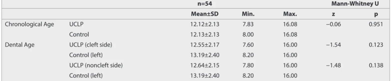
No significant difference was found between the UCLP and con-trol groups in terms of chronological age or between dental age (cleft and noncleft sides) in the UCLP group and left-side dental age in the control group (Table 3).
When age groups were evaluated separately, we found that chronological age was lower than dental age (cleft side and non-cleft side) in the UCLP group aged 7–12 years (p<0.05), where-as chronological age wwhere-as lower than the left and right dental ages in the control group aged 7-12 (p<0.01) and 12–16 years (p<0.05) (Table 4).
Table 1. Distribution of the cleft side by age and sex in individuals with unilateral cleft lip and palate
Age Gender Right Left
UCLP 7-12 Male 4 18
Female 3 4
12-16 Male 4 11
Female 4 6
Total 15 39
Table 3. Comparison of chronological age and dental age in unilateral cleft lip and palate and control groups
n=54 Mann-Whitney U
Mean±SD Min. Max. z p
Chronological Age UCLP 12.12±2.13 7.83 16.08 −0.06 0.951
Control 12.13±2.13 8.00 16.08
Dental Age UCLP (cleft side) 12.55±2.17 7.60 16.00 −1.54 0.123
Control (left) 13.19±2.40 8.20 16.00
UCLP (noncleft side) 12.64±2.15 7.80 16.00 −1.48 0.138
Control (left) 13.19±2.40 8.20 16.00
Min: minimum; Max: maximum; SD: standard deviation (Mann-Whitney U Test)
Table 2. Intragroup comparison of chronological and dental ages in the unilateral cleft lip and palate and control groups
n=54 Kruskall-Wallis H
Mean±SD Min. Max. H p
UCLP Chronological Age 12.12±2.13 7.83 16.08 2.3 0.309
-Dental Age (cleft side) 12.55±2.17 7.60 16.00
Dental Age (noncleft side) 12.64±2.15 7.80 16.00
Control Chronological Age 12.13±2.13 8.00 16.08 9.3 0.009** 1-2
1-3
Dental Age (right) 13.16±2.38 8.20 16.00
Dental Age (left) 13.19±2.40 8.20 16.00
Min: minimum; Max: maximum; SD: standard deviation (Kruskal-Wallis H test; **p<0.01)
Table 4. Intragroup comparison of chronological and dental ages for the 7-12 and 12-16 year age subgroups in the unilateral cleft lip and palate and control groups
n Mean±SD Min. Max. Kruskall-Wallis H
H p
UCLP 7-12 Chronological Age 29 10.44±1.02 7.83 11.83 6.9 0.031* 1-2
1-3
Dental Age (cleft side) 29 11.23±1.77 7.60 14.60
Dental Age (noncleft side) 29 11.26±1.72 7.80 14.60
12-16 Chronological Age 25 14.06±1.22 12.00 16.08 0.246 0.884
-Dental Age (cleft side) 25 14.08±1.50 10.80 16.00
Dental Age (noncleft side) 25 14.24±1.33 11.60 16.00
Control 7-12 Chronological Age 29 10.46±1.03 8.00 11.92 12.03 0.002** 1-2
1-3
Dental Age (right) 29 11.74±2.04 8.20 15.20
Dental Age (left) 29 11.79±2.16 8.20 15.70
12-16 Chronological Age 25 14.06±1.23 12.00 16.08 6.7 0.033* 1-2
1-3
Dental Age (right) 25 14.82±1.53 11.60 16.00
Dental Age (left) 25 14.81±1.48 11.60 16.00
Min: minimum; Max: maximum; SD: standard deviation (Kruskal-Wallis H test; **p<0.01)
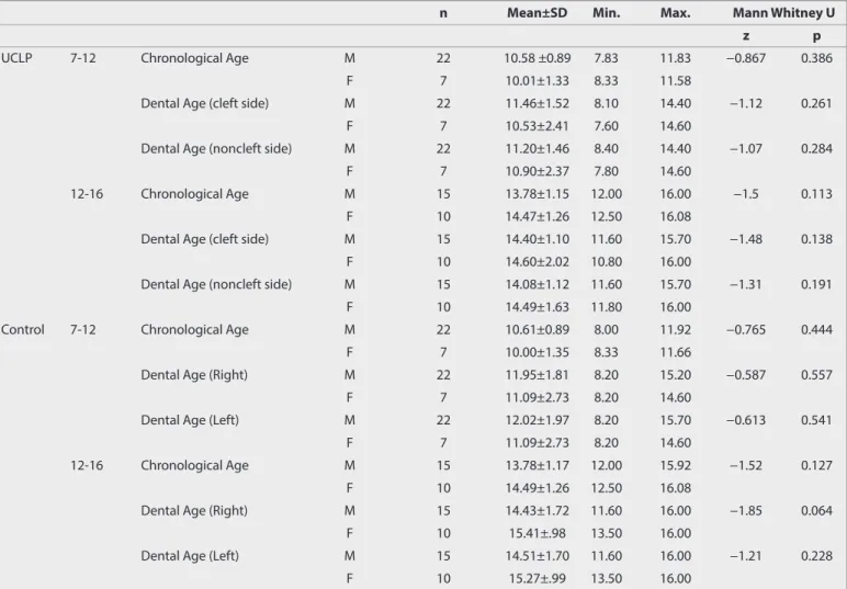
Table 5. Sex-based comparison of chronological age and dental age between the unilateral cleft lip and palate and control groups in individuals aged 7-12 and 12-16 years
n Mean±SD Min. Max. Mann Whitney U
z p
UCLP 7-12 Chronological Age M 22 10.58 ±0.89 7.83 11.83 −0.867 0.386
F 7 10.01±1.33 8.33 11.58
Dental Age (cleft side) M 22 11.46±1.52 8.10 14.40 −1.12 0.261
F 7 10.53±2.41 7.60 14.60
Dental Age (noncleft side) M 22 11.20±1.46 8.40 14.40 −1.07 0.284
F 7 10.90±2.37 7.80 14.60
12-16 Chronological Age M 15 13.78±1.15 12.00 16.00 −1.5 0.113
F 10 14.47±1.26 12.50 16.08
Dental Age (cleft side) M 15 14.40±1.10 11.60 15.70 −1.48 0.138
F 10 14.60±2.02 10.80 16.00
Dental Age (noncleft side) M 15 14.08±1.12 11.60 15.70 −1.31 0.191
F 10 14.49±1.63 11.80 16.00
Control 7-12 Chronological Age M 22 10.61±0.89 8.00 11.92 −0.765 0.444
F 7 10.00±1.35 8.33 11.66
Dental Age (Right) M 22 11.95±1.81 8.20 15.20 −0.587 0.557
F 7 11.09±2.73 8.20 14.60
Dental Age (Left) M 22 12.02±1.97 8.20 15.70 −0.613 0.541
F 7 11.09±2.73 8.20 14.60
12-16 Chronological Age M 15 13.78±1.17 12.00 15.92 −1.52 0.127
F 10 14.49±1.26 12.50 16.08
Dental Age (Right) M 15 14.43±1.72 11.60 16.00 −1.85 0.064
F 10 15.41±.98 13.50 16.00
Dental Age (Left) M 15 14.51±1.70 11.60 16.00 −1.21 0.228
F 10 15.27±.99 13.50 16.00
Min: minimum; Max: maximum; SD: standard deviation; M: male; F: female (Mann- Whitney U test)
106
Sex-based comparisons of the age groups revealed no signifi-cant differences in terms of chronological or dental ages in the UCLP and control groups (Table 5).
DISCUSSION
Although there are many studies in literature that have investi-gated the correlation between CLP and tooth development, to the best of our knowledge, no studies have been conducted in the Turkish population. In general, previous studies have in-dicated that there can be differences between ethnicities with respect to dental age or development or both (7-11). Therefore, our study aimed to assess the correlation between dental age of patients and asymmetric dental development in different age groups and to compare our findings with individuals without CLP.
Although delays in dental development in children with CLP was reported to decrease after the age of nine, several studies have shown that this delay may be greater for permanent teeth in old-er age groups (23, 24). Thold-erefore, we set 12 years as the limit (an inclusion criterion) because permanent dentition is completed by that age. Nonetheless, future studies could be planned to ex-plore different age intervals to reveal more detailed information. Tan et al. (18) investigated dental development in Singaporean children with UCLP and found that dental age was delayed by 0.55 years compared with the control group, and that asymmet-rical dental development was significantly more common in these patients. Asymmetric delays in dental development ob-served in these individuals were primarily attributed to genetic factors, malnourishment, and insufficient space for tooth growth on the cleft side, as well as reduced blood circulation due to fi-brous tissue formation following surgical repair of the cleft and consequent developmental damage to the dental buds. Eerens et al. (16) compared hypodontia, dental age, and asymmetric tooth development between 54 individuals with CLP, 63 of their siblings without cleft, and 250 unrelated individuals without cleft. According to the results of the study, the prevalence of hy-podontia and asymmetric tooth development was significantly higher in individuals with CLP and their siblings than in individ-uals in the control group. No significant difference in dental age was revealed among the three groups. This underlines a close correlation between cleft formation and dental development. In the present study, there was no difference in the dental ages be-tween the right and left sides in individuals with UCLP or those in the control group (Table 2).
Despite the fact that we identified a small degree of dental age retardation on the cleft and noncleft sides in the UCLP group compared with that on the left side in the control group, the dif-ference was not significant (Table 3). Hazza’a et al. (17) reported a significant deviation in dental age between individuals with uni-lateral and biuni-lateral CLP compared with individuals with no cleft. The authors found that the proportion of dental age retardation was significantly greater in males than in females. According to the researchers, environmental and postnatal developmental factors such as nutritional problems, recurrent respiratory
infec-tions, and surgical procedures cause retardation in tooth devel-opment in individuals with CLP. Moreover, many genes related to tooth development have been implicated to the development of craniofacial structures. This suggests that genetic mutations that cause CLP may also cause dental development disorders. Altuğ et al. (25) reported that the frequency of the MSX1 c.*6C>T polymorphism was higher in individuals with CLP and in patients with congenital absence of maxillary lateral teeth than in the normal population; hence, it was speculated that the two condi-tions may be related.
Several studies have reported that delay in dental development may be greater in children with CLP because of the increasing number of missing teeth (13, 21). According to Lai et al. (21), the etiology of hypodontia alone cannot explain dental delays in children with cleft, because delays in tooth formation have been observed in both cleft and noncleft subjects without hy-podontia. Furthermore, Topolski et al. (20) state that the etiologic factors of delayed dental development of individuals with clefts seem to be the same factors responsible for the occurrence of dental anomalies in these individuals, as well as for the manifes-tation of the cleft itself. However, and although the authors did not observe delayed dental development in the individuals with CLP, this study excluded subjects with agenesis in the mandible. Therefore, the notable absence of delayed dental development in our study and the study by Topoplski et al. (20), as opposed to other studies performed in literature, may be explained by this exclusion criterion.
On the other hand, Pöyry et al. (15) evaluated 131 Finnish chil-dren with and without CLP using Demirjian’s method and found that 3-9 year old children had a 6-month delay in tooth devel-opment, a delay that decreased to 2 months in 8–14 year old children. According to their study, a 2-month delay was also detected in children with cleft lip alone, whereas the authors reported 6.5 and 7 months delays in individuals with UCLP and bilateral CLP, respectively. In our study, we divided the UCLP pa-tients into two age groups: 7–12 and 12–16 years, and we did not observe any differences in dental age between the UCLP and control groups (Table 4). Our findings further revealed no associ-ation between dental age and sex in these age groups (Table 5). A recent study has identified retardation of dental age compared with the control group after analyzing the dental age of 108 chil-dren with UCLP using Demirjian’s and Willems’ methods (19). However, both methods revealed discrepancies in determining chronological age, particularly in sex-based comparisons. Although several previous studies have reported delays in den-tal development, we failed to detect a significant difference between UCLP and control subjects (15, 17, 18, 21). In line with our findings, Topolski et al. (20) have also reported no significant retardation in dental age in CLP patients compared to control subjects. Furthermore, the authors underline that the differenc-es observed in their study may be explained by the methodolog-ical design adopted pertaining to the selected sample size, pair-ing of the sample, blindpair-ing, number of examiners and number of radiograph evaluations per examiner. In addition, we believe that one of the main reasons propelling these differences may
be attributed to ethnic and racial differences among study pop-ulations. Although the method developed by Demirjian et al. (4) allows a user-friendly determination of the dental age, their model is based on data obtained from French–Canadian chil-dren. Consequently, this disputes the applicability and reliability of the method in other ethnicities and has triggered a great deal of controversy in current literature (7-11). Studies conducted in Turkey that investigated dental age in different regions have suggested that Demirjian’s method is not a reliable model in the Turkish population. For instance, Özveren & Serindere (11) used the Willems’ and Demirjian’s methods to assess the dental ages of children in the Aegean region and reported that Demirjian’s method was less reliable than the Willems’ method in determin-ing the participants’ dental age. In addition, Sen Tunç & Koyutürk (8) in their study on children from Northern Turkey and Çelikoğlu et al. (9) in their study on children from Eastern Turkey have both stated that Demirjian’s method provided exaggerated results in Turkish children; thus, it may not be suitable for this population. Similarly, we also found that chronological age was lower than the dental age in the control group, and assessments performed in age subgroups showed that chronological age was signifi-cantly lower than dental age in individuals with UCLP aged 7-12 years and in control subjects aged 7-12 and 12-16 years.
One of the main limitations of the present study was the unbal-anced distribution of gender among groups that can be consid-ered as a prevalent confounding factor. The retrospective nature of our study allowed us to use the panoramic radiographs of pa-tients admitted to our hospital. However, data obtained from a more balanced sample could reflect related differences in a more accurate manner. Our study included 108 individuals, 54 with UCLP and 54 healthy controls. Considering the notable statisti-cally non-significant retardation of dental age in individuals with UCLP, further studies with larger study groups that also investi gate individuals with bilateral CLP will be beneficial. In addition, further studies with equal numbers of subjects are required to reach definitive conclusions.
CONCLUSION
Based on the results of this study, there was no asymmetric man-dibular tooth development detected on the cleft or noncleft side in individuals with UCLP. Moreover, we found no differences in the 7-12 year age group, 12-16 year age group, or between the sexes while comparing the dental ages of these individuals with those of individuals without cleft.
Ethics Committee Approval: Ethics committee approval was received for this study from the Ethics Committee of İstanbul Medipol University School of Dentistry (approval number: 543).
Informed Consent: Written informed consent was obtained from the patients who participated in this study.
Peer-review: Externally peer-reviewed.
Author Contributions: Concept - E.C. ; Design - E.C. ; Supervision - A.T.A., A.P.M.; Materials -A.T.A., A.P.M; Data Collection and/or Processing
- E.C., C.A.; Analysis and/or Interpretation - E.C., C.A.; Literature Search - E.C., C.A., A.T.A., A.P.M; Writing Manuscript - E.C.; Critical Review - A.T.A. Conflict of Interest: The authors have no conflict of interest to declare. Financial Disclosure: The authors declared that this study has received
no financial support.
REFERENCES
1. Menezes R, Vieira AR. Dental anomalies as part of the cleft spec-trum. Cleft Palate Craniofac J 2008; 45: 414-9. [CrossRef]
2. Tortora C, Meazzini MC, Garattini G, Brusati R. Prevalence of abnor-malities in dental structure, position, and eruption pattern in a pop-ulation of unilateral and bilateral cleft lip and palate patients. Cleft Palate Craniofac J 2008; 45: 154-62. [CrossRef]
3. Kaul R, Jain P, Saha S, Sarkar S. Cleft lip and cleft palate: Role of a pediatric dentist in its management. Int J Pedod Rehabil 2017; 2: 1-6. [CrossRef]
4. Demirjian A, Goldstein H, Tanner JM. A new system of dental age assessment. Hum Biol 1973; 45: 211-27.
5. Hägg U, Taranger J. Maturation indicators and the pubertal growth spurt. Am J Orthod 1982; 82: 299-309. [CrossRef]
6. Willems G, Van Olmen A, Spiessens B, Carels C. Dental age estima-tion in Belgian children: Demirjian’s technique revisited. J Forensic Sci 2001; 46: 893-5. [CrossRef]
7. Leurs IH, Wattel E, Aartman IH, Etty E, Prahl-Andersen B. Dental age in Dutch children. Eur J Orthod 2005; 27: 309-4. [CrossRef]
8. Sen Tunc E, Koyutürk AE. Dental age assessment using Demirjian’s method on northern Turkish children. Forensic Sci Int 2008; 175: 23-6. [CrossRef]
9. Celikoglu M, Cantekin K, Ceylan I. Dental age assessment: the appli-cability of Demirjian method in eastern Turkish children. J Forensic Sci 2011; 56: 220-2. [CrossRef]
10. Kırzıoğlu Z, Ceyhan D. Accuracy of different dental age estima-tion methods on Turkish children. Forensic Sci Int 2012; 216: 61-7.
[CrossRef]
11. Ozveren N, Serindere G. Comparison of the applicability of Demir-jian and Willems methods for dental age estimation in children from the Thrace region, Turkey. Forensic Sci Int 2018; 285: 38-43.
[CrossRef]
12. Tonge CH. Identification of cell patterns in human tooth differentia-tion. J Dent Res 1967; 46: 876-8. [CrossRef]
13. Ranta R. Comparison of tooth formation in noncleft and cleft affect-ed children with and without hypodontia. ASDC J Dent Child 1982; 49: 197-9.
14. Ranta R. A review of tooth formation in children with cleft lip/pal-ate. Am J Orthod Dentofacial Orthop 1986; 90:11-8. [CrossRef]
15. Pöyry M, Nyström M, Ranta R. Tooth development in children with cleft lip and palate: a longitudinal study from birth to adolescence. Eur J Orthod 1989; 11: 125-30. [CrossRef]
16. Eerens K, Vlietinck R, Heidbüchel K, Van Olmen A, Derom C, Willems G, Carels C. Hypodontia and tooth formation in groups of children with cleft, siblings without cleft, and nonrelated controls. Cleft Pal-ate Craniofac J 2001; 38: 374-8. [CrossRef]
17. Hazza’a AM, Rawashdeh MA, Al-Jamal AL, Al-Nimri KS. Dental de-velopment in children with cleft lip and palate: a comparison be-tween unilateral and bilateral clefts. Eur J Paediatric Dent 2009; 10: 90-4.
18. Tan EL, Yow M, Kuek MC, Wong HC. Dental maturation of uni-lateral cleft lip and palate. Ann Maxillofac Surg 2012; 2: 158-62. [CrossRef]
19. Almotairy N, Pegelow M. Dental age comparison in patients born with unilateral cleft lip and palate to a control sample
ing Demirjian and Willems methods. Eur J Orthod 2018; 40: 74-81.
[CrossRef]
20. Topolski F, de Souza RB, Franco A, Cuoghi OA, da Silva Assunção LR, Fernandes A. Dental development of children and adolescents with cleft lip and palate. Braz J Oral Sci 2014; 13: 319-24. [CrossRef]
21. Lai MC, King NM, Wong HM. Dental development of Chinese children with cleft lip and palate. Cleft Palate Craniofac J 2008; 45: 289-96.
[CrossRef]
22. Cesur E, Altug AT, Toygar-Memikoglu U, Gumru-Celikel D, Tagrikulu B, Erbay E. Assessment of sella turcica area and skeletal maturation
patterns of children with unilateral cleft lip and palate. Orthod Cra-niofac Res 2018; 21: 78-83. [CrossRef]
23. Prahl-Andersen B. The dental development in patients with cleft lip and palate. Trans Eur Orthod Soc 1976; 52: 155-60.
24. Bailit HL, Sung B. Maternal effects on the developing dentition. Arch Oral Biol. 1968; 13: 155-61. [CrossRef]
25. Altug AT, Senol A, Ozkepir NO, Dogan H, Ceylaner S, Ozdiler E. Eval-uation of MSX1 gene as the common candidate gene of nonsyn-dromic congenital hypodontia and cleft lip and palate. J Cleft Lip Palate Craniofac Anomal 2017; 4: 31-7. [CrossRef]