Introduction
A healthy ovulation is critical in terms of health and fer-tility of women in the reproductive period. Whereas, in in-fertility which affects approximately 15% of the couples, one fourth of the problems is caused by the ovulatory fac-tors [1-2]. Clomiphene citrate (CC) is usually used as the first choice for anovulation treatment for a long time. More-over, CC is very inexpensive, easily accessible, and well-tolerated by patients [3-6].
There may be some confounding factors that affect suc-cess in the use of CC such as obesity, alcohol use, and smoking [7]. Smoking is seen in about 25-33% of women in the reproductive age [8, 9]. Active smoking may reduce natural conception by about 80% [10]. Cigarette contains over 4,000 chemicals and it is not clear which of these are particularly effective on ovulation [11, 12]. Furthermore, levels of these toxicants in target organs may be different from their serum levels [13, 14].
Nicotine, one of the most studied components of ciga-rette, partially inhibit follicular growth during all stages [15, 16]. In a study by Mailhes et al. with mouse oocytes, nicotine was reported to increase the rate of aneuploidy in
the in vitro media and to cause premature separation of sis-ter chromatids [17]. Liu et al. also showed high likelihood of aneuploidy due to deterioration of meiosis in bovine oocytes exposed to in vitro nicotine [18].
The effect of nicotine on steroidogenesis may be dose-dependent, such as on oxidative stress [16]. However, steroidogenesis is impaired due to granulosa cell apoptosis produced by nicotine rather than angiogenesis disruption and accordingly, the production of androgen and proges-terone decreases [16, 19, 20]. Although previous studies have shown that smoking may inhibit ovulation because of the impaired angiogenesis [21], Bordel et al. demonstrated that nicotine did not disrupt neovascularization [16].
Some dysplastic changes may develop in the ovary that are a result of excessive proliferation and increased angio-genesis when CC is used for induction of ovulation [22]. In this context, both smoking and CC use can cause syner-gistic dysplastic changes in ovarian tissue and negatively affect follicular growth. Ki-67, the best proliferation marker which can detect these changes [23] and CD34 which is used as a panendothelial cell marker in the assessment of angiogenesis [24] may show the changes occurring in the
Revised manuscript accepted for publication January 11, 2018
7847050 Canada Inc. www.irog.net
Clin. Exp. Obstet. Gynecol. - ISSN: 0390-6663 XLVI, n. 3, 2019
doi: 10.12891/ceog4550.2019
Effects of nicotine exposure on clomiphene citrate induced rats:
morphological and immunohistochemical analysis in the ovaries
N. Karaca1, Y.K. Akpak2, A. Cakır3, M. Marasli1, H. Aktun4
1Department of Obsterics and Gynaecology, School of Medicine, Bezmialem Vakif University, Istanbul
2Department of Obstetrics and Gynaecology, Tepecik Training and Research Hospital, University of Health Sciences, Izmir 3Department of Pathology, School of Medicine, Medipol University Hospital, Istanbul
4Department of Obsterics and Gynaecology, School of Medicine, Medipol University, Istanbul (Turkey)
Summary
Objective: The most common use of ovulation induction in clomiphene citrate (CC) administered rats is to investigate whether there is any morphologically and immunohistochemically difference in nicotine exposure between rats not exposed to nicotine and with no CC. Materials and Methods: A total of 24 healthy rats were randomly divided into three groups: Group 1 was the group that received transdermal nicotine patched followed by clomiphene citrate. Group 2 was the only intraperitoneally CC applied group. Group 3 was the normal saline administered intraperitoneally group. On the fourth day of the cycle animals were sacrificed and bilateral salpingo-oophorectomy was performed. Sections were taken and stained with standard haematoxylin for histopathological examination. For im-munohistochemical evaluation, sections were stained with Ki-67 and CD34. Results: There were no significant differences between the groups in terms of ovarian follicular number and types, and follicle-stroma immunohistochemical staining Ki-67 expression. There was also no significant difference in the thickness of granulosa cell between the groups. However, among the groups, CD34 expressions in group 1 were statistically less in the secondary follicle (p = 0.000), corpus luteum (p = 0.012), and more in the ovarian stroma (p = 0.001). Conclusion: In the CC-stimulated animal model, the authors did not observe that transdermal nicotine exposure was morphologically deleterious to the follicular count and types. They also could not detect the thickness of granulosa cell. Perhaps the effect on over-stim-ulation with CC may be less than expected, depending on the route and dose of nicotine administered.
N. Karaca, Y.K. Akpak, A. Cakır, M. Marasli, H. Aktun
388
ovaries due to both ovulation induction and smoking. Based on this information, the objective of this study is to investigate whether there is any morphological or im-munohistochemical differences between the rats adminis-tered CC exposed to nicotine, and the rats that were not given CC and not exposed to nicotine. In addition, the au-thors aimed to shed light on how follicular growth is af-fected by the complex association between smoking and ovulation induction.
Materials and Methods
This study was conducted at the animal laboratory of the Veterinary Faculty of Bezmialem Foundation University in 2016. The ethics committee of the same university approved the study in June 2016.
A total of 24 healthy rats were used. The reason for choos-ing this type of rats was that they have been used in similar previous studies [25, 26]. All animals weighed 200-250 grams on average and had regular four-day estrous cycles. The ani-mals were kept at 12 hours light/dark cycle at 22°C and they had free access to food and water.
Rats were randomly divided into three groups. Group 1: The group which firstly received nicotine and then CC; a total of eight rats were shaved from the nuchal area and 1 mg/kg/day nicotine band was applied by changing every day for three con-secutive cycles before the use of CC. As demonstrated in ear-lier acute toxicity studies, it has been reported that application of nicotine at this dose is non-toxic [26]. No nicotine-depen-dent side effects were observed in the rats. Then, rats were in-traperitoneally injected with 1 mg/kg/day CC diluted in saline, starting from the first day of the cycle for 3 days. Group 2: The group which received CC only; again, eight rats were in-traperitoneally administered 1 mg/kg/day CC from the first day of the cycle for three days. Group 3: Randomly selected eight rats were intraperitoneally given normal saline as 2.5 mL/kg/ day in the first three days of the cycle for three days.
All rats were killed by 60 mg/kg xilazyne (100 mg/ml) fol-lowing intraperitoneal injection of 2 ml/kg ketamine (50 mg/ml) on the forth day of the cycle. Each rat was laid on its back and bilateral salpingo-oophorectomy was performed. The ovaries were placed in a 10% formol solution and sent to the Medipol University Medical School, Pathology Laboratory for examination.
Ovaries of each material were sampled. The samples were embedded into paraffin blocks after tissue processing. The numbers of total primordial follicles (the follicle where the oocyte is surrounded by with the flattened granulosa cells), pri-mary follicles (the follicle where the oocyte is surrounded by a single layer of granulosa cells), preantral-antral follicles (the follicle surrounded by two or more layers of granulosa cells and containing antral cavity) and the corpus luteum were noted on 4-micron thickness hematoxylin eosin stained sections under light microscope at ×200 magnification. The thicknesses of the granulosa cells in corpus luteum were measured in mi-crometers with microscope using NIS-elements program.
Four micrometer thick ovarian sections were immunohisto-chemically stained with CD34 and Ki67 antibodies using an automated device. Diaminobenzydine was used as a chro-mogen. The Ki67 immunoreactivity was expressed as percent-age (%) in the area in follicles and stroma where it had the highest value. CD34 where the endothelial cells were stained were calculated by counting the vessels at 1 mm2in the
sec-ondary follicle, corpus luteum, and ovarian stroma.
Statistical analyses were performed using SPSS v20. Con-tinuous variables are presented as mean ± standard deviation (SD) or median (range) while categorical variables were ex-pressed as frequencies (%). Multi-group comparisons of con-tinuous variables were performed by one-way ANOVA test, and Dunnett’s post hoc test was run to confirm where the dif-ferences occurred between groups when it was shown an over-all statisticover-ally significant difference in group means. A two tailed p-value < 0.05 was considered statistically significant.
Results
There was no statistically significant difference between the groups in terms of the number of follicular types and the thickness of the granulosa cells in the follicles. In the groups administered CC, the mean number of corpus lu-teum was higher than the groups which did not receive CC, the difference was not statistically significant (p=0.093) (Table1, Figures 1 and 2).
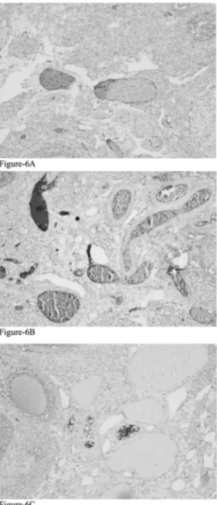
Immunohistochemical examination revealed that, al-though Ki67 expression was higher in the follicles than in the stroma; difference between the groups was not sta-tistically significant (p = 0.68, p = 0.61; respectively) (Figure 3). CD34 expression in the secondary follicles was significantly different among all groups (p = 0.000). In the detailed analysis of the differences between the groups, CD34 expression was found to be significantly lower group 1 compared to both groups 2 (p = 0.008) and 3 (p = 0.000) (Figure 4). There were again statistically significant difference between the groups in terms of CD34 expression in the corpus luteum (p = 0.012). Al-though not statistically significant, CD34 expression in the corpus luteum was found to be lower in group 1 than in group 2 (p = 0.735). However, when compared with the group 3, CD34 expression in the corpus luteum was quite significantly lower in group-1 (p = 0.009) (Figure 5). In addition, differences between the groups in terms of CD34 expressions in the stroma had statistical significance (p = 0.001). Here also group 1 was the group which caused the difference. When groups 1 and 3 were compared, although the difference between them did not reach statistical sig-nificance, stroma showed higher CD34 expression in favour of group 1 (p = 0.572). However, the authors found that CD34 expression in the stroma was statistically sig-nificantly higher in group 1 compared to group 2 (p = 0.001) (Tables 1 and 2, Figure 6).
Discussion
In has been found in the CC-stimulated animal model that previous exposure to transdermal nicotine does not have a significant negative effect on the number of the follicular types in the ovary, and the thickness of the granulosa cells in corpus luteum was not changed by transdermal nicotine band application. When examined immunohistochemically,
it has been determined that the use of nicotine band did not change the expression of Ki67 which is the known best pro-liferation marker. However, it has been found that with the use of transdermal nicotine band, expression of CD34 which is a panendothelial cell marker, is low in the
sec-ondary follicle and corpus luteum but high in the stroma. The ovulation process, one of the most important stages of fertility, is highly sensitive to potentially toxic agents. Smoking may lead to a decrease in the number of ovarian follicles in smokers compared to non-smokers [27]. Dif-Figure 1. — Ovarian sections showing
fol-licles in group 3 (A), group 2 (B), and group 1 (C). (HE×40)
Figure 2. — Granulosa cell thickness in group 3 (A), group 2 (B), and group 1 (C). (HE×40)
Figure 3. — Ki67 expressions in follicles and stroma of the ovaries in group 3 (A), group 2 (B), and group 1 (C). (Ki67×100)
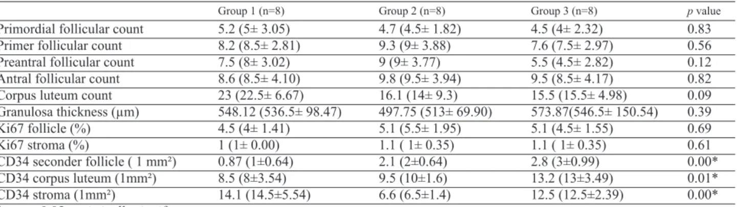
Table 1. — Comparison of follicular and corpus luteum counts, the thicknesses of granulosa cells, and immunohisto-chemical findings among groups.
Group 1 (n=8) Group 2 (n=8) Group 3 (n=8) p value
Primordial follicular count 5.2 (5± 3.05) 4.7 (4.5± 1.82) 4.5 (4± 2.32) 0.83 Primer follicular count 8.2 (8.5± 2.81) 9.3 (9± 3.88) 7.6 (7.5± 2.97) 0.56 Preantral follicular count 7.5 (8± 3.02) 9 (9± 3.77) 5.5 (4.5± 2.82) 0.12 Antral follicular count 8.6 (8.5± 4.10) 9.8 (9.5± 3.94) 9.5 (8.5± 4.17) 0.82 Corpus luteum count 23 (22.5± 6.67) 16.1 (14± 9.3) 15.5 (15.5± 4.98) 0.09 Granulosa thickness (µm) 548.12 (536.5± 98.47) 497.75 (513± 69.90) 573.87(546.5± 150.54) 0.39 Ki67 follicle (%) 4.5 (4± 1.41) 5.1 (5.5± 1.95) 5.1 (4.5± 1.55) 0.69 Ki67 stroma (%) 1 (1± 0.00) 1.1 ( 1± 0.35) 1.1 ( 1± 0.35) 0.61 CD34 seconder follicle ( 1 mm²) 0.87 (1±0.64) 2.1 (2±0.64) 2.8 (3±0.99) 0.00* CD34 corpus luteum (1mm²) 8.5 (8±3.54) 9.5 (10±1.6) 13.2 (13±3.49) 0.01* CD34 stroma (1mm²) 14.1 (14.5±5.54) 6.6 (6.5±1.4) 12.5 (12.5±2.39) 0.00* * = p ≤ 0.05 statistically significant
N. Karaca, Y.K. Akpak, A. Cakır, M. Marasli, H. Aktun
390
ferent animal studies supporting these findings have been conducted. Mohammadghasemi et al. examined follicle numbers by exposing mouse ovaries to nicotine and con-trary to the present study, they reported that the number of follicles, especially other than primordial follicles, were greatly reduced by the effect of nicotine [28]. They noted that this situation might be a result of the apoptotic effect of on the granulosa cells, and thus its negative effect was seen on the more growing follicles. Likewise, Tuttle et al. showed that smoking causes follicular loss in the mouse ovaries [29]. However, unlike this information in the pres-ent study the authors determined that the number and types of follicles were not affected by nicotine exposure. More-over, transdermal nicotine administration did not change the number of growing follicles affected by CC. This may be related to the dose and administration way that the pres-ent authors used in this study. In addition, since thousands of toxic substances (carbon monoxide, cadmium, benza-pyrine, etc.) in the cigarette are not found in the nicotine bands, as reported by studies on nicotine replacement
ther-apy, some of the harmful effects of cigarette smoking re-ported in the literature might not be observed in this study [30].
As we know, nicotine acetylcholine receptors (nAChR-2) have been identified in granulosa cells [31]. Both clinical and animal studies have reported that smoking has delete-rious effects on granulosa cells through these receptors. Freour et al. found that active smoking disrupts granulosa cell function, reducing ovarian reserve and IVF outcomes [27]. Bordel et al. also showed in rats administered subcu-taneous nicotine that dose-dependent apoptosis occurs in granulosa cells. They found that the involvement of anti-PCNA (proliferating cell nuclear antigen) antibody, which they used as an apoptotic cell parameter, was quite higher in rats given high doses of nicotine compared to the rats administered a low dose or no subcutaneous nicotine. Thus, they emphasized the fact that nicotine stimulates the gran-ulosa cell apoptosis in a dose-dependent manner [16]. Al-though the present authors did not use any apoptotic marker in this study, they did not find a detrimental effect of nico-Figure 4. — CD 34 staining in the
sec-ondary follicles of group 3 (A), group 2 (B), and group 1 (C). (CD34 ×200)
Figure 5. — CD34 staining in the corpus luteum of group 3 (A), group 2 (B), and group 1 (C). (CD34×100)
Figure 6. — CD34 expression in the stroma of the ovaries in group 3 (A), group 2 (B), and group 1 (C). (CD34×100)
tine exposure in the thickness of granulosa cells in the cor-pus luteum in the rats they administered CC, possibly due to the dose and adminstration way of nicotine. Furthermore, contrary to previous studies, the present authors determined that the number of follicles and thickness of granulosa cells did not change also in rats with ovaries induced with CC alone. Chaube et al. reported that CC treatment caused atre-sia-like changes in rat ovaries. They indicated that this atresic effect caused the disruption of steroidogenesis in granulosa cells by CC [32]. Another study conducted by the same group one year later showed that atresic effects in granulosa cells were less when estradiol was co-adminis-tered with CC [33]. However, CC doses used by these au-thors in rats were 10 mg/kg in both studies, which is quite high compared to the dose used in the present study. In the present study, granulosa cells were not affected in both groups, possibly due to the dose we applied.
Ki67 antigen, one of the known most important markers to show cell proliferation, is expressed in all active phases of the cell cycle [23]. Therefore, it shows a good correlation with the mitotic indices of the cells [34]. In the present study, they examined this expression to investigate whether nicotine bands had any effect on cell proliferation. The au-thors were determined that nicotine did not affect Ki-67 ex-pression in both the stroma and follicles of the ovary. However, Bordel et al. reported inhibition at every phase of follicular growth, especially in small-diameter follicles [16]. Although nicotine doses were the same with the pres-ent study, in that study subcutaneous administration way
was preferred. The authors already reported in their studies that this administration method is not applicable for nor-mal nicotine exposure and should be taken into account in interpreting the results [16]. In addition, Bordel et al. used PCNA as the marker of cell proliferation, different from the present. In the present study, Ki-67 expression, which the authors used as a proliferation marker, was not different be-tween the groups, and might be due to the way the nicotine was administered. Interestingly, Ki67 expressions that were not statistically significant compared to the control group were determined in the present group-2. However, Lima et al. reported that Ki67 expressions after stimulation with CC were significantly higher than the control groups with more marked at the stromal levels [35]. Whereas, the dose and administration way of CC were the same in the present and their studies. But in their study, both carried out CC stimu-lation for two days and used 100 IU / kg hCG afterwards. Perhaps, rather than CC, what caused the changes was hCG effect or combined use of CC and hCG.
CD34 is a panendothelial cell marker specific to usually pre-existing vascular structures [34, 36]. Therefore, CD34 is of great importance in assessment of angiogenesis char-acterized by the formation of new capillaries in existing blood vessels. Since the physiological events such as fol-liculogenesis and corpus luteum formation in the female reproductive system are associated with angiogenesis, CD34 may provide important information of the ovary [37]. Smoking may have harmful effects by influencing the nAChR receptors on the endothelium and decreases the Table 2. — Post-hoc analysis of groups and variables.
Dependant variable Groups Std. Error 95% CI p value
Primordial follicular count Group-3 Group-1 -0.75 ア 1.229 -3.66 2.16 0.769 Group-2 Group-1 -0.50 ア 1.229 -3.41 2.41 0.888 Primer follicular count Group-3 Group-1 -0.62 ア 1.630 -4.48 3.23 0.899
Group-2 Group-1 1.12 ア 1.630 -2.73 4.98 0.717 Preantral follicular count Group-3 Group-1 -2.00 ア 1.618 -5.83 1.83 0.375
Group-2 Group-1 1.50 ア 1.618 -2.33 5.33 0.560 Antral follicular count Group-3 Group-1 0.87 ア 2.037 -3.95 5.70 0.876
Group-2 Group-1 1.25 ア 2.037 -3.57 6.07 0.767
Corpus luteum count Group-3 Group-1 -7.50 ア 3.607 -16.04 1.04 0.089
Group-2 Group-1 -6.87 ア 3.607 -15.42 1.67 0.124 Granulosa cell thickness (µm) Group-3 Group-1 25.75 ア 55.71 -106.30 157.80 0.858
Group-2 Group-1 -50.37 ア 55.71 -182.42 81.67 0.575
Ki67 follicle (%) Group-3 Group-1 0.62 ア 0.82 -1.34 2.59 0.675
Group-2 Group-1 0.62 ア 0.82 -1.34 2.59 0.675
Ki67 stroma (%) Group-3 Group-1 0.12 ア 0.14 -0.21 0.46 0.600
Group-2 Group-1 0.12 ア 0.14 -0.21 0.46 0.600 CD34 second follicle (1 mm²) Group-3 Group-1 2.00 ア 0.38 1.08 2.91 0.000*
Group-2 Group-1 1.25 ア 0.38 0.33 2.16 0.008* CD34 corpus luteum (1 mm²) Group-3 Group-1 4.74 ア 1.50 1.17 8.32 0.009*
Group-2 Group-1 1.00 ア 1.50 -2.57 4.57 0.735 CD34 stroma (1 mm²) Group-3 Group-1 -1.62 ア 1.78 -5.86 2.61 0.572
Group-2 Group-1 -7.50 ア 1.78 -11.73 -3.26 0.001* * = p ≤ 0.05 statistically significant
N. Karaca, Y.K. Akpak, A. Cakır, M. Marasli, H. Aktun
392
numbers of vessels in the corpus luteum and follicles [38]. In the present study also CD34 expression was significantly decreased in the secondary follicle and corpus luteum of the rats which received transdermal nicotine compared to those did not receive it. The harmful effects of nicotine on normal folliculogenesis were also observed during CC in-duction. However, Bordel et al. reported that examining the follicular structures with microcirculation analyses after subcutaneous administration of nicotine at the same dose with that of the present authors, reported that this applica-tion had no significant effect on new vascular formaapplica-tion [16]. In fact, these different results might be due to the an-giogenesis assessment method of that study that differed from the present. In addition, the present authors found that CD34 expression in the ovarian stroma was significantly higher in the rats given transdermal nicotine compared to those that did not receive it. As pointed out in previous stud-ies, this supports the fact that it has a dose-dependent an-giogenesis effect which is associated with microenvironment in the different regions of the ovary [16, 39].
In conclusion, it can be stated that the number of folli-cles was not affected, but the angiogenesis in the follifolli-cles was suppressed in the ovaries of the rats exposed to trans-dermal nicotine prior to over-stimulation with CC. How-ever, the fact that thickness of granulosa cells in the corpus luteum was not affected by nicotine exposure may indicate that nicotine may have a more limited effect than expected during CC over-stimulation, perhaps due to the nicotine dose and the way it is administered. Finally, it can be stated that this study may shed a different light in explaining the complex relations between nicotine and ovarian function during stimulation.
References
[1] Chandra A., Copen C.E., Stephen E.H.: “Infertility and impaired fe-cundity in the United States, 1982–2010: data from the National Sur-vey of Family Growth”. Natl. Health Stat. Rep., 2013, 67, 1. [2] Practice Committee of American Society for Reproductive
Medi-cine: “Diagnostic evaluation of the infertile female: a committee opinion”. Fertil Steril., 2012, 98, 302.
[3] National Collaborating Centre for Women’s and Children’s Health: “Fertility: assessment and treatment for people with fertility prob-lems. London, United Kingdom: National Institute for Health and Clinical Excellence (NICE), February 2013, 1, 63. (Clinical guide-line no. 156).
[4] Veltman-Verhulst S.M., Cohlen B.J., Hughes E., Heineman M.J.: “Intra-uterine insemination for unexplained subfertility”. Cochrane
Database Syst. Rev., 2012, 9, CD001838.
[5] Vause T.D., Cheung A.P., Sierra S., Claman P., Graham J., Guillemin J.A., Society of Obstetricians and Gynecologists of Canada: “Ovu-lation induction in polycystic ovary syndrome”. Int. J. Gynaecol.
Obstet., 2010, 111, 95.
[6] Barbieri R.L.: “Infertility”. In: Yen S.S.C., Jaffe R.B., Barbieri R.L., (eds). Reproductive endocrinology, physiology, pathophysiology, and
clinical management. 4thed. Philadelphia, PA: WB Saunders, 1999,
562.
[7] Homan G., Litt J., Norman R.J.: “The FAST study: fertility
assess-ment and advice targeting lifestyle choices and behaviours: a pilot study”. Hum. Reprod., 2012, 27, 2396.
[8] Huisman M., Kunst A.E., Mackenbach J.P.: “Educational inequalities in smoking among men and women aged 16 years and older in 11 European countries:”. Tob. Control, 2005, 14, 106.
[9] CDC, CfDCaP.: “Smoking prevalence among women of reproduc-tive age—United States, 2006”. Morb. Mortal. Wkly. Rep., 2008, 57, 849.
[10] Hughes E.G., Brennan B.G.: “Does cigarette smoking impair natu-ral or assisted fecundity?” Fertil. Steril., 1996, 66, 679.
[11] Benedict M.D., Missmer S.A., Vahratian A., Berry K.F., Vitonis A.F., Cramer D.W., Meeker J.D.: “Secondhand tobacco smoke exposure is associated with increased risk of failed implantation and reduced IVF success”. Hum. Reprod., 2011, 26, 2525.
[12] Lindbohm M.L., Sallmen M., Taskinen H.: “Effects of exposure to environmental tobacco smoke on reproductive health”. Scand. J.
Work Environ. Health, 2002, 28, 84.
[13] McLachlan J.A., Dames N.M., Sieber S.M., Fabro S.: “Accumula-tion of nicotine in the uterine fluid of the six-day pregnant rabbit”.
Fertil. Steril., 1976, 27, 1204.
[14] Paszkowski T.: “Concentration gradient of cotinine between blood serum and preovulatory follicular fluid”. Ginekol. Pol., 1998, 69, 1131. [In Polish].
[15] Dwyer J.B., Broide R.S., Leslie F.M.: “Nicotine and brain develop-ment”. Birth Defects Res. C. Embryo Today, 2008, 84, 30. [16] Bordel R., Laschke M.W., Menger M.D., Vollmar B.: “Nicotine does
not affect vascularization but inhibits growth of freely transplanted ovarian follicles by inducing granulosa cell apoptosis”. Hum.
Re-prod., 2006, 21, 610.
[17] Mailhes J.B., Young D., Caldito G., London S.N.: “Sensitivity of mouse oocytes to nicotine-induced perturbations during oocyte mei-otic maturation and aneuploidy in vivo and in vitro”. Mol. Hum.
Re-prod., 2000, 6, 232.
[18] Liu Y., Li G.P., Sessions B.R., Rickords L.F., White K.L., Bunch T.D.: “Nicotine induces multinuclear formation and causes aberrant em-bryonic development in bovine”. Mol. Reprod. Dev., 2008, 75, 801. [19] Vrsanska S., Nagyova E., Mlynarcikova A., Fickova M., Kolena J.: “Components of cigarette smoke inhibit expansion of oocyte-cumu-lus complexes from porcine follicles”. Physiol. Res., 2003, 52, 383. [20] Sanders S.R., Cuneo S.P., Turzillo A.M.: “Effects of nicotine and co-tinine on bovine theca interna and granulosa cells”. Reprod.
Toxi-col., 2002, 16, 795.
[21] Shiverick K.T., Salafia C.: “Cigarette smoking and pregnancy I: ovarian, uterine and placental effects”. Placenta, 1999, 20, 265. [22] Lacoste C.R., Clemenson A., Lima S., Lecointre R., Peoc'h M.,
Chene G.: “Tubo-ovarian dysplasia in relationship with ovulation in-duction in rats”. Fertil. Steril., 2013, 99, 1768.
[23] Gerdes J.: “Ki-67 and other proliferation markers useful for im-munohistological diagnostic and prognostic evaluations in human malignancies”. Semin. Cancer Biol., 1990, 1, 199.
[24] Weidner N.: “Intratumor microvessel density as a prognostic factor in cancer”. Am. J. Pathol., 1995, 147, 9.
[25] Akpak Y.K., Çekmez Y., Erdoğan Çakır A., Karaca N., Batmaz G., Gülşen S., Tuştaş Haberal E.: “An animal model of effects of nico-tine exposure on endometrial receptivity and embryo implantation in pregnancy”. J. Matern. Fetal Neonatal Med., 2016, 20, 1. [26] Mohsenzadeh Y., Rahmani A., Cheraghi J., Pyrani M., Asadollahi
K.: “Prenatal exposure to nicotine in pregnant rat increased inflam-matory marker in newborn rat”. Mediators Inflamm., 2014, 2014, 274048.
[27] Freour T., Masson D., Mirallie S., Jean M., Bach K., Dejoie T., Bar-riere P.: “Active smoking compromises IVF outcome and affects ovarian reserve”. Reprod. Biomed Online, 2008, 16, 96.
[28] Mohammadghasemi F., Jahromi S.K., Hajizadeh H., Homafar M.A., Saadat N.: “The Protective Effects of Exogenous Melatonin on Nico-tine-induced Changes in Mouse Ovarian Follicles”. J. Reprod.
In-fertil., 2012, 13, 143.
fol-licle loss in mice ovaries at concentrations representative of human exposure”. Hum. Reprod., 2009, 24, 1452.
[30] Hukkanen J., Jacob P., Benowitz N.L.: “Metabolism and disposition kinetics of nicotine”. Pharmacol. Rev., 2005, 57, 79.
[31] Petrik J.J., Gerstein H.C., Cesta C.E., Kellenberger L.D., Alfaidy N., Holloway A.C.: “Effects of rosiglitazone on ovarian function and fertility in animals with reduced fertility following fetal and neona-tal exposure to nicotine”. Endocrine, 2009, 36, 281.
[32] Chaube S.K., Prasad P.V., Thakur S.C., Srivastava T.G.: “Estradiol protects clomiphene citrate induced apoptosis in rat ovarian follicles and superovulated cumulus oocyte complexes”. Fertil. Steril., 2005,
84, 1163.
[33] Chaube S.K., Prasad P.V., Tripathi V., Phil. M., Shrivastav T.G.: “Clomiphene citrate inhibits gonadotropin-induced ovulation by re-ducing cyclic adenosine 3=,5=-cyclic monophosphate and prostaglandin E2 levels in rat ovary”. Fertil. Steril., 2006, 86, 1106. [34] Celik O., Hascalik S., Elter K., Tagluk M.E., Gurates B., Aydin N.E.:
“Combating endometriosis by blocking proteasome and nuclear fac-tor-kB pathways”. Hum. Reprod., 2008, 23, 2458.
[35] Lima S., Clemenson A., Trombert B., Lecointre R., Lacoste C.R., Peoc’h M., Chene G.: “Morphological and immunohisto-chemical analysis in ovaries and fallopian tubes of tamoxifen, letro-zole and clomiphene-treated rats”. Arch. Gynecol. Obstet., 2014, 290, 553.
[36] Czekierdowska S., Stachowicz N., Chróściel M., Czekierdowski A.: “Proliferation and maturation of intratumoral blood vessels in women with malignant ovarian tumors assessed with cancer stem cells marker nestin and platelet derived growth factor PDGF-B”.
Ginekol. Pol., 2017, 88, 120.
[37] Tamanini C., De Ambrogi M.: “Angiogenesis in developing follicle and corpus luteum”. Reprod. Domest. Anim., 2004, 39, 206. [38] Magers T., Talbot P., Dıcarlantonıo G., Knoll M., Demers D., Tsaı I.,
Hoodbhoy T.: “Cigarette smoke inhalation affects the reproductive sys-tem of female hamsters”. Reprod. Toxicol., 1995, 9, 513.
[39] Villablanca A.C.: “Nicotine stimulates DNA synthesis and prolifer-ation in vascular endothelial cells in vitro”. J. Appl. Physiol., 1998, 84, 2089.
Corresponding Author: N. KARACA, M.D.
Department of Obstetrics and Gynaecology Bezmilaem University, School of Medicine Istanbul, 34200 (Turkey)