Could there be an association between chronic brucellosis and endothelial
damage?
Turhan Togan1, Ozgur Ciftci2, Hale Turan1, Huseyin Narci3, Hakan Gullu2, Hande Arslan1
1 Department of Infectious Diseases and Clinical Microbiology, Faculty of Medicine, Baskent University, Ankara, Turkey
2 Department of Cardiology, Faculty of Medicine, Baskent University, Ankara, Turkey 3 Department of Emergency Faculty of Medicine, Baskent University, Ankara, Turkey
Abstract
Introduction: In this study, we examined the effects of Brucella infection on endothelial dysfunction. Flow-mediated dilatation (FMD) measurement is indicator of the endothelial function, and abnormal values indicating endothelial dysfunction are accepted as the first stage of atherosclerosis.
Methodology: Twenty-four patients who had been treated for acute brucellosis two years before, and who had had no relapses in the follow-up, were prospectively included in the study, along with 30 healthy individuals in the control group. Results: While the highly sensitive C-reactive protein (hs-CRP) value was 2.42 ± 1.45 in the patient group, it was 1.72 ± 0.61 in the control group (p = 0.025). While the FMD value was 3.50 ± 1.58 in the patient group, it was 5.88 ± 1.88 in the control group (p < 0.001). While the percentage increase in FMD was 9.88 ± 4.92 in the patient group, it was 17.49 ± 6.3 in the control group (p < 0.001). It was observed that FMD value, the percentage increase in FMD, and basal radius were correlated with hs-CRP (r = -0.644, p < 0.001; r = - 0.558, p = 0.002; r = 0.444, p = 0.018, respectively). The carotid artery intima media thickness (IMT) value was found to be 0.61 ± 0.17 in the patient group and 0.49 ± 0.12 in the control group (p = 0.004).
Conclusions: The abnormal FMD and IMT values observed in brucellosis patients might be an indicator of more frequent arterial dysfunction, increased cardiovascular risk, and atherosclerosis.
Key words:brucellosis; atherosclerosis; endothelial dysfunction; hs-CRP.
J Infect Dev Ctries 2015; 9(1):048-054. doi:10.3855/jidc.4345 (Received 22 October 2013 – Accepted 07 October 2014)
Copyright © 2015 Togan et al. This is an open-access article distributed under the Creative Commons Attribution License, which permits unrestricted use, distribution, and reproduction in any medium, provided the original work is properly cited.
Introduction
Brucellosis is a zoonotic disease, frequently encountered in developing countries, that constitutes an important public health problem [1]. It can infect people especially through contaminated meat, milk, or dairy products, as well as through direct contact with the excrements and secretions of infected animals [2].
Brucella are small, Gram-negative and facultative,
intracellular, pathogenous bacteria that invade mononuclear phagositic system cells to increase in them [3]. Brucellosis is an inflammatory disease that may involve any organ or system in the body [4]. It can lead to serious cardiovascular complications by affecting endothelial cells and provoking a strong inflammatory response. The secretion of adhesion molecules and proinflammatory chemokines during a
Brucella infection plays an important role in the
activation of the endothelial system. This inflammatory response, as well as the immunological
reactions that develop against the Brucella antigens in the vessel wall, are responsible for the pathogenesis of the damage that results in the vascular system [5]. The dysfunction observed in the endothelial system is one of the first changes that occur in the pathogenesis of cardiovascular diseases, and is encountered in various clinical conditions [6]. The measurement technique of flow-mediated dilatation (FMD) has been developed in the last decade to assess the endothelium-dependent function of the vascular system following an occlusion in the brachial artery.
Nitric oxide (NO) is secreted and vasodilatation develops as a result of the stimulation of the endothelium, a situation that is considered to be a sign of vasomotor function. Ultrasound imaging of the reactive hyperaemia in the brachial artery is used to calculate the endothelium-dependent vasomotor function [7].
The state of the carotid arteries gives an idea about the state of the coronary arteries. In most of the patients with carotid artery obstruction, a stenosis is detected in the coronary arteries as well. For this reason, it is possible to get an idea about atherosclerosis through IMT measurement, a non-invasive and highly reproducible technique [8,9]. This means that the atherosclerotic changes in the carotid artery constitute an indicator of changes in the coronary artery as well. Carotid IMT measurement is recommended for people over the age of 45, and gives an idea about the risk of heart attack, coronary artery disease, or stroke that could possibly occur in this age group [10,11].
Abnormal endothelium-dependent vasodilatation is a diffuse process; the removal of the protective effect provided against atherosclerosis by a normal endothelium leads to abnormalities in the regulation of the vascular tonus. Moreover, this situation might be a sign of increased risk of cardiovascular disease. Endothelial dysfunction develops as the first stage of the atherosclerotic process [12].
A tight association between brucellosis and cardiovascular diseases is already known. Indeed, cardiovascular involvement is the main cause of mortality due to infection with Brucella spp., and most commonly manifests as endocarditis, peripheral and cerebrovascular aneurysms, or vascular complications such as arterial and venous thromboses. In this study, we investigated the effect of brucellosis in atherosclerosis progression. By using FMD and IMT results, we attempted to investigate the long-term effects of brucellosis on the endothelial functions of patients who have had this disease. In the literature, we did not encounter any previous studies dealing with the long-term links of brucellosis with endothelial dysfunction and the development of atherosclerosis. Methodology
Study protocol
This prospective randomized clinical study was conducted in accordance with the Ethical Principles for Medical Research Involving Human Subjects as presented in the Declaration of Helsinki. It was carried out in the Application and Research Center of Baskent University, Konya, Turkey, after approval of the ethics board of the university was received. A total of 24 patients (15 males and 9 females) who had been treated for acute brucellosis two years before, and who had had no relapses in the follow-up, were prospectively included in the study, along with 30 healthy individuals (20 males and 10 females) in the
control group. Patients between 18 and 50 years of age were included in the study between April 2010 and August 2012. After a full medical history was obtained for all patients, they were physically examined and their body mass indices (BMIs) were calculated. The exclusion criteria included a history of alcohol use, smoking, coronary artery disease in family anamnesis, intravenous drug addiction, pregnancy, use of antioxidants, hypertension, hyperlipidemia, diabetes mellitus, kidney or liver disease, rheumatological diseases, lung disease, and active infection.
Laboratory analysis
In the study, blood samples were received from the patients as well as from the control group to measure fasting blood sugar, alanine transaminase (ALT), creatinine, hemogram, highly sensitive C-reactive protein (hs-CRP), and the lipid profile, which comprised the total cholesterol, high-density lipoprotein (HDL), low density lipoprotein (LDL), and triglyceride levels. The blood samples were obtained after a night of fasting. They were collected in empty tubes and immediately stored on ice at 4°C. Following this, the serum samples were separated from the blood cells by being placed in the centrifuge at 3,000 rpm for 10 minutes. The diagnoses of brucellosis were made by standard tube agglutination test. A possible correlation between the percentage increase in FMD and IMT was sought on the one hand, and between BMI, systolic and diastolic blood pressure, fasting blood sugar, and leukocyte count on the other.
Brachial artery measurements
FMD measurements were made in both groups. FMD measurement is an indicator of the endothelial function in the brachial artery, and abnormal values indicating endothelial dysfunction are accepted as the first stage of atherosclerosis.
In this study, a high-resolution 7.5-MHz line-array ultrasound transducer (attached to a Hitachi EUB 6500, Osaka, Japan) was used. All the measurements were made by a specialist who was uninformed about the clinical data. The patients lay prone in a silent room with an interior temperature of 22°C. All the patients were prohibited from having drinks with caffeine and from smoking in the last two hours preceding the measurements.
For the imaging of the brachial artery, each patient’s arm was immobilized in a relaxed, extended position, and the artery was scanned longitudinally 3– 5 cm above the antecubital fossa.
The gain and depth adjustments were optimized to identify the lumen-vessel wall interface. After the transducer was placed in position, the skin area was marked as a reference point for later adjustments and the arm was held in the same position. Once all the measurements were completed, the inner diameter of the brachial artery was calculated, taking into account the QRS complex at the end of diastole; the average of the three consecutive measurements obtained during the consecutive cardiac cycle was also calculated and recorded. The basal measurements of the brachial artery were recorded. The sleeve was then placed on the proximal section of the brachial artery. The sleeve was inflated up to a pressure of 200 mm Hg (or up to 50 mm Hg above the diastolic blood pressure), and held at that pressure for a duration of five minutes, inducing ischemia in the forearm.
After this, the sleeve was deflated and the arterial diameter was measured 60 seconds after deflation. All these measurements were then recorded in a cassette for later analysis. The measurements were repeated while the diameter was greatest. The difference in percentage between the diameter measured after reactive hyperaemia and the basal diameter was taken as the FMD (FMD = 100 x [diameter after reactive hyperaemia - basal diameter] / basal diameter).
The data was independently analyzed by two specialists at the end of the study. The intra-observer variabilities for the measurements of brachial artery diameter and flow remained below 5%. The intra-observer intraclass correlation coefficient for the FMD value was 0.912. Endothelium-dependent dilatation was expressed in the form of a percentage change in the diameter of the brachial artery from baseline to post-reactive hyperaemia.
The measurement of carotid IMT
Carotid IMT was measured in this study by a previously described method, i.e., by means of high-resolution, two-dimensional ultrasound images obtained with a 7.5 MHz linear-array transducer attached to an ultrasound machine (Hitachi EUB 6500, Osaka, Japan) [8]. While the subject lay in a supine position, longitudinal scanning was carried out from the common carotid artery to the bifurcation point. After confirming the bifurcation of the common carotid artery, the measurement of carotid IMT was performed from the far wall of the right carotid artery within 10 mm proximal to the bifurcation. On each scan, three points were measured, synchronized with the R-wave peaks on the electrocardiogram (ECG) to avoid errors that might possibly occur as a result of
variable arterial compliance. Mean carotid IMT was calculated using the six measurements made from two scans. All the measurements were performed by a researcher who was not informed about the subjects’ clinical data. For offline analysis, the ultrasound images obtained were recorded on an S-VHS videotape. The intraobserver intraclass correlation coefficient for the measurement of carotid IMT was 0.937, and the interobserver coefficient was 0.912.
Statistical analysis
Statistical analysis was done using SPSS software version 10. The t-test was used for the independent samplings, and the Chi-square test was used for the continuous variables. In comparing the FMD values, the Mann-Whitney U test was used for the non-homogenously distributed data. The numerical values were expressed in the form of mean ± standard deviation (SD). Pearson’s test was used to determine the correlation between FMD, IMT, and the other variables. The differences were accepted as statistically significant whenever p < 0.05.
Results
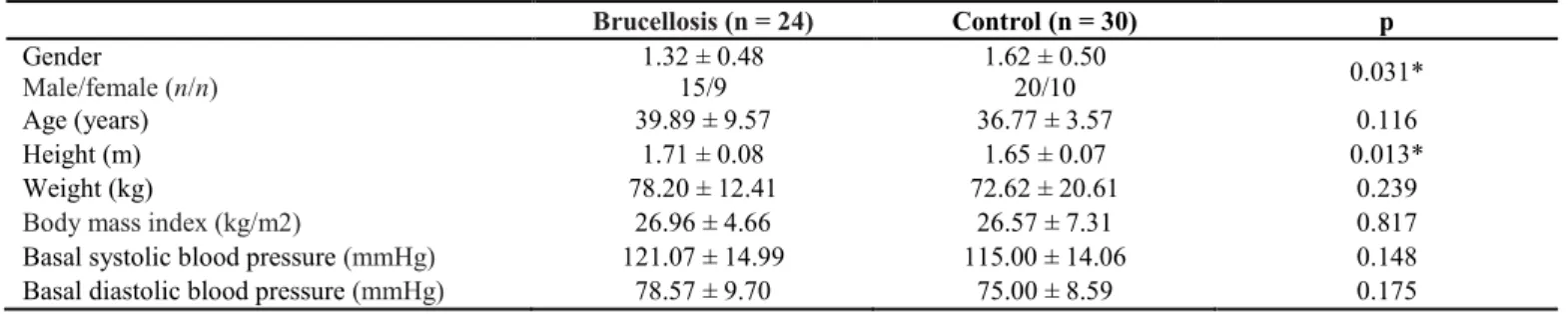
In this study, 54 patients were assessed; 24 of them had brucellosis and the remaining 30 were in the control group. There was no statistically significant difference (p > 0.05) in terms of gender or age between the patients in the brucellosis group and those in the control group (Table 1).
Similarly, there was no statistically significant difference between the two groups in terms of the total cholesterol, glucose, creatinine, ALT, LDL, HDL, BMI, basal systolic blood pressure, and ejection fraction (EF) values (Tables 1 and 2). Again, no statistically significant difference was found between the brucellosis and control group patients as far as leukocyte cell distribution (neutrophils, lymphocytes, and monocytes) was concerned.
As for the statistically significant variables, the hs-CRP value was 2.42 ± 1.45 in the patient group, while it was 1.72 ± 0.61 in the control group (p = 0.025). Similarly, the triglyceride value was 156.64 ± 63.89 in the patient group, while it was 101.73 ± 48.96 in the control group (p = 0.001). The HDL value was 40.68 ± 9.56 in the patient group, while it was 47.65 ± 10.05 in the control group (p = 0.012). The height value was 1.71 ± 0.08 in the patient group, while it was 1.65 ± 0.07 in the control group (p = 0.013). The FMD value was 3.50 ± 1.58 in the patient group, while it was 5.88 ± 1.88 in the control group (p < 0.001). The percentage increase in FMD was 9.88 ± 4.92 in the
patient group, while it was 17.49 ± 6.37 in the control group (p < 0.001). The IMT value turned out to be 0.61 ± 0.17 in the patient group, and 0.49 ± 0.12 in the control group (p = 0.004). All the data were found to be statistically significant (Tables 1 and 2). Furthermore, the FMD value, the percentage increase in FMD, and the basal radius were found to be correlated with hsCRP (r = 0.644, p < 0.001; r = -0.558, p = 0.002; r = -0.444, p = 0.018, respectively). The IMT values were also highly correlated with hs-CRP (r = 0.466, p = 0.014).
Discussion
There is increasing evidence of the existence of a link between infection/inflammation and atherosclerosis [16]. Previous studies have focused on
infections with Chlamydia pneumoniae,
Cytomegalovirus, herpes simplex virus, Helicobacter pylori, and periodontitis in this context [13,14]. In
contrast to these, various other studies have challenged the causal role of infectious agents in atherogenesis [15,16]. What current evidence does suggest, however, is that atherosclerosis may indeed develop in response to inflammatory stimuli. Accordingly, common or
uncommon infections could well represent a risk factor in this regard. Some of the mechanisms that might be involved in cases of atherogenesis induced by infectious agents include a local increase of proinflammatory cells, a local effusion of endotoxins, an autoimmune reaction, a systemic cytokine release, or changes in the lipid metabolism [17]. The cytokine-induced changes that take place in the lipid and lipoprotein metabolisms on account of infection and inflammation are similar [18].
An inflammatory response or an immune reaction against the Brucella antigens on the vessel wall have come under consideration as possible mechanisms in the pathogenesis of vessel involvement. The activation of the endothelium in response to the Brucella infection, as a result of the stimulation of the secretion of adhesion molecules and proinflammatory chemokines, may also play a role in the pathogenesis. Brucellosis may lead to cardiovascular complications, affecting the endothelial cells and provoking a strong inflammatory response [5].
In the development of atherogenesis, arterial endothelial dysfunction is one of the early-stage events leading to the development of structural Table 1. Demographic characteristics of the patients
Brucellosis (n = 24) Control (n = 30) p Gender Male/female (n/n) 1.32 ± 0.48 15/9 1.62 ± 0.50 20/10 0.031* Age (years) 39.89 ± 9.57 36.77 ± 3.57 0.116 Height (m) 1.71 ± 0.08 1.65 ± 0.07 0.013* Weight (kg) 78.20 ± 12.41 72.62 ± 20.61 0.239
Body mass index (kg/m2) 26.96 ± 4.66 26.57 ± 7.31 0.817
Basal systolic blood pressure (mmHg) 121.07 ± 14.99 115.00 ± 14.06 0.148
Basal diastolic blood pressure (mmHg) 78.57 ± 9.70 75.00 ± 8.59 0.175
Table 2. Patients’ laboratory and echochardiographic results
Brucellosis (n = 24) Control (n = 30) p Total cholesterol (mg/dL) 182.25 ± 34.65 173.81 ± 26.06 0.314 Triglyceride (mg/dL) 156.64 ± 63.89 101.73 ± 48.96 0.001* HDL (mg/dL) 40.68 ± 9.56 47.65 ± 10.05 0.012* LDL (mg/dL) 110.39 ± 31.07 109.54 ± 23.60 0.909 Glucose (mg/dL) 98.46 ± 13.99 94.04 ± 6.62 0.141 Creatinine (mg/dL) 0.79 ± 0.12 0.80 ± 0.13 0.639 ALT (mg/dL) 26.14 ± 17.53 19.85 ± 11.01 0.118 Hs-CRP (mg/L) 2.42 ± 1.45 1.72 ± 0.61 0.025* FMD 3.50 ± 1.58 5.88 ± 1.88 < 0.001* IMT 0.61 ± 0.17 0.49 ± 0.12 0.004* FMD (%) 9.88 ± 4.92 17.49 ± 6.37 < 0.001* Basal diameter 36.64 ± 5.70 34.19 ± 4.23 0.078 Peak diameter 40.14 ± 5.78 40.08 ± 4.60 0.963 EF (%) 67.03 ± 8.86 68.38 ± 4.69 0.490
ALT: alanine aminotransferase; EF: ejection fraction; FMD: flow-mediated dilatation; HDL: high-density lipoprotein; hs-CRP: high-sensitivity C-reactive protein; IMT: carotid intima media thickness; LDL: low-density lipoprotein.
atherosclerotic changes. This predisposition to vasoconstriction and/or thrombosis is important for later development of obstructive atherosclerosis [19].
Endothelium-dependent vasodilatation is realized through vasodilator agents such as NO. Abnormal NO activity is linked to atherosclerosis and vasculopathy [20]. The changes in NO levels may be determined through the measurement of the nitrite/nitrate levels in the plasma or indirectly through a non-invasive technique such as FMD measurement [21]. This measurement determines the vasodilator reaction linked to the production of endothelial NO that develops in reaction to the increased shear stress. FMD is used to detect endothelial dysfunction in cardiovascular diseases as well as in the presence of the risk factors of such diseases [7,22]. IMT is measured by FMD. It has been shown in previous studies that IMT is correlated with FMD, which is a good indicator of the endothelial function [23].
Carotid IMT can be considered a general measure of the degree of severity of atherosclerosis, and increased IMT is associated with generalized atherosclerosis [24]. The link between carotid IMT and coronary atherosclerotic status has been demonstrated in several studies [25]. Carotid IMT stands today as the most established surrogate marker of coronary atherosclerosis. It is also the only noninvasive method that is acknowledged by the American Heart Association (AHA) to be an adequate surrogate marker of coronary atherosclerosis [10]. It was also suggested in a recently published study that FMD and carotid IMT taken together have still more predictive power as a surrogate marker of coronary atherosclerosis [26]. The IMT and FMD measurements in our study also give a good idea about the risk of atherosclerosis.
The link between brachial FMD and carotid IMT
It has been demonstrated by Hashimato et al. that there exists a significant negative correlation between carotid IMT and brachial endothelium-dependent dilation (EDD) in patients with atherosclerosis [27]. A recently published study by Kobayashi et al. also indicated that there is a significant correlation between carotid IMT and brachial FMD (r = -0.343, p < 0.0001). Moreover, when FMD and carotid IMT are taken together, they have yet greater predictive power as a surrogate marker of coronary atherosclerosis [26]. Similarly, Paradossi et al. have found out that there is a significant association between carotid IMT and brachial FMD (r = -0.28, p = 0.002), and that it remains significant after multivariate analysis (r =
-0.24, p < 0.01) [28]. The IMT and FMD values obtained in our study corroborate the results of these studies.
The hs-CRP is an acute-phase protein that is synthesized in the liver when the organ is stimulated by various cytokines, including tumor necrosis factor-alpha and interleukin-6 [29]. Increased concentrations of hs-CRP in the blood suggests the presence of a bacterial infection and high levels of hs-CRP have been reported as an early indicator of such infections [30,31]. Moreover, there are other circumstances that cause important changes in hs-CRP concentrations. Normally, the hs-CRP levels begin to rise two to three days after the stimulus, reach a peak approximately 50 hours after it, and then begin to fall again. The exact timing depends on how fast the inflammatory process develops [32].
Considering that no such study had been conducted before on brucellosis patients, we measured the hs-CRP levels and FMD simultaneously. Our study revealed the existence of a significant link between the hs-CRP and FMD values. The fact that our study revealed a difference in the hs-CRP values between the patient and control groups, with higher values observed in the former group, is an indicator of the ongoing inflammatory process. The study also showed that the endothelium-dependent vasodilatation values, as measured by FMD, were abnormal. It was observed further that FMD, one of the noninvasive means of assessing atherosclerosis, as well as the basal percentage increase and the basal diameter, were correlated with high hs-CRP values.
There is very limited information on the serum lipid profile of brucellosis patients [33]. It has been demonstrated in previous studies that the lipids are altered in infected patients. The major part of the studies on humans focused on patients with sepsis [34].
It is well known that cholesterol levels decrease during infections, but the mechanisms underlying this phenomenon are unclear. Several studies have demonstrated that there is a correlation between the reduction in cholesterol levels as well as the alterations in apolipoprotein concentrations on the one hand, and certain cytokine and acute phase protein levels on the other. Various studies have shown the existence of such correlations in patients with sepsis [35] and in neutropenic patients with fever as well as in normal volunteers after a single intravenous endotoxin injection [36]. In addition, studies of patients with AIDS [37] and critically ill surgical patients [38] have also revealed such associations. Among these are
reductions in the serum levels of total cholesterol, HDL and LDL, as well as increases in triglycerides [39]. According to current evidence, the response of the host to infection and inflammation leads to an increase in oxidized lipids in serum, and brings about LDL oxidation in vivo. Oxidative modification of LDL is one of the important events leading to the development of atherosclerosis [37,40]. According to the study of Apostolou et al., the total cholesterol and triglyceride levels significantly increased, the HDL levels were suppressed, and the magnitude of these changes were observed to remain high over a four-month time period [40]. In our study, the triglyceride value was found to be high in patients who had had brucellosis, while the HDL value was found to be low. In contrast to the study of Apostolou et al., we found that total cholesterol was normal.
The evidence at hand suggests that Brucella infection might be linked with an atherogenic lipid profile. Considering the lipid profile values of the two groups compared in our study, it was observed that the differences in the triglyceride and HDL values were correlated with the abnormalities in FMD measurements, and constituted determining factors in atherosclerosis.
Study limitations
In this study, the number of patients was not sufficient. Moreover, interleukin-6, tumor necrosis factor, catecholamines, and reactive oxygen species could not be taken into consideration. To test the validity of our study, we recommend epidemiological studies focusing on the link between brucellosis and coronary artery disease.
Conclusions
Brucellosis was found to be a risk factor for coronary artery disease on account of the peripheral endothelial dysfunctions that develop in patients with this infection. The abnormal FMD and IMT values observed in brucellosis patients might be an indicator of more frequent arterial dysfunction, increased cardiovascular risk, and atherosclerosis. Moreover, we believe that brucellosis contributes to the development of coronary artery disease by altering the lipid profile (reduced HDL, increased triglycerides). This suggests that brucellosis is a preparative factor in the development of atherosclerosis. When the known and unknown risk factors of atherosclerosis are taken into account, it appears that FMD and IMT measurement might constitute a sensible method for identifying
individuals at a high risk of cardiovascular disease and for taking active preventive measures.
References
1. Ertek M, Yazgı H, Kadanalı A, Özden K, Taşyaran MA (2006) Complications of Brucella Infection among Adults: An 18-Year Retrospective Evaluation. Turk J Med Sci 36: 377-381.
2. Pappas G, Akritidis N, Bosilovski M, Tsianos E (2005) Brucellosis. N Engl J Med 352: 2325-2336.
3. Colmenero JD, Reguera JM, Martos F, Sánchez-De-Mora D, Delgado M, Causse M, Martín-Farfán A, Juárez C (1996) Complications Associated with Brucella melitensis Infection: A Study of 530 Cases. Medicine (Baltimore) 75: 195-211. 4. Karahocagil MK, Aslan M, Ceylan MR, Cıkman A,
Sunnetcioglu M, Kucukoglu ME, Taskın A (2012) Serum myeloperoxidase activity and oxidative stress in patients with acute Brucellosis. Clin Biochem 45: 733-736.
5. Ferrero MC, Bregante J, Delpino MV, Barrionuevo P, Fossati CA, Giambartolomei GH, Baldi PC (2011) Proinflammatory response of human endothelial cells to Brucella infection. Microbes and Infect 13: 852-861.
6. Obad A, Marinovic J, Ljubkovic M, Breskovic T, Modun D, Boban M, Dujic Z (2010) Successive deep dives impair endothelial function and enhance oxidative stress in man. Clin Physiol Funct Imaging 30: 432-438.
7. Corretti MC, Anderson TJ, Benjamin EJ, Celermajer D, Charbonneau F, Creager MA, Deanfield J, Drexler H, Gerhard-Herman M, Herrington D, Vallance P, Vita J, Vogel R (2002) Guidelines for the ultrasound assessment of endothelial-dependent flow-mediated vasodilation of the brachial artery: a report of the International Brachial Artery Reactivity Task Force. J Am Coll Cardiol 39: 257-265. 8. Stein JH (2004) Carotid intima-media thickness and vascular
age: you are only as old as your arteries look. J Am Soc Echocardiogr 17: 686-689.
9. O’Leary DH, Polak JF (2002) Intima-media thickness: A tool for atherosclerosis imaging and event prediction. Am J Cardiol 90: 18-21.
10. Greenland P, Abrams J, Aurigemma GP, Bond MG, Clark LT, Criqui MH, Crouse JR 3rd, Friedman L, Fuster V, Herrington DM, Kuller LH, Ridker PM, Roberts WC, Stanford W, Stone N, Swan HJ, Taubert KA, Wexler L (2000) Prevention Conference V: Beyond secondary prevention: identifying the high-risk patient for primary prevention: noninvasive tests of atherosclerotic burden: Writing Group III. Circulation 101: E16-E22.
11. O'Leary DH, Polak JF, Kronmal RA, Manolio TA, Burke GL, Wolfson SK Jr (1999) Carotid-artery intima and media thickness as a risk factor for myocardial infarction and stroke in older adults. Cardiovascular Health Study Collaborative Research Group. N Engl J Med 340: 14-22.
12. Caliskan M, Erdogan D, Gullu H, Tok D, Bilgi M, Muderrisoglu H (2007) Low serum bilirubin concentrations are associated with impaired aortic elastic properties, but not impaired left ventricular diastolic function. Int J Clin Pract 61: 218-224.
13. Rufail ML, Schenkein HA, Barbour SE. Tew JG, van Antwerpen R (2005) Altered lipoprotein subclass distribution and PAF-AH activity in subjects with generalized aggressive periodontitis. J Lipid Res 46: 2752-2760.
14. Adiloglu AK, Can R, Nazli C, Ocal A, Ergene O, Tinaz G, Kisioglu N (2005). Ectasia and severe atherosclerosis:
relationships with Chlamydia pneumoniae, Helicobacter pylori and inflammatory markers. Tex Heart Inst J 32: 21-27. 15. Ferrari M, Werner GS , Richartz BM, Oehme A, Straube E,
Figulla HR (2005) Lack of association between Chlamydia pneumoniae serology and endothelial dysfunction of coronary arteries. Cardiovasc Ultrasound 3: 12.
16. Haider AW, Wilson PW, Larson MG, Evans JC, Michelson EL, Wolf PA, O’Donnell CJ, Levy D (2002) The association of seropositivity to Helicobacter pylori, Chlamydia pneumoniae and Cytomegalovirus with risk of cardiovascular disease: a prospective study. J Am Coll Cardiol 40: 1408-1413.
17. Fong IW (2000) Emerging relations between infectious diseases and coronary artery disease and atherosclerosis. CMAJ 163: 49-56.
18. Khovidhunkit W, Kim MS, Memon RA, Shigenaga JK, Moser AH, Feingold KR, Grunfeld C (2004) Effects of infection and inflammation on lipid and lipoprotein metabolism: mechanisms and consequences to the host. J Lipid Res 45: 1169-1196.
19. Raitakari OT, Celermajer DS (2000) Flow-mediated dilatation. Br J Clin Pharmacol 50: 397-404.
20. Furumoto T, Fujii S, Saito N, Mikami T, Kitabatake A (2002) Relationships between brachial artery flow mediated dilation and carotid artery intima-media thickness in patients with suspected coronary artery disease. Jpn Heart J 43: 117-125. 21. Freestone B, Chong AY, Nuttall S, Lip GY (2008) Impaired
flow mediated dilatation as evidence of endothelial dysfunction in chronic atrial fibrillation: relationship to plasma von Willebrand factor and soluble E-selectin levels. Thromb Res 122: 85-90.
22. Doshi SN, Naka KK, Payne N, Jones CJ, Ashton M, Lewis MJ, Goodfellow J (2001) Flow-mediated dilatation following wrist and upper arm occlusion in humans: the contribution of nitric oxide. Clin Sci 101: 629-635.
23. Korkmaz H, Akbulut M, Ozbay Y, Koc M (2010) The relation of intima-media thickness with endothelial. Anadolu Kardiyol Derg 10: 220-225.
24. Takiuchi S, Rakugi H, Fujii H, Kamide K, Horio T, Nakatani S, Kawano Y, Higaki J, Ogihara T (2003) Carotid intima-media thickness is correlated with impairment of coronary flow reserve in hypertensive patients without coronary artery disease. Hypertens Res 26: 945-951.
25. Chambless LE, Heiss G, Folsom AR, Rosamond W, Szklo M, Sharrett AR, Clegg LX. (1997) Association of coronary heart disease incidence with carotid arterial wall thickness and major risk factors: the Atherosclerosis Risk in Communities (ARIC) Study, 1987-1993. Am J Epidemiol 15: 483-494. 26. Kobayashi K, Akishita M, Yu W, Hashimoto M, Ohni M,
Toba K (2004) Interrelationship between non-invasive measurements of atherosclerosis: flow-mediated dilation of brachial artery, carotid intima-media thickness and pulse wave velocity. Atherosclerosis 173: 13-18.
27. Hashimoto M, Eto M, Akishita M, Kozaki K, Ako J, Iijima K, Kim S, Toba K, Yoshizumi M, Ouchi Y (1999) Correlation between flow-mediated vasodilatation of the brachial artery and intima-media thickness in the carotid artery in men. Arterioscler Thromb Vasc Biol 19: 2795-2800.
28. Paradossi U, Ciofini E, Clerico A, Botto N, Biagini A, Colombo MG (2004) Endothelial function and carotid intima-media thickness in young healthy subjects among endothelial nitric oxide synthase Glu298->Asp and T-786->C polymorphisms. Stroke 35: 1305-1309.
29. Gabay C, Kushner I (1999) Acute-phase protein and other systemic responses to inflammation. N Engl J Med 340: 448-454.
30. Sierra R, Rello J, Bailén MA, Benítez E, Gordillo A, León C, Pedraza S (2004) C-reactive protein used an early indicator of infection in patients with systemic inflammatory response syndrome. Intensive Care Med 30: 2038-2045.
31. Castelli GP, Pognani C, Meisner M, Stuani A, Bellomi D, Sgarbi L (2004) Procalcitonin and C-reactive protein during systemic inflammatory response syndrome, sepsis and organ dysfunction. Crit Care 8: R234-R242.
32. Povoa P (2002). C reactive protein: a valuable marker of sepsis. Intensive Care Med 28: 235-243.
33. Memon RA, Staprans I., Noor M., Holleran WM, Uchida Y, Moser AH, Feingold KR, Grunfeld C (2000) Infection and inflammation induce LDL oxidation in vivo. Arterioscler Thromb Vasc Biol 20: 1536-1542.
34. Fraunberger P, Pilz G, Cremer P, Werdan K, Walli AK (1998) Association of serum tumor necrosis factor levels with decrease of cholesterol during septic shock. Shock 10: 359-363.
35. Van Leeuwen HJ, Heezius EC, Dallinga GM, van Strijp JA, Verhoef J, van Kessel KP (2003) Lipoprotein metabolism in patients with severe sepsis. Crit Care Med 31: 1359-1366. 36. Hudgins LC, Parker TS, Levine DM, Gordon BR, Saal SD,
Jiang XC, Seidman CE., Tremaroli JD, Lai J, Rubin A (2003) A single intravenous dose of endotoxin rapidly alters serum lipoproteins and lipid transfer proteins in normal volunteers. J Lipid Res 44: 1489-1498.
37. Grunfeld C, Pang M, Doerrler W, Shigenaga JK, Jensen P, Feingold KR (1992) Lipids, lipoproteins, triglyceride clearance, and cytokines in human immunodeficiency virus infection and the acquired immunodeficiency syndrome. J Clin Endocrinol Metab 74: 1045-1052.
38. Gordon BR, Parker TS, Levine DM, Saal SD, Wang JC, Sloan BJ, Barie PS, Rubin AL (2001) Relationship of hypolipidemia to cytokine concentrations and outcomes in critically ill surgical patients. Crit Care Med 29: 1563-1568. 39. Khovidhunkit W, Kim MS, Memon RA, Shigenaga JK,
Moser AH, Feingold KR, Grunfeld C (2004) Effects of infection and inflammation on lipid and lipoprotein metabolism: mechanisms and consequences to the host. J Lipid Res 45: 1169-1196.
40. Apostolou F, Gazi IF, Kostoula A, Tellis CC, Tselepis AD, Elisaf M, Liberopoulos EN (2009) Persistence of an atherogenic lipid profile after treatment of acute infection with Brucella. J Lipid Res 50: 2532-2539.
Corresponding author
Hale Turan, MD
Baskent University Konya Medical and Research Center Hocacihan mahallesi. Saray caddesi No: 1
Selçuklu 42080 Konya, Turkey Phone: +90 332 2570606 ext: 2506 Fax: +90 332 2570637
Email: [email protected]