Ankara Üniv Vet Fak Derg, 62, 303-306, 2015
Evaluation of serum cystatinC concentrations in dogs infected with
Dirofilaria immitis
Didem PEKMEZCİ
1, Murat GÜZEL
1, Alparslan YILDIRIM
2, Gülay ÇİFTCİ
3,
Gökmen Zafer PEKMEZCİ
4, Mehmet TÜTÜNCÜ
1, Abdullah İNCİ
21
Department of Internal Medicine, Faculty of Veterinary Medicine, University of Ondokuz Mayıs, Samsun; 2Department of Parasitology, Faculty of Veterinary Medicine, University of Erciyes, Kayseri; 3Department of Biochemistry, Faculty of Veterinary Medicine, University of Ondokuz Mayıs, Samsun; 4
Department of Preclinical Science, Faculty of Veterinary Medicine, University of Ondokuz Mayıs, Samsun, Turkey.
Summary: To assess glomerular filtration rate (GFR) in dogs naturally infected by D. immitis new renal marker serum cystatinC (sCysC) was measured with canine CysC ELISA kit. Twenty infected microfilaremic-seropositive and 20 amicrofilaremic and seronegative dogs evaluated in the study. Serum urea and creatinine (sCre) concentrations were measured in both groups. Mean sCysC concentrations in infected dogs were detected as 2.40 mg/L and 2.30 mg/L in controls. Mean serum urea 9.97 mmol/L and sCre 116.24 µmol/L concentrations in infected and in the uninfected group detected as 11.92 mmol/L and 128.62 µmol/L, respectively. Including mean sCysC and sCre concentrations with mean serum Urea:Cre ratio were not statistically differed between the groups. On the other hand, serum urea concentrations were statistically differed between groups. Based on the results of the present study, it is concluded that, sCysC concentrations seemed to be not altered in dogs with D. immitis infection.
Key words: Dirofilaria immitis, dog, serum cystatinC.
Dirofilaria immitis enfekte köpeklerde serum cystatinC konsantrasyonlarının değerlendirilmesi
Özet: Glomerular filtrasyon hızının değerlendirilmesi amacı ile Dirofilaria immitis ile doğal enfekte köpeklerde yeni bir renal markır olan serum cystatinC (sCysC) konsantrasyonu kanin CysC ELISA kiti ile ölçüldü. Çalışmada yirmi mikrofilaremik-seropozitif ve 20 amikrofilaremik-seronegatif köpek değerlendirildi. Her iki grupta serum üre ve kreatinin (sCre) konsantrasyonları ölçüldü. Enfekte köpeklerde ortalama sCysC konsantrasyonu 2.40 mg/L iken, kontrol grubunda 2.30 mg/L olarak bulundu. Enfekte köpeklerin ortalama serum üre konsantrasyonu 9.97 mmol/L, sCre 116.24 µmol/L bulunurken, kontrol grubunda değerler sırasıyla 11.92 mmol/L ve 128.62 µmol/L olarak tespit edildi. Gruplar arasında sCysC, sCre ile serum Urea:Cre oranları bakımından istatistiksel olarak farklılık bulunmadı. Diğer taraftan, serum üre konsantrasyonu gruplar arasında istatistiksel olarak farklı bulundu. Bu çalışma ile D. immitis ile enfekte köpeklerde sCysC konsantrasyonları değişmez gibi göründü.
Anahtar sözcükler: Dirofilaria immitis, köpek, serum cystatinC.
Introduction
Dirofilaria (D.) immitis is readily transmitted
among certain mammals by mosquitoes; most commonly infects dogs and causing canine heartworm disease (26). Many of the studies including the last decades indicate the pathologies of D. immitis in various organs including the lung, heart, liver, and kidney in dogs (19). Kidney damage, such as glomerulonephritis is described in dogs with heartworm disease (19). Despite of the other ultra-structural changes, most common changes are occurred in the glomerular basement membrane (GBM), such as thickening (7, 27)and electron dense deposits (1) in the
D. immitis infected dogs. It is reported that to better
clarify kidney pathophysiology in dirofilariasis, it is necessary to detail the glomerular injury as well as to study the role(s) of microfilariae, immature heartworms,
and adult worms in the pathogenesis (19). The early determination of renal function (RF) in D. immitis infected dogs is crucial. Beside its histopathological investigations some blood parameters with its non-invasive route could also be affirm the glomerulonephropathy with determining the RF in dogs.
In veterinary clinical practice, serum urea and sCre concentrations are widely used as endogenous markers for evaluating RF in dogs and cats because they are easy and inexpensive to perform (16). Serum urea is more likely to increase due to prerenal factors than sCre, whereas both parameters are equally likely to increase due to renal diseases (8). On the other hand, sCre usually only becomes elevated once two-thirds of RF is lost.
Since, serum urea and sCre concentrations can also be influenced by extra-renal factors. Over several years
Didem Pekmezci - Murat Güzel - Alparslan Yıldırım - Gülay Çiftci - Gökmen Zafer Pekmezci - Mehmet Tütüncü - Abdullah İnci
304
has led to consider serum concentration of CysC as a significant marker for filtrating rate, and the blood protein sCysC is believed to be an important endogenous marker of RF (3, 22). Therefore, sensitive markers are necessary for the early diagnosis of renal dysfunction in canine dirofilariasis. CysC is a low molecular weight (14 kD) basic protein of the cystatin super family, inhibitors of cystein-proteases, which is produced at a constant rate by all nucleated cells (4). Moreover, it is freely filtered by the renal glomerulus, and entirely catabolized in the proximal tubule (6). Its filtration is unchanged in kidney tubular diseases where its urinary excretion is increased (6). CysC concentration in plasma is thus mainly dependent on the GFR (23).
Therefore, the aim of the current study was to assess GFR in dogs naturally infected by D. immitis using serum indirect markers plus a new renal marker sCysC.
Material and Methods
Animals: Forty mixed breed, 1 to 6 years old dogs
with both sex, and weighed between 16 and 40 kg were selected for this study. The study included 20 dogs naturally infected with D. immitis and 20 control dogs that detected as amicrofilaremic and seronegative for D.
immitis. All of the infected dogs were designated as
being always outdoors by their owners or dog shelter attendants. None of the control dogs had a history or clinical signs consisted with any of the diseases prior to the sampling. Kidney biopsy as an invasive procedure being other diagnostic modality was not performed in dogs of this study. The treatment procedures were approved with the certificated number (2012/47) by the Local Ethical Committee for Animal Studies.
Samples: Venous blood was taken from the cephalic
vein, with 2 ml evacuated into a plain additive tube with K3EDTA (7.5% 0.040 ml) and 10 ml into a vacutainer
without anticoagulant for biochemical analysis. The latter sample was centrifuged at 3000g for 10 min at room temperature. Serum samples were separated and stored at -80◦C until analysis.
Parasitological examinations: The detection of
circulating D. immitis microfilariae in blood samples was performed with polycarbonate membrane filtration technique (2, 29). Identification of microfilariae was performed by Naphtol AS-OL-phosphoric acid histochemical staining (18, 20) and PCR-based methods (25, 28). The sera were examined for circulating antigen from sexually-mature female of D. immitis using a commercial antigen ELISA test kit (DiroCHEK®, Synbiotics Corp., USA) following the manufacturer’s instruction.
Serum biochemical analyses: Serum CysC concentrations were measured by sandwich enzyme immunoassay method using ELISA kit (BioVendor®, Canine Cystatin C ELISA Cat. No.: RD491009100R, Czech Republic) according to the manufacturer’s instructions. All samples were calculated with a spectrophotometer (Digital and Analog Systems S.R.L.) at 450 nm. Serum urea concentrations were analyzed within urease UV principle Spinreact® kits (Cat. No.: 1001332, Spain) and sCre concentrations were analyzed within Jaffe Reaction principle Spinreact® kits (Cat. No.: 1001110, Spain) in Tokyo Boeki TMS 1024 model automate biochemical analyzer.
Statistical analyses: Serum urea, sCre, Urea:Cre
ratio and sCysC biochemical data for control and Dirofilaria groups were non-normally distributed. Therefore, Mann Whitney U-tests were used for comparison of the difference between the control and Dirofilaria groups using the SPSS 15.0 program. Data are expressed as mean, standard deviation (SD), range (min-max). P<0.05 is considered statistically significant.
Results
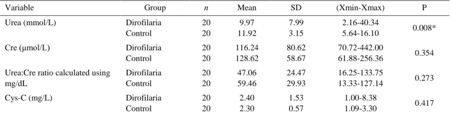
A summary of the descriptive statistics of the serum urea, sCre, Urea:Cre ratio and sCysC, data is provided in Table 1. Mean serum urea concentrations were 9.97 mmol/L in the Dirofilaria group with the presence of mean sCre (116.24 µmol/L) and sCysC (2.40 mg/L) concentrations. Mean serum Urea:Cre ratio was detected
Table 1. Serum biochemistry data in D. immitis infected and control dogs.
Tablo 1. D. immitis ile enfekte ve kontrol köpeklerindeki serum biyokimyasal veriler.
Variable Group n Mean SD (Xmin-Xmax) P
Urea (mmol/L) Dirofilaria Control 20 20 9.97 11.92 7.99 3.15 2.16-40.34 5.64-16.10 0.008*
Cre (μmol/L) Dirofilaria
Control 20 20 116.24 128.62 80.62 58.67 70.72-442.00 61.88-256.36 0.354 Urea:Cre ratio calculated using
mg/dL Dirofilaria Control 20 20 47.06 59.46 24.47 29.93 16.25-133.75 13.33-127.14 0.273 Cys-C (mg/L) Dirofilaria Control 20 20 2.40 2.30 1.53 0.57 1.00-8.38 1.09-3.30 0.417 * P<0.05 is considered statistically significant.
Ankara Üniv Vet Fak Derg, 62, 2015 305
in Dirofilaria group as 47.06 mg/dL. Control dogs’ urea concentrations were 11.92 mmol/L with mean sCre (128.62 µmol/L) and sCysC (2.30 mg/L) concentrations. Mean serum Urea:Cre ratio were detected as 59.46 mg/dL in control group. Mean sCysC concentrations with sCre, and serum Urea:Cre ratio did not differ significantly between groups. Only mean serum urea concentrations were found statistically different (P=0.008, Mann Whitney U-test).
Discussion and Conclusion
Various literatures indicate the pathogenity of filarial nematodes in the kidney as well as changes occurred in the GBM, such as thickening (7, 27) and electron dense deposits (1) in D. immitis infected dogs. Almost of these studies accepted the pathogenity of D.
immitis are mainly related to circulating immune
complexes. Microfilaria and adult worm antigens (17) which have an important role in the process through immune-mediated mechanisms as well (1, 7, 13). Furthermore, it is reported that even though immune complexes may appear in the kidneys of heartworm-infected dogs, only a small percentage of the animals would be expected to develop clinical renal disease (7). In the current study, it is therefore not surprising that RF did not alter in dogs with D. immitis measured with serum indirect markers plus sCysC.
Traditionally, biochemical evidence of renal insufficiency is sought from elevations in serum urea and sCre concentrations (10, 12). Their reciprocal plots roughly correlate with GFR (12). Both urea and sCre concentrations require about 60-75% loss of nephron function before they became elevated (12). To increase their specificity, serum urea and sCre are often tested concurrently (9). In renal disease they are expected to behave similarly with both parameters increasing as GFR decreases (9).
In the current study mean serum urea concentration (9.97 mmol/L) were above the reference limit (2.65-8.96 mmol/L) in the Dirofilaria group with the presence of normal mean sCre (116.24 µmol/L) and sCysC (2.40 mg/L) concentrations. Moreover, there was not found a statistical significance between the groups for sCre, sCysC concentrations and Urea:Cre ratio. On the other hand, serum urea concentrations were statistically differed between groups. However, mean serum Urea:Cre ratio (47 mg/dL) was also above the reference limit (10-15 mg/dL) in Dirofilaria group. Control dogs also had a higher mean serum Urea:Cre ratio (59.4 mg/dL). In human medicine a serum Urea:Cre ratio in azotemic patients of ≥20 indicates prerenal azotemia, whereas a serum Urea:Cre ratio of <20 presents intrinsic renal disease (5). Researchers hypothesized that some dogs with high serum urea and low or normal sCre
concentrations may have had renal azotemia (9). However, in a following study concluded that elevated serum urea concentrations in canine babesiosis in the absence of elevated sCre concentrations were not likely to be of renal origin, since sCysC concentrations were not elevated in dogs with normal sCre concentrations (8). In the present study, mean sCysC concentrations were not found statistically significant in Dirofilaria group (2.40 mg/L) when compared to the healthy controls (2.30 mg/L). Same as, with sCysC considered free of the perturbations that sCre are subject to an elevated serum urea concentrations in the Dirofilaria and control dogs in the absence of an elevated sCre concentration does not reflect significant renal dysfunction.
Therefore, it is concluded that serum urea is often elevated due to non-renal factors in canine dirofilariasis same as canine babesiosis infections (8, 15), which cause an elevated serum Urea:Cre ratio. However, in the present study the cause of elevated Urea:Cre ratio remains undetermined. Recent meal may be a reason of this hyperureagenesis. While, other ammonia loading reasons such as, haemolysis, blood transfusions, and gastro-intestinal haemorrhage (24) were all absent in the study dogs. On the other hand, in humans, cardiac disease has been reported to a major contributor of an increased serum Urea:Cre ratio (11). Cardiac evaluations were however not conducted and further studies are required to investigate the potential contribution of myocardial injury to the Urea:Cre ratio in dogs with D.
immitis.
Same as, cardiac evaluations, there are some other several limitations of the present study. One of the limitation is that the present study did not investigate the stage of the dirofilariasis as either asymptomatic or mild (stage I), moderate (stage II), or severe (stage III) (21). Unfortunately, enrolled cases also were not evaluated for co-infections anaplasmosis, leishmaniasis, lyme diseases which may lead renal failure. Dogs are should be fasted for at least 12 hours before taking blood samples to measure sCysC concentration (14). However, the authors in the present study could not evaluate the blood collecting and feeding time with physical activity index, age, breed, weight of the dogs that may also affect the sCysC concentrations. Therefore, further investigations are required to evaluate the sCysC concentrations in canine dirofilariasis with considering those spoken factors.
Consequently, this manuscript as an initial study represents the mean sCysC levels in dogs with D. immitis for the first time. Furthermore, it is concluded that, GFR were not altered in the dogs infected group rather than uninfected group, and sCysC concentrations seemed to be not altered in dogs with D. immitis infection.
Didem Pekmezci - Murat Güzel - Alparslan Yıldırım - Gülay Çiftci - Gökmen Zafer Pekmezci - Mehmet Tütüncü - Abdullah İnci
306
References
1. Abramowsky CR, Powers KG, Aikawa M, Swinehart G (1981): Dirofilaria immitis. 5. Immunopathology of filarial
nephropathy in dogs. Am J Pathol, 104, 1-12.
2. Acevedo RA, Ciencias L, Theis JH, et al. (1981):
Combination of filtration and histochemical stain for detection and differentiation of Dirofilaria immitis and Dipetalonema reconditum in the dog. Am J Vet Res, 42,
537-540.
3. Antognoni MT, Siepi D, Porciello F, Fruganti G (2005):
Use of serum cytatin C determination as a marker of renal function in the dog. Vet Res Commun, 29, 265-267.
4. Antognoni MT, Siepi D, Porciello F, et al. (2007): Serum
cystatin-C evaluation in dogs affected by different diseases associated or not with renal insufficiency. Vet Res
Commun, 31, 269-271.
5. Brady HR, Brenner BM (1994): Acute renal failure. 1265-1274. In: Isselbaher KJ, Braunwald E, Wilson JD (Ed), Harrison’s Principles of Internal Medicine. McGraw-Hill, Columbus.
6. Braun JP, Perxachs A, Pechereau D, De La Farge F (2002): Plasma cystatin C in the dog: reference values and
variations with renal failure. Comp Clin Pathol, 11, 44-49.
7. Casey HW, Splitter GA (1975): Membranous glomerulonephritis in dogs infected with Dirofilaria immitis. Vet Pathol, 12, 111-117.
8. De Scally MP, Leisewitz AL, Lobetti RG, Thompson PN (2006): The elevated serum urea:creatinine ratio in
canine babesiosis in South Africa is not of renal origin. J S
Afr Vet Assoc, 77, 175-178.
9. De Scally MP, Lobetti RG, Reyers F, Humphris D (2004): Are urea and creatinine values reliable indicators
of azotaemia in canine babesiosis? J S Afr Vet Assoc, 75,
121-124.
10. Di Bartola SP (2000): Clinical approach and laboratory
evaluation of renal disease. 1600-1614. In: Ettinger SJ,
Feldman EC (Ed), Textbook of Veterinary Internal Medicine (5th ed). WB Saunders, Philadelphia.
11. Feinfeld DA, Bargouthi H, Niaz Q, Carvounis CP (2002): Massive and disproportionate elevation of blood
urea nitrogen in acute azotemia. Int Urol Nephrol, 34,
143-145.
12. Finco DR (1997): Evaluation of renal function. 216-229. In: Osborne CA, Finco DR (Ed), Canine and Feline Nephrology and Urology. Williams and Wilkins, Philadelphia, USA.
13. Grauer GF, Culham CA, Bowman DD, et al. (1988):
Parasite excretory-secretory antigen and antibody to excretory-secretory antigen in body fluids and kidney tissue of Dirofilaria immitis infected dogs. Am J Trop Med
Hyg, 39, 380-387.
14. Ghys L, Paepe D, Smets P, et al. (2014): Cystatin C: A
new renal marker and its potential use in small animal medicine. J Vet Intern Med, doi: 10.1111/jvim.12366.
15. Lobetti R (2012): Changes in the serum urea: creatinine
ratio in dogs with babesiosis, haemolytic anaemia, and experimental haemoglobinaemia. Vet J, 191, 253-256.
16. Miyagawa Y, Takemura N, Hirose H (2009): Evaluation
of the measurement of serum cystatin C by an enzyme-linked immunosorbent assay for humans as a marker of the
glomerular filtration rate in dogs. J Vet Med Sci, 71,
1169-1176.
17. Nakagaki K, Nogami S, Hayashi Y, et al. (1993):
Dirofilaria immitis: detection of parasite-specific antigen by monoclonal antibodies in glomerulonephitis in infected dogs. Parasitol Res, 79, 49-54.
18. Oge H, Doganay A, Oge S, Yildirim A (2003):
Prevalence and distribution of Dirofilaria immitis in domestic dogs from Ankara and vicinity in Turkey. Dtsch
Tierarztl Wochenschr, 110, 69-72.
19. Paes-de-Almeida EC, Ferreira AMR, Labarthe NV, et al. (2003): Kidney ultrastructural lesions in dogs
experimentally infected with Dirofilaria immitis (Leidy, 1856). Vet Parasitol, 113, 157-168.
20. Peribáňez MA, Lucientes J, Arce S, et al. (2001):
Histochemical differentiation of Dirofilaria immitis, Dirofilaria repens and Acanthocheilonema dracunculoides microfilariae by staining with a commercial kit,
Leucognost-SP®. Vet Parasitol, 102, 173-175.
21. Polizopoulou ZS, Koutinas AF, Saridomichelakis MN, et al. (2000): Clinical and laboratory observations in 91
dogs infected with Dirofilaria immitis in northern Greece.
Vet Rec, 146, 466-469.
22. Randers E, Erlandsen EJ (1999): Serum cystatin C as an
endogenous marker of the renal function – A Review. Clin
Chem Lab Med, 37, 389-395.
23. Randers E, Kristensen JH, Erlandsen EJ, Danielsen H (1998): Serum cystatin C as a marker of renal function. Scand J Clin Lab Investig, 58, 585-592.
24. Reyers F (1992): Is azotaemia in canine babesiosis an
indication of renal disease? In: Proceedings of the 9th
Faculty Day. University of Pretoria, Faculty of Veterinary Science, 17.
25. Rishniw M, Barr SC, Simpson KW, et al. (2006):
Discrimination between six species of canine microfilariae by a single polymerase chain reaction. Vet Parasitol, 135,
303-314.
26. Simon F, Kramer LH, Román A, et al. (2007):
Immunopathology of Dirofilaria immitis infection. Vet Res
Commun, 31, 161-171.
27. Sutton RH (1988): Pathology and pathogenesis of
dirofilariasis. 99-132. In: Boreham PFF, Atwell RB (Ed),
Dirofilariasis. Florida, CRC Press.
28. Watts KJ, Courteny CH, Reddy GR (1999):
Development of a PCR- and probe-based test for the sensitive and specific detection of the dog heartworm, Dirofilaria immitis, in its mosquito intermediate host. Mol
Cell Probes, 14, 425-430.
29. Yildirim A (2004): The prevalence of filarial agents in
dogs in Ankara and vicinity. Ankara Univ Vet Fak Der, 51,
35-40.
Geliş tarihi: 03.07.2014/ Kabul tarihi: 10.02.2015
Yazışma Adresi:
Yrd.Doç.Dr. Didem Pekmezci Ondokuz Mayıs Üniversitesi Veteriner Fakültesi
İç Hastalıkları Anabilim dalı, Samsun, Türkiye e-posta: [email protected]