Ranunculus sericeus Banks & Sol. Extract Fractions Possess Antibacterial and
Antifungal Activity
HAKAN ATCI1 AND YALÇIN KARAGÖZ1
1Agri Ibrahim Cecen University, Faculty of Pharmacy, Department of Pharmaceutical Botany Abstract
Ranunculus sericeus, collected from Ağrı, was
successively extracted with n-hexane, chloroform, acetone and methanol, in a Soxhlet extractor. Obtained extracts were tested for antibacterial activity against human and plant pathogenic bacteria Pseudomonas
aeruginosa, Bacillus subtilis, Staphylococcus aureus, Enterococcus faecalis, Xanthomonas axonopodis pv. vitians, Enterobacter cloacae, Burkholderia sepasia, Pantoea ananatis, and for antifungal activity against
fungi Rhizoctonia sp., Alternaria sp., and Fusarium sp. Minimum Inhibitory Concentration (MIC) values were determined. Our results indicate that acetone and methanol fractions have moderate antibacterial and antifungal activity.
Keywords: Ranunculus sericeus, antibacterial activity, antifungal activity, extract fractions
Introduction
Since ancient times, people have frequently been utilizing plants for health benefits. It also provides insights for raw materials, mostly bioactive molecules, to be used in pharmaceutical industry. Therefore, researchers all over the world investigate biological actions of plant extracts and isolated substances. An important stimulus on these investigations is the increase in number and prevalence of multiple drug resistant bacteria. World Health Organization called Received: 08.04.2018
Revised: 18.04.2018 Accepted:21.04.2018
Corresponding author: Yalçın Karagöz, PhD Agri Ibrahim Cecen University, Faculty of Pharmacy Department of Pharmaceutical Botany, Agrı Turkey E-mail: [email protected]
Cite this article as: H. Atcı and Y. Karagöz, Ranunculus sericeus Banks & Sol. Extract Fractions Possess Antibacterial and Antifungal Activity, Eastern Anatolian Journal of Science, Vol. 4, Issue 1, 9-15,2018.
for immediate action against these organisms early in 2017 (WHO 2017). The search, however, is not restricted to new antibiotics against already resistant bacteria, as any pathogen is a potential resistant organism.
Turkey, concerning plant species number and diversity, has a rich flora of some 9500 species (DAVIS et al. 1965). Turkish government agencies declare 93 plant species as medicinal plants (TİTCK 2018). Almost half of them are not native to Turkey. Given the number of plant species above and the long history of modern human existence in Turkey, one can easily speculate that there should be more medicinal plants in the country. This clearly reveals the need for more investigations on biological actions of plants in Turkey. Also needed are more ethnobotanical studies to shed light on use of plants by people as remedies in the country. One of the few ethnobotanical studies (SEZIK et al. 1997) reports use of Ranunculus sericeus Banks & Sol. as an external poultice to reduce inflammation in rheumatism, in Ağrı province. Ranunculus genus is a controversial taxon in the family Ranunculaceae, which is associated with several cases of dermatitis (POLAT et al. 2007; KOSE et al. 2008; AKBULUT et al. 2011; CALKA et al. 2011; OZKOL et al. 2014; UCMAK et al. 2014; MILANESI et al. 2015; POLAT 2016) and also a cure for dermatitis (PRIETO et al. 2008). Members of the genus have been found to possess anti-inflammatory (CAO et al. 1992; FOSTOK et al. 2009; AKKOL et al. 2012), antimicrobial (MISRA AND DIXIT 1978; LORIMER et al. 1996; BARBOUR et al. 2004; DENG et al. 2013; BHATTI et al. 2015b; KHAN et al. 2016), antioxidant (LV et al. 2010; BHATTI et al. 2015a; KHAN et al. 2016; RAZIQ et al. 2017; BOROOMAND et al. 2018), and antitumor (BHATTI et al. 2015b) activities. The reader is referred to ASLAM et al. (2012), for an excellent review of the genus.
Ranunculus sericeus has not been investigated for
biological acitivities so far.
Considering all above factors, Ranunculus sericeus seems to be a good target for screening antimicrobial substances. The aim of this study is investigation of antimicrobial and antifungal activity of the herba of
Ranunculus sericeus.
Materials and methods
Solvents, media and devices
All solvents used in this study were analytical grade. N-hexane and acetone were purchased from Merck (Germany). Chloroform was purchased from TEKKİM (İstanbul, Turkey). Methanol was purchased from Sigma (USA). The rotary evaporator was IKA Labortek (Switzerland). Tryptic Soy Agar (TSA) medium was purchased from Sigma (USA, catalog no 22091). Nutrient Broth (Catalog no CM0001) and Potato Dextrose Agar (PDA, Catalog no CM0139) media were purchased from Oxoid (UK).
Plant material
Ranunculus sericeus was collected from Sarıcan
village of Eleşkirt county, Ağrı, Turkey, during its flowering period (May-June) in 2017. It was identified according to DAVIS et al. (1965). A voucher specimen is kept in the private collection of Y.K., with accession number YK-2017-008. Plant material was dried in the shade at room temperature, and aboveground parts were ground into a fine powder using liquid nitrogen with the aid of a mortar and pestle.
Extraction
Fifty grams of ground material was successively extracted, for 12 hours each, with n-hexane, chloroform, acetone and methanol, respectively, in a Soxhlet apparatus. Solvents were evaporated under reduced pressure at 40 ± 5 °C in rotary evaporator. This procedure resulted in 4 extracts, which were labeled H for hexane, C for chloroform, A for acetone and M for methanol.
Test microorganisms
All test organisms were courtesy of Dr. Kenan KARAGÖZ of Agri Ibrahim Cecen University, Faculty of Science and Letters, Department of Molecular Biology and Genetics, from his personal collection.
Agar disc diffusion test
Assays were performed with TSA media, according to Murray (MURRAY 1995) with a minor modification. Tetracycline and kanamycin were used as positive controls. The fractions and antibiotics were prepared in 10% Dimethyl sulfoxide (DMSO) or sterile distilled water (sdH2O) at a concentration of 50 mg/ml. Bacterial suspension (100 μl) containing ~1x10⁸ CFU / ml (adjusted by 0,5 McFarland standard turbidity) of bacteria spread by a sterile swab on TSA medium. The discs (6 mm in diameter) were impregnated with 10 μl (the final amount on one disk for each fraction was 0.5 mg) of the fractions or antibiotic solutions and put in the middle of the inoculated plates. The bacterial cultures were incubated at 37 °C for 48 h, and then inhibition zones were measured in diameter (mm) around the discs. 10% DMSO and sdH2O were used as negative control. The assays were performed with three replicates.
MIC tests
MIC values of fractions and antibiotics were determined for the microorganisms by microdilution assay (KARAMAN et al. 2003; SAHIN et al. 2003). The 96-well microtiter plates were used for this test. Consecutive wells containing fractions or antibiotics at the concentration range of 9.75 -2500 µg/ml was prepared in nutrient broth. Then, 5 µl bacterial suspensions at the concentration of ~1x10⁸ CFU / ml were added each well and total volume reached 200 µl with the addition of nutrient broth. The plate was covered with a sterile plate sealer. The contents of each well were mixed on a microtiter plate shaker at 300 rpm for 20 s and then incubated at 37 °C for 24 h. Microbial growth was determined by absorbance values at 600 nm using plate reader and confirmed by plating 5 µl samples from clear wells on nutrient agar media. The fractions and antibiotics tested in this study
were screened three times for each organism and 10 % DMSO and sdH2O were used as negative control. The MIC was defined as the lowest concentration of the compounds or antibiotics to inhibit the growth of microorganisms.
Antifungal activity test
Antifungal activity test was performed as previously described (TORRES et al. 2017), with minor modifications. Briefly, 25 μl of fractions (50 mg/ml) were inoculated in wells, which were carved in the middle of PDA filled 9 cm diameter petri dishes. 4 mm plugs of 7-day-old culture of each fungal organism were placed in the center of each dish. Plates without fractions were used as negative controls. Cycloheximide (50 mg/ml) was used as positive control. After 7 days of incubation at 28 °C the mycelial growth diameter of each fungus was measured and the percentage of fungal inhibition (FI)
was calculated as follows; FI (%) = 100 × ((mycelial growth diameter of control – mycelial growth diameter of tested fraction) ÷ mycelial growth diameter of control).
Results and Discussion
H, C, A, and M yielded 246, 850, 985, and 12905 mg extract fractions, respectively. This represents a very high yield of approximately 30%, in total. The roots were excluded from the study, which means substance amount per individual plant could be higher.
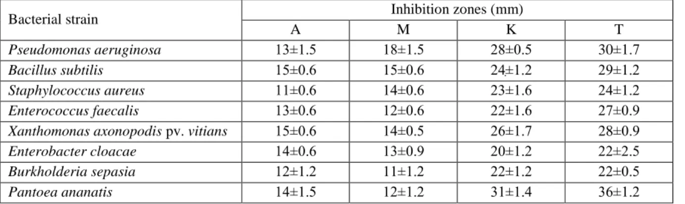
Hexane and chloroform fractions had no activity in agar disc diffusion assay. However, acetone and methanol fractions had moderate antibacterial activity against all tested organisms. Negative controls had no effect on bacterial growth. The results are summarized in Table 1. Hexane and chloroform fraction data and negative controls are not presented to save space.
Table 1. Antibacterial activity of Ranunculus sericeus, expressed as inhibition zone (mm). A: Acetone fraction, M: Methanol fraction, K: Kanamycin, T: Tetracycline. Values are represented as avergae ± standard deviation of three replicates.
Bacterial strain Inhibition zones (mm)
A M K T
Pseudomonas aeruginosa 13±1.5 18±1.5 28±0.5 30±1.7
Bacillus subtilis 15±0.6 15±0.6 24ׅ±1.2 29±1.2
Staphylococcus aureus 11±0.6 14±0.6 23±1.6 24±1.2 Enterococcus faecalis 13±0.6 12±0.6 22±1.6 27±0.9 Xanthomonas axonopodis pv. vitians 15±0.6 14±0.5 26±1.7 28±0.9
Enterobacter cloacae 14±0.6 13±0.9 20±1.2 22±2.5 Burkholderia sepasia 12±1.2 11±1.2 22±1.2 22±0.5
Pantoea ananatis 14±1.5 12±1.2 31±1.4 36±1.2
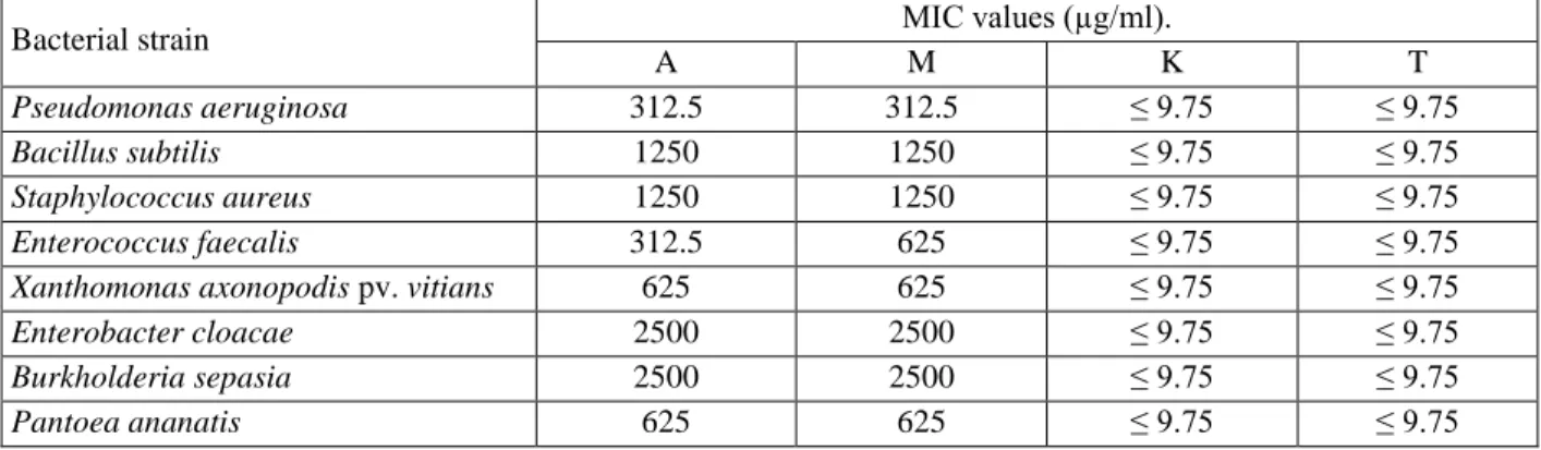
Hexane and chloroform fractions did not show any antibacterial activity. Hence, they were not evaluated in terms of MIC. Negative controls had no activity. Acetone and methanol fractions had considerably low MIC values, given the fact that they are mixtures. MIC values are for A, M, kanamycin and tetracycline are presented in Table 2.
Hexane and chloroform fractions did not show any antifungal activity against any of the fungi. All four fractions were ineffective against Rhizoctonia sp. Acetone fraction inhibited growth of Alternaria sp. and
Fusarium sp., while methanol fraction was active
against Alternaria sp. Growth diameter and percent inhibition data for A, M, and cycloheximide are presented in Table 3. H and C fraction data were excluded to save space
Table 2. MIC value of the extract fractions and antibiotics against the pathogens, expressed as concentration (µg/ml). A: Acetone fraction, M: Methanol fraction, K: Kanamycin, T: Tetracycline.
Bacterial strain MIC values (µg/ml).
A M K T
Pseudomonas aeruginosa 312.5 312.5 ≤ 9.75 ≤ 9.75
Bacillus subtilis 1250 1250 ≤ 9.75 ≤ 9.75
Staphylococcus aureus 1250 1250 ≤ 9.75 ≤ 9.75
Enterococcus faecalis 312.5 625 ≤ 9.75 ≤ 9.75
Xanthomonas axonopodis pv. vitians 625 625 ≤ 9.75 ≤ 9.75
Enterobacter cloacae 2500 2500 ≤ 9.75 ≤ 9.75
Burkholderia sepasia 2500 2500 ≤ 9.75 ≤ 9.75
Pantoea ananatis 625 625 ≤ 9.75 ≤ 9.75
.
Table 3. Antifungal activity of fractions represented as growth diameter and fungal inhibiton. D: growth diameter expressed as avarege ± standard deviation (where available) of three replicates, FI: fungal inhibition expressed as percent, CH: cycloheximide, A: acetone fraction, M: methanol fraction, —: no activity.
Fungi Control D (mm) CH A M D (mm) FI (%) D (mm) FI (%) D (mm) FI (%) Rhizoctonia sp. 60.3±0.5 40.3±1.9 33.15 — 0 — 0 Alternaria sp. 60 — 0 50.3±0.5 16.11 51.3±0.5 14.44 Fusarium sp. 60 28.0±2.2 53.33 50.7±0.5 15.56 — 0
This study deals with the antibacterial and antifungal activities of extract fractions of Ranunculus sericeus. As far as we know this is the first report on biological actions of R. sericeus. According to the records investigation of antimicrobial activity of Ranunculus genus started as early as some 50 years ago (BUKOWIECKI AND ZAREBSKA 1966a, b). Authors isolated protoanemonin and demonstrated its antibiotic activity. Other substances isolated from Ranunculus species include flavonoids, phytosterols, coumarin derivatives, lactone derivatives, triterpenes, fatty acids, and saponins (ASLAM et al. 2012).
There are many studies in the literature dealing with biological actions and detrimental effects of other Ranunculus species (see Introduction). In general, this genus is considered poisonous to humans. However, people keep using members of it, especially in Asia (UMAIR et al. 2017), for alleged health benefits. On the other hand, Ranunculus ternatus, a well-established species used in traditional Chinese medicine is reported to have various activities on human health. These include Tumor Necrosis Factor-α induction in cultured tumor cells (ZHOU et al. 1995), anti-tuberculosis activity (DENG et al. 2013), and promotion
of immune cell proliferation and phagocytosis (LV et al. 2010). Another species, Ranunculus japonicus was found to be protective against myocardial ischemic-reperfusion injury in isolated rat hearts (GAO et al. 2014) through prevention of hypertrophy in cardiomyocytes via alleviating chronic Ca2+ overload (DAI et al. 2015), and to possess anti-inflammatory and analgesic effects in mice (CAO et al. 1992). In light of these facts, it is difficult to assert the whole genus as detrimental to human health.
The lack of studies on Ranunculus sericeus dictates comparison of the obtained data to other Ranunculus species. In a study by MISRA AND DIXIT (1978), the authors evaluated the antifungal potential of
Ranunculus sceleratus against various fungal pathogens, including Alternaria tenuis, Alternaria
solani, and Fusarium nivale. They concluded that
water extract of the species inhibited – in fact, annihilated – fungal growth, and was promising as an antifungal agent, particularly for plant pathogens. They also emphasized the importance of using fresh plant samples, as dried plant lost a great deal of antifungal power. It may account for the extent of antifungal power we found in this study. Also, it might be inferred
that the same issue may be valid for antibacterial activity, too; fresh plant samples may exhibit much stronger antibacterial activity.
In a study by BARBOUR et al. (2004), researchers found
Ranunculus myosuroudes to possess strong antibacterial activity against Pseudomonas aeruginosa and Staphylococcus aureus. However, the dosage of the plant extract is vague and a comparison seems impossible. In a more recent study by BHATTI et al. (2015b), authors concluded that water extract of
Ranunculus arvensis exhibited weak antimicrobial
activity and no effect against the fungus Fusarium
solani. In another study (KHAN et al. 2016), root, stem, and leaf extracts of Ranunculus muricatus were tested against Bacillus subtilis, Pseudomonas aeruginosa, and Staphylococcus aureus. Researchers reported similar inhibition zones to our results, for acetone and methanol extracts but they did not reveal the extract concentrations. Therefore, a comparison is again impossible.
Conclusion
In conclusion, this is the first report or biological actions of Ranunculus sericeus, demonstrating it may have some potential of providing antimicrobial, particularly antifungal, compounds. More studies in the future definitely will help to elucidate the compounds responsible for this, and perhaps their modes of actions.
Acknowledgements
This study was excerpted from the dissertation thesis of H.A. Authors express their gratitude to Dr. Kenan KARAGÖZ for his priceless help in conduction of antimicrobial tests and interpretation of test results.
References
AKBULUT, S., SEMUR, H., KOSE, O., OZHASENEKLER, A., CELIKTAS, M., BASBUG, M. AND YAGMUR, Y. (2011), Phytocontact dermatitis
due to Ranunculus arvensis mimicking burn injury: report of three cases and literature review, Int J Emerg Med, 4, 7.
AKKOL, E. K., SUNTAR, I., ERDOGAN, T. F., KELES, H., GONENC, T. M. AND KIVCAK, B. (2012),
Wound healing and anti-inflammatory properties of Ranunculus pedatus and Ranunculus constantinapolitanus: a comparative study, J Ethnopharmacol, 139,
478-484.
ASLAM, M., A. CHOUDHARY, B., UZAIR, M. AND SUBHAN IJAZ, A. (2012), The genus
Ranunculus: A phytochemical and ethnopharmacological review, International
Journal of Pharmacy and Pharmaceutical Sciences, 4, 15-22.
BARBOUR, E. K., AL SHARIF, M., SAGHERIAN, V. K., HABRE, A. N., TALHOUK, R. S. AND TALHOUK, S. N. (2004), Screening of selected
indigenous plants of Lebanon for antimicrobial activity, J Ethnopharmacol, 93,
1-7.
BHATTI, M. Z., ALI, A., AHMAD, A., SAEED, A. AND MALIK, S. A. (2015a), Antioxidant and
phytochemical analysis of Ranunculus arvensis L. extracts, BMC Res Notes, 8, 279.
BHATTI, M. Z., ALI, A., SAEED, A., SAEED, A. AND MALIK, S. A. (2015b), Antimicrobial,
antitumor and brine shrimp lethality assay of Ranunculus arvensis L. extracts, Pak J Pharm
Sci, 28, 945-949.
BOROOMAND, N., SADAT-HOSSEINI, M., MOGHBELI,
M. AND FARAJPOUR, M. (2018),
Phytochemical components, total phenol and mineral contents and antioxidant activity of six major medicinal plants from Rayen, Iran,
Nat Prod Res, 32, 564-567.
BUKOWIECKI, H. AND ZAREBSKA, I. (1966a),
[Antibiotic activity of protoanemonin in extracts of some lowland species of Ranunculus L], Acta Pol Pharm, 23, 163-167.
BUKOWIECKI, H. AND ZAREBSKA, I. (1966b),
[Structure of the vegetative organs of the species Ranunculus lanuginosus L. and of some ranunculi plants with antibiotic activity], Acta Pol Pharm, 23, 159-162.
CALKA, O., AKDENIZ, N., OZKOL, H. U., KARADAG, A. S. AND BEHCET, L. (2011), Irritant contact
dermatitis caused by Ranunculus kotschyi Boiss in 6 cases, Contact Dermatitis, 64,
CAO, B. J., MENG, Q. Y. AND JI, N. (1992), Analgesic
and anti-inflammatory effects of Ranunculus japonicus extract, Planta Med, 58, 496-498.
DAI, H. L., JIA, G. Z. AND ZHAO, S. (2015), Total
glycosides of Ranunculus japonius prevent hypertrophy in cardiomyocytes via alleviating chronic Ca(2+) overload, Chin
Med Sci J, 30, 37-43.
DAVIS, P. H., CULLEN, J. AND COODE, M. J. E. (1965),
Flora of Turkey and the East Aegean Islands
(Edinburgh,: University Press)
DENG, K. Z., XIONG, Y., ZHOU, B., GUAN, Y. M. AND LUO, Y. M. (2013), Chemical constituents
from the roots of Ranunculus ternatus and their inhibitory effects on Mycobacterium tuberculosis, Molecules, 18, 11859-11865.
FOSTOK, S. F., EZZEDDINE, R. A., HOMAIDAN, F. R., AL-SAGHIR, J. A., SALLOUM, R. G., SALIBA, N. A. AND TALHOUK, R. S. (2009),
Interleukin-6 and Cyclooxygenase-2 downregulation by fatty-acid fractions of Ranunculus constantinopolitanus, BMC Complement Altern Med, 9, 44.
GAO, X. W., LIU, Y., YANG, Z. C. AND TAN, Y. Z. (2014), [Protective effect of total glycosides
of Ranunculus japonicus on myocardial ischemic-reperfusion injury in isolated rat hearts], Zhong Yao Cai, 37, 1429-1433.
KARAMAN, I., SAHIN, F., GULLUCE, M., OGUTCU, H., SENGUL, M. AND ADIGUZEL, A. (2003),
Antimicrobial activity of aqueous and methanol extracts of Juniperus oxycedrus L, J
Ethnopharmacol, 85, 231-235.
KHAN, F. A., ZAHOOR, M. AND KHAN, E. (2016),
Chemical and biological evaluation of Ranunculus muricatus, Pak J Pharm Sci, 29,
503-510.
KOSE, R., OKUR, M. I., BINGOL, I. AND CETIN, H. (2008), Phytocontact dermatitis mimicking a
burn injury due to Ranunculus constantinopolitanus, Contact Dermatitis, 59,
249-250.
LORIMER, S. D., BARNS, G., EVANS, A. C., FOSTER, L. M., MAY, B. C., PERRY, N. B. AND TANGNEY, R. S. (1996), Cytotoxicity and antimicrobial
activity of plants from New Zealand's subantarctic islands, Phytomedicine, 2,
317-323.
LV, X., WANG, H., HAN, H., LV, S. AND QIN, D. (2010),
[Effects of polysaccharide of radix ranunculi ternati on immunomodulation and anti-oxidation], Zhongguo Zhong Yao Za Zhi, 35,
1862-1865.
MILANESI, N., D'ERME, A. M. AND GOLA, M. (2015),
De rerum natura: a case of irritant phytodermatitis from Ranunculus bulbosus,
Int J Dermatol, 54, 202-203.
MISRA, S. B. AND DIXIT, S. N. (1978), Antifungal
properties of leaf extract of Ranunculus sceleratus L, Experientia, 34, 1442-1443.
MURRAY, P. R. (1995), Manual of clinical
microbiology (6th ed.; Washington, D.C.:
ASM Press)
OZKOL, H. U., CALKA, O., AKDENIZ, N. AND PINAR, S. M. (2014), Phytodermatitis in eastern
Turkey: a retrospective, observational study,
Dermatitis, 25, 140-146.
POLAT, M. (2016), A case of phytodermatitis due to
Ranunculus arvensis used as an herbal remedy, Int J Dermatol, 55, e37-38.
POLAT, M., OZTAS, P., YALCIN, B., TAMER, E., GUR, G. AND ALLI, N. (2007), Contact dermatitis due
to Allivum sativum and Ranunculus illyricus: two cases, Contact Dermatitis, 57, 279-280.
PRIETO, J. M., RECIO, M. C., GINER, R. M., SCHINELLA, G. R., MANEZ, S. AND RIOS, J. L. (2008), In
vitro and in vivo effects of Ranunculus peltatus subsp. baudotii methanol extract on models of eicosanoid production and contact dermatitis, Phytother Res, 22, 297-302.
RAZIQ, N., SAEED, M., ALI, M. S., ZAFAR, S., SHAHID, M. AND LATEEF, M. (2017), A new glycosidic
antioxidant from Ranunculus muricatus L. (Ranunculaceae) exhibited lipoxygenasae and xanthine oxidase inhibition properties,
Nat Prod Res, 31, 1251-1257.
SAHIN, F., KARAMAN, I., GULLUCE, M., OGUTCU, H., SENGUL, M., ADIGUZEL, A., OZTURK, S. AND
KOTAN, R. (2003), Evaluation of
antimicrobial activities of Satureja hortensis L, J Ethnopharmacol, 87, 61-65.
SEZIK, E., YEŞILADA, E., TABATA, M., GISHO, H., TAKAISHI, Y., FUJITA, T., TOSHIHIRO, T. AND TAKEDA, Y. (1997), Traditional Medicine in
Turkey VIII. Folk Medicine in East Anatolia; Erzurum, Erzincan, Ağri, Kars, Iğdir Provinces, Economic Botany, 51, 195-211.
Türkiye İlaç ve Tıbbi Cihaz Kurumu, Tıbbi Bitki
Listesi. Accessed: 06.04.2018.
http://www.titck.gov.tr/T%C4%B1bbiBitkiL istesi?PageNo=10.
TORRES, M. J., BRANDAN, C. P., SABATE, D. C., PETROSELLI, G., ERRA-BALSELLS, R. AND AUDISIO, M. C. (2017), Biological activity of
the lipopeptide-producing Bacillus amyloliquefaciens PGPBacCA1 on common bean Phaseolus vulgaris L. pathogens,
UCMAK, D., AYHAN, E., MELTEM AKKURT, Z. AND HAYDAR, U. (2014), Presentation of three
cases with phyto contact dermatitis caused by Ranunculus and Anthemis genera, J Dermatolog Treat, 25, 467-469.
UMAIR, M., ALTAF, M. AND ABBASI, A. M. (2017), An
ethnobotanical survey of indigenous medicinal plants in Hafizabad district, Punjab-Pakistan, PLoS One, 12, e0177912.
WHO, WHO publishes list of bacteria for which new antibiotics are urgently needed. Accessed: 06.04.2018.
http://www.who.int/mediacentre/news/releas es/2017/bacteria-antibiotics-needed/en/. ZHOU, L., ZHANG, W. AND XU, J. (1995), [Effect of the
active components of Ranunculus ternatus Thunb. on the inductive production of tumor necrosis factors by macrophages], Zhongguo