www.nature.com/leu
Frequent demonstration of human herpesvirus 8 (HHV-8) in bone marrow biopsy
samples from Turkish patients with multiple myeloma (MM)
M Beksac1, M Ma3, C Akyerli2, M DerDanielian3, L Zhang3, J Liu3, M Arat1, N Konuk1, H Koc1, T Ozcelik2, R Vescio3and JR Berenson3
1Ibni Sina Hospital, Department of Hematology, Ankara University School of Medicine, Ankara;2Bilkent University, Department of Molecular Biology and Genetics, Ankara, Turkey; and3Cedars Sinai Medical Center, Jonsson Comprehensive Cancer Center, UCLA School of Medicine, Los Angeles, CA, USA
In order to investigate the frequency of HHV-8 in MM patients from another geographic location, we obtained fresh bone mar-row (BM) biopsies from Turkish patients with MM (n = 21), monoclonal gammopathy of undetermined significance (MGUS) (n= 2), plasmacytoma (n = 1) with BM plasma cell infil-tration, various hematological disorders (n= 6), and five heal-thy Turkish controls. The frequency of HHV-8 was analyzed by polymerase chain reaction (PCR) in two independent labora-tories in the USA and in Turkey. Using fresh BM biopsies, 17/21 MM patients were positive for HHV-8 whereas all five healthy controls, and six patients with other hematological disorders were negative. Two patients with MGUS, and one patient with a solitary plasmacytoma were also negative. The data from the two laboratories were completely concordant. Also using primer pairs for v IRF and v IL-8R confirmed the results observed with the KS330233primers. Furthermore, sequence
analysis demonstrated a C3 strain pattern in the ORF26 region which was also found in MM patients from the US. Thus, HHV-8 is present in the majority of Turkish MM patients, and the absence of the virus in healthy controls further supports its role in the pathogenesis of MM. Leukemia (2001) 15, 1268–1273. Keywords: HHV-8; multiple myeloma; frequency and sequence;
Turkish patients
Introduction
HHV-8 is a new member of the gamma herpesvirus family originally discovered in tumor tissue derived from patients with Kaposi’s sarcoma.1The virus is known to produce homo-logues to human cytokines such as IL-6 and IL-8R that could potentially promote tumor cell growth.2 Recently, the virus was identified in nonmalignant stromal cells cultured from the bone marrow (BM) of patients with multiple myeloma (MM).3 Subsequent analysis of fresh BM biopsies and peripheral blood mononuclear cells enriched for the dendritic markers CD68 and CD83 also revealed HHV-8 DNA in most of these patients.4–6Similar cultures and samples derived from healthy controls and patients with other hematological malignancies only rarely showed detectable virus. These initial findings have been confirmed by other investigators within the USA and France7,8but refuted by other groups from the UK, France and Sweden.9–11These conflicting results may be the conse-quence of technical problems or epidemiological differences in the patient populations studied. Thus, in order to clarify the role of epidemiological differences, we studied a patient population with a different ethnic origin. For this purpose, samples from MM cases and normal subjects living in Turkey were selected.
Correspondence: M Beksac, Ankara University School of Medicine, Ibni Sina Hospital, Department of Hematology, Sihhiye 06100, Ankara, Turkey; Fax:+90 313 2355763
Received 14 August 2000, accepted 9 April 2001
Patients and methods Patients
Twenty-four patients with monoclonal gammopathies referred from different geographical regions and admitted to the Department of Hematology at Ibni Sina Hospital, Ankara Uni-versity School of Medicine were included in the study. Sam-pling was done at the time of diagnosis in 18 patients, and following treatment in six patients. All patients with MM (n= 21) had an increased frequency of clonal plasma cells in the BM except for a patient who was in remission following auto-logous peripheral blood stem cell transplantation (PBSCT). In addition, uninvolved marrow from a patient with a solitary plasmacytoma, and two patients with monoclonal gammopa-thy of undetermined significance (MGUS) were also included in the study. Previously treated patients (n= 6) had received either melphalan and prednisolone or VAD but were either refractory or had relapsed at the time of analysis.
As controls, five individuals (related donors for BM trans-plants (n= 3) and healthy subjects following treatment of iron deficiency anemia (n = 2)) were analyzed. In addition, six patients with other hematological disorders were included in the study: one patient each with polyclonal gammopathy, nephrotic syndrome and monoclonal gammopathy, ITP, CML in second chronic phase, and two patients with non-Hodgkin’s lymphoma.
Methods
BM biopsies were obtained from the posterior iliac crest in all subjects, transferred in RPMI 1640 medium containing 10% DMSO, and stored in liquid nitrogen. Samples were shipped in dry ice to the West LA VA Medical Center where molecular studies were performed in a blinded fashion. Furthermore, 14 of the same samples were also analyzed in the Department of Molecular Biology and Genetics at Bilkent University in a double-blind fashion. Since the remaining 15 DNA samples were sent to the US without a back-up aliquot they could not be studied in Turkey.
HHV-8 detection by DNA-PCR
Extraction of nucleic acids: DNA was extracted by using the Easy DNA kit (Invitrogen, La Jolla, CA, USA), and the con-centration was measured by a fluorometer (Hoeffer Scientific, San Francisco, CA, USA). Concentration of the DNA was adjusted to 50 ng/l. RNA was prepared with RNAzol (Tel-Test, Friendswood, TX, USA) according to the manufacturer’s instructions, and measured by a spectrophotometer.
1269
DNA-PCR
For detection of KS 330233(ORF 26), a forward primer (5⬘-AGC CGA AAG GAT TCC ACC AT-3⬘) and a reverse primer (5⬘-TCC GTG TTG TCT ACG TCC AG-3⬘) was used. We evaluated 200 ng of DNA (equivalent to the amount of DNA from 30 000 cells). A total of 45 cycles of PCR amplification was performed on each sample. The PCR mixture consisted of Buffer-B (PCR Optimizer Kit; Invitrogen, San Diego, CA, USA), Taq polymerase (Perkin Elmer, Norwalk, CT, USA), and Taq antibody (Clontech, Palo Alto, CA, USA). Each PCR cycle consisted of 1 min at 95°C, 90 s at 55°C, 2 min at 72°C, after an initial denaturation of 5 min at 95°C. The positive control was genomic DNA from the HHV-8-containing pleural effusion lymphoma cell line KS-1. To evaluate whether gen-omic DNA was intact and in adequate amounts, PCR was per-formed for all the specimens using either beta actin primers (Stratagene, La Jolla, CA, USA) (forward primer, 5⬘-TGA CGG GGT CAC CCA CAC TGT GCC CAT CTA-3⬘; reverse primer, 5⬘-CTA GAA GCA TTT GCG GTG GAC GAT GGA GGG-3⬘), or hMLH1 (human mut-L homologue 1, a DNA mismatch repair gene) exon 14 primers (forward primer, 5⬘-TGGTG TCTCTAGTTCTGG-3⬘; reverse primer, 5⬘-CATTGTTGTAG-TAGCTCTGC-3⬘). Two sets of primers were used to amplify ORF74 (vIL-8R) (forward primer, 5⬘-CAGCACTAGGTTA GGTTGAA-3⬘; reverse primer, 5⬘-GAACGGGAGGCTAGAT-TAAA-3⬘) to generate a 533 base pair product and ORFK9 (vIRF) (forward primer, 5⬘-TGT GGT GGC TGC GAT AAC-3⬘; reverse primer, 5⬘-TCT TCG TCT TCC CAC TCT A-3⬘) to give a PCR product of 147 bp. To eliminate bias in our results, the PCR assays were performed randomly and in a blinded fashion in both institutions. In Turkey, ORFK9 and hMLH1 primers were used for confirmation. Negative controls were either a PCR mix without DNA or with placental DNA (Sigma, St Louis, MO, USA). Amplified PCR products (10 l) were electrophoresed on a 1% agarose gel impregnated with ethid-ium bromide and then photographed. In order to determine the sensitivity of our PCR assay to detect HHV-8, plasmid con-taining these HHV-8 primers was serially diluted with normal human granulocyte DNA, and PCR amplification was perfor-med. The results showed that the sensitivity of the PCR assay could detect approximately one to two copies of vIRF or ORF26.12
RT-PCR
Oneg of total RNA was used in reverse transcription. RNA was extracted from BM biopsies of MM and MGUS patients as well as controls. First-strand synthesis was performed at 42°C for 1 h with 1g of total RNA in 0.1 mMDTT, and 50 Mdownstream primer after an initial incubation at 65°C for 10 min. Following denaturation at 95°C for 5 min, cDNA was used as template to run PCR in the presence of Taq buffer, upstream primer and Taq polymerase. The PCR protocol was as follows: initial denaturation at 95°C for 5 min, 44 cycles of 94°C for 1 min, 58°C for 1 min, 72°C for 1.5 min, with a final 5 min extension step at 72°C. The ORFK9 primers were used in RT-PCR. To ensure that the samples were not contami-nated with DNA, they were treated with RNAase-free DNAase before performing RT-PCR.
Sequence analysis
The KS330233PCR product was cloned into the pCR 2.1 vector (Invitrogen, San Diego, CA, USA) and sequenced with the Sequenase Version 2.0 Sequencing Kit (Amersham Life Science, Cleveland, OH, USA) according to the manufac-turer’s instructions, and developed on X-ray film.
Data were analyzed using the DNAsis program (Hitachi, San Bruno, CA, USA), and compared with the GenBank library data for 8. For comparison we used two HHV-8-containing cell lines, KS-1 and BC-1.13
Results
In this study, BM biopsy samples from patients with MM (n= 21), MGUS (n= 2), plasmacytoma (n = 1), and other hematol-ogical disorders (n = 6), in addition to five healthy control subjects, were evaluated for the presence of HHV-8. An amplified product specific for KS330233primers was observed in 17 of 21 MM patients. Specifically, four of the six pre-viously treated patients, and 13 of the 15 newly diagnosed MM samples revealed viral presence (Figures 1 and 2) (Table 1). Results with three sets of primers (ORF26, ORFK9, and
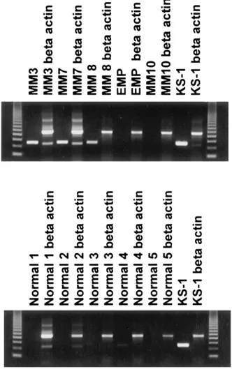
Figure 1 Detection of KSHV gene sequences in DNA isolated from bone marrow core biopsy of patients (upper lane: MM 3, 7, 8 and 10, and extramedullary plasmacytoma (EMP)), normal controls (lower lane) and as a positive control KS-1 cell line by DNA-PCR using ORF26 (KS330233) and beta-actin primers.
1270
Figure 2 DNA-PCR amplification results on DNA samples obtained from BM core biopsy from patients with MM (MM 11, 16, 17, 18 and 6; MGUS: MM 12, 19, 5 and 20), other hematological disorders such as ITP, CML and lymphoma and DNA from KS-1 cell line (positive control) using the vIRF primer (ORF K9) (upper lane) and beta actin control (lower lane).
Table 1 Patient characteristics and the results of the DNA PCR with three pairs of primers
Patient No. Stage Age M type Year of dx KS330233 v IRF v IL-8R
(ORF26) (ORF K9) (ORF74)
Previously treated patients
1 IA 55 Lambda 1996 + + +
2 IIA 54 IgA lambda 1996 − ND ND
3 IIIB 58 IgG lambda 1998 + + ND
4 IIIA 47 IgG kappa 1996 + + +
5 IIIA 50 Kappa 1998 − − ND
6 IIIA 46 IgG 1999 + + ND
Untreated patients
7 IIA 65 IgG kappa 1998 + + +
8 IIB 72 IgA 1998 + + +
9 66 Plasmacytoma 1998 − − −
10 IIIA 56 IgG 1998 − − −
11 MGUS 43 IgG kappa 1998 − − −
12 MGUS 60 IgG 1998 − − ND
13 IIIA 78 IgG 1998 + + +
14 IIA 62 Kappa 1998 + + +
15 IIIB 58 IgA 1998 + + +
16 IIIA 38 Lambda 1998 + + −
17 IIA 70 IgA kappa 1999 + + +
18 IIA 51 IgG lambda 1999 + + +
19 IIA 69 IgG kappa 1999 + + +
20 IIA 66 IgG lambda 1999 + + +
21 IIIB 79 IgG 1998 + + ND
22 IIIA 71 IgG kappa 2000 ND + ND
23 IIA 50 Lambda 2000 ND + ND
24 IA 61 Lambda 1998 ND + ND
ND, not done.
ORF74) were in agreement as shown in Figure 3 and Table 1. Fourteen of the 29 samples including patients with plasma cell disorders, and controls were independently analyzed in Turkey. These analyses were done blindly and results were completely in agreement with the results obtained in the US Laboratory (Figure 4). Interestingly, the MM patient (patient 2) who was in remission following autologous PBSCT showed the absence of the virus. In addition, using these same three sets of HHV-8 primers, no amplified product was obtained from the patients with CML, ITP, lymphoma, and polyclonal gammopathy (Figure 2). BM biopsy samples from the five nor-mal subjects were also negative for the presence of HHV-8 in both laboratories (Figures 1 and 4).
Figure 3 Comparison of three different primers: ORF 74 (vIL-8R), ORFK9 (vIRF) and ORF 26 (KS330233) on the same samples (MM 13,
1271
Figure 4 DNA-PCR amplification results on samples from patients with MM (MM 1, 7, 8, 12, 16, 22, 23, 24); EMP and five normal controls performed in an independent laboratory in Turkey using the vIRF(ORFK9) and hMLH1(human mismatch repair gene) primers.
RT-PCR results
As demonstrated by amplification with beta actin primers, intact RNA was obtained from seven individuals (Table 2). Four patients with monoclonal gammopathies (myeloma, n= 3; MGUS, n= 1), and three controls were analyzed. Using the ORFK9 primer pair, the two MM patients showing viral RNA also contained HHV-8 DNA (Table 2). Importantly, in two patients (MM and MGUS) without DNA evidence of HHV-8, there was also no amplified product with RT-PCR. In the three normal subjects’ BM biopsies, no viral RNA was found consistent with the DNA results.
Table 2 DNA and RT-PCR results using primer ORFK9 (vIRF)
Patient No. DNA-PCR RT-PCR
1 (MM) + + 8 (MM) + + 10 (MM) − − 12 (MGUS) − − Control 1 − − Control 2 − − Control 3 − − HHV-8 strain
Sequence analysis of ORF26 showed an HHV-8 strain pattern consistent with a C3 subtype (Table 3) which has also been found in MM patients from the United States.14
Discussion
Following the detection of HHV-8 in Kaposi’s sarcoma, its association with AIDS-related body cavity-based lymphoma, and multicentric Castleman’s disease has been reported.15,16 Since the initial observation demonstrating HHV-8 in nonma-ligant stromal cells from patients with MM,3conflicting reports have been published.3–7,10,11,16–24 The HHV-8 genome enco-des homologues of IL-6, IL-8R, viral interferon regulatory fac-tor (vIRF), macrophage inflammafac-tory proteins, bcl-2, and cyclin D1.25These molecules may be involved in both pro-moting HHV-8 infection as well as directly or indirectly con-tributing to malignant transformation and growth. Since HHV-8 infects the supportive nonmalignant cells in the myeloma bone marrow, the role of this virus is probably due to an indirect influence in this malignant disease.
This study provides further evidence for the presence of this virus in myeloma patients from another part of the world, sup-porting its importance in myeloma pathogenesis. In this study, we observed HHV-8 presence in 17 of the 21 Turkish patients with MM. In contrast, none of the healthy subjects or patients with other hematological disorders showed the presence of HHV-8. Patients with solitary plasmacytoma (uninvolved marrow) or MGUS also did not show evidence of the virus in this study. One of the treated MM patients in this study with-out HHV-8 was observed in a patient in remission following autologous PBSCT. This type of treatment may reduce the viral load below the sensitivity of this PCR assay. The inability to detect virus following PBSCT was reported previously and was regarded as evidence against the role of the virus in MM.18,23,26However, as demonstrated in this study, the lack of detectable HHV-8 in this group of patients may not be uncommon, especially in responding patients. In addition, the absence of the virus in circulating cells is not sufficient to rule out the presence of HHV-8 in MM patients.11,24,26In support of this, the phenotype of cells examined for viral presence by other groups differed from the cells that have been shown by our group and others to contain HHV-8 in myeloma patients’ blood samples. Certainly, the frequency of virally infected cir-culating cells may be less than the sensitivity of the method of detection which can also vary greatly between laboratories. Thirteen of the 15 previously untreated MM patients demon-strated the presence of the virus. BM biopsy material was essential for the demonstration of the virus. In contrast, BM aspirates showed viral presence in only one sample in this
1272 Table 3 Sequence from 233 base pair fragment within ORF26
DNA Country Dx 475899 595 614 621 631 632 647 653 654 664 668 688 691 705 719 731 738 750 756 757 798 47788 Sub-group source Changet al KS C G A C C C T C G G A T G C C A A A T C T G A KS USA KS C C G A A A KS-1 C-L A T T A C B BC-1 C-L C T G A A A MM USA MM A T T G C C3 MM 1 TR MM A T T T G C C C3 MM 4 TR MM A T T C G C C3 MM 7 TR MM A T T G C C3 MM 18 TR MM A T T G C C3 MM 19 TR MM A T T G C C3 Tr, Turkish.
study (data not shown) consistent with our previous findings in US MM specimens.3 Since fresh BM aspirate is likely to contain a lower proportion of dendritic cells than biopsy material, a reduced ability to detect HHV-8 in these samples is not surprising.17
Previous reports on the presence of HHV-8 in MM have yielded conflicting results. Investigators have focused on the detection of molecular evidence in a variety of tissues, includ-ing BM aspiration or biopsy material, cultured dendritic cells from leukapheresis products or BM samples.3–11,17,18,23–30 Others searched for serological evidence by antibodies against the virus.19–22 All of these studies have been reviewed recently.17,23 The most frequent method used in the studies that failed to demonstrate viral presence in MM were PCR assays with peripheral blood dendritic cells obtained after long-term culture.23,24,26The types of cells obtained in these negative studies were highly variable, and contained different cell populations than those studies showing HHV-8 in these patients.3,7In addition, the PCR assays including the types of primers and positive and negative controls for detecting HHV-8 were highly variable in these studies.22–24,27 Other groups from the United States and Europe have identified HHV-8 DNA and RNA in MM patients consistent with this study.3–5,7 With an attempt to eliminate this uncertainty, we used the same primer sets in two independent laboratories in a blind fashion, and the results were completely concordant. In addition, the presence of the same strain, C3, in both US and Turkish myeloma patients further supports the presence of a specific HHV-8 subtype in this B cell malignancy.14
In conclusion, this study demonstrates the frequent pres-ence of HHV-8, confirmed by two independent laboratories, in MM patients with a specific viral strain in another geo-graphic location; and, thus, provides further support for the important role of this type of HHV-8 in the pathogenesis of multiple myeloma.
Acknowledgements
M Beksac is a member and is partially supported by the Turk-ish Academy of Sciences.
References
1 Chang Y, Cesarman E, Pessin MS, Lee F, Culpepper J, Knowles DM, Moore PS. Identification of herpesvirus-like DNA sequences in AIDS-associated Kaposi’s sarcoma. Science 1994; 266: 1865 –1869.
2 Burger R, Neipel F, Fleckenstein B, Savino R, Ciliberto G, Kalden
JR, Gramatzki M. Human herpesvirus type 8 interleukin-6 homol-ogue is functionally active on human myeloma cells. Blood 1998;
91: 1858–1863.
3 Rettig MB, Ma HJ, Vescio RA, Pold M, Schiller G, Belson D, Sav-age A, Nishikubo C, Wu J, Fraser J, Said JW, Berenson JR. Kaposi’s sarcoma-associated herpesvirus infection of bone marrow den-dritic cells from multiple myeloma patients. Science 1997; 276: 1851–1854.
4 Rettig M, Vescio R, Ma H, Schiller G, Savage A, Berenson J. Sequence variability of HHV-8 from dendritic cells of multiple myeloma patients. Blood 1997; 90: 86a.
5 Rettig MB, Vescio RA, Moss TJ, Ma HJ, Schiller G, Berenson JR. Detection of Kaposi’s sarcoma-associated herpesvirus in the per-ipheral blood of multiple myeloma patients. Blood 1997; 90: 587a.
6 Said JW, Rettig MR, Heppner K, Vescio RA, Schiller G, Ma HJ, Belson D, Savage A, Shintaku P, Koeffler HP, Asou H, Pinkus G, Pinkus J, Schrage M, Green E, Berenson JR. Localization of Kapo-si’s sarcoma-associated herpesvirus in bone marrow biopsy samples from patients with multiple myeloma. Blood 1997; 90: 4278–4282.
7 Raje N, Gong J, Chauhan D, Teoh G, Avigan D, Wu Z, Chen D, Treon SP, Kufe DW, Anderson KC. Bone marrow and peripheral blood dendritic cells from patients with multiple myeloma are phenotypically and functionally normal despite the detection of Kaposi’s sarcoma herpesvirus gene sequences. Blood 1999; 93: 1487–1495.
8 Brousset P, Meggetto F, Attal M, Delsol G. Kaposi’s sarcoma-asso-ciated herpesvirus infection and multiple myeloma. Science 1997;
278: 1972.
9 Cull GM, Timms JM, Haynes AP, Russel NH, Irving WL, Ball JK, Thomson BJ. Dendritic cells cultured from mononuclear cells and CD34 cells in myeloma do not harbour human herpesvirus 8. Br
J Haematol 1998; 100: 793–796.
10 Bouscary D, Dupin N, Fichelson S, Grandadam M, Fontenay-Roupe M, Marcelin AG, Blanche P, Picard F, Freyssinier JM, Ravaud P, Dreyfus F, Calvez V. Lack of evidence of an association between HHV-8 and multiple myeloma. Leukemia 1998; 12: 1840–1841.
11 Yi Q, Ekman M, Anton D, Bergenbrant S, O¨ sterborg A, Georgii-Hemming P, Holm G, Nilsson K, Biberfeld P. Blood dendritic cells from myeloma patients are not infected with Kaposi’s sarcoma-associated herpesvirus. Blood 1998; 92: 402–404.
12 Shak-Shie NN, Vescio RA, Berenson JR. HHV-8 infection and mul-tiple myeloma. J Leuk Biol 1999; 66: 357–360.
13 Cesarman E, Moore PS, Rao P, Inghirami G, Knowles DM, Chang Y. In vitro establishment and characterization of two acquired immmunodeficiency syndrome-related lymphoma cell-lines (BC-1 and BC-2) containing Kaposi‡s sarcoma-associated herpes virus-like (KSHV) DNA sequences. Blood 1995; 86: 2708–2714. 14 Ma HJ, Sjak-Shie NN, Vescio RA, Kaminsky M, Mikail A, Pold M,
Parker K, Beksac M, Belson D, Moss JJ, Wu CH, Zhou J, Zhang L, Chen G, Said JW, Berenson JR. Human herpes virus 8 open reading frame 26 and open reading frame 65 sequences from mul-tiple myeloma patients: a shared pattern not found in Kaposi’s sarcoma or primary effusion lymphoma. Clin Cancer Res. 2000;
1273 15 Cesarman E, Chang Y, Moore PS, Said JW, Knowles DM. Kaposi’s
sarcoma-associated herpesvirus-like DNA sequences in AIDS-related body cavity-based lymphomas. N Engl J Med 1995; 32: 1186–1191.
16 Soulier J, Grollet L, Oksenhendler E, Cacoub P, Cazals-Hatem D, Babinet P, D’Agay MF, Clauvel JP, Raphael M, Degos L, Sigaux F. Kaposi’s sarcoma-associated herpesvirus-like DNA sequences in multicentric Castleman’s disease. Blood 1995; 86: 1276–1280. 17 Berenson JR, Vescio RA. HHV-8 is present in multiple myeloma
patients. Blood 1999; 93: 3157–3166.
18 Tisdale JE, Stewart AK, Dickstein B, Dube ID, Cappe D, Dunbar CE, Brown KE. Human herpesvirus 8 in patients with multiple myeloma. Blood 1997; 90: 588a.
19 Luppi M, Barozzi P, Marasca R, Ferari MG, Torelli G. Human Herpesvirus 8 strain variability in clinical conditions other than Kaposi’s sarcoma. J Virol 1997; 71: 8082–8083.
20 Massod R, Zheng T, Tulpule A, Arora N. Kaposi’s sarcoma-asso-ciated herpesvirus infection and multiple myeloma. Science 1997;
278: 1970–1971.
21 Olsen SJ, Tarte K, Sherman W, Hale EE, Weisse MT, Orazi A, Klein B, Chang Y. Evidence against KSHV infection in the pathogenesis of multiple myeloma. Virus Res 1998; 57: 197–202.
22 Parravicini C, Lauri E, Baldini L, Neri A, Poli F, Sirchia G, Moroni M, Galli M, Corbellino M. Kaposi’s sarcoma-associated herpesvi-rus infection and multiple myeloma. Science 1997; 278: 1969– 1970.
23 Tarte K, Chang Y, Klein B. Kaposi’s sarcoma-associated herpesvi-rus and multiple myeloma: lack of criteria for causality. Blood 1999; 93: 3159–3166.
24 Tarte K, Olsen SJ, Lu ZY, Legouffe E, Rossi JF, Chang Y, Klein B. Clinical grade functional dendritic cells from patients with mul-tiple myeloma are not infected with Kaposi’s sarcoma-associated herpesvirus. Blood 1998; 91: 1852–1857.
25 Ma H, Vescio R, DerDanielian M, Schiller G, Berenson JR. The HHV-8 IL-8R homologue and interferon regulatory factor genes are frequently expressed in myeloma bone marrow biopsies whereas the vIL-6 is rarely found. Blood 1998; 92: 515a. 26 Degreef C, Bakkus M, Heirman C, Schots R, Laacon P, De Weale
M, Van Camp B, Van Riet I. The absence of Kaposi’s sarcoma-associated herpesvirus DNA sequences in leukapheresis products and ex-vivo expanded CD34-positive cells in multiple myeloma patients. Blood 1997; 90: 86a.
27 Agbalika F, Mariette X, Marolleau JP, Fermand JP, Brouet JC. Detection of human herpesvirus-8 DNA in bone marrow biopsies from patients with multiple myeloma and Waldenstrom’s macrog-lobulinemia. Blood 1998; 91: 4393–4394.
28 Cathomas G, Stalder A, Kurrer MO, Regamey N, Erb P, Joller-Jemelka HI. Multiple myeloma and HHV-8 infection. Blood 1998;
91: 4391–4392.
29 Schonrich G, Raftery M, Schnitzler P, Rohr U, Goldschmidt H. Absence of a correlation between Kaposi’s sarcoma-associated herpesvirus and multiple myeloma. Blood 1998; 92: 3474–3491. 30 Tisdale JF, Stewart AK, Dickstein B, Little RF, Dube I, Cappe D, Dunbar CE, Brown KE. Molecular and serological examination of the relationship of human herpesvirus 8 to multiple myeloma: ORF26 sequences in the bone marrow stroma are not restricted to myeloma patients and other regions of the genome are not detected. Blood 1998; 92: 2681–2687.