Full Terms & Conditions of access and use can be found at
https://www.tandfonline.com/action/journalInformation?journalCode=ibih20
Biotechnic & Histochemistry
ISSN: 1052-0295 (Print) 1473-7760 (Online) Journal homepage: https://www.tandfonline.com/loi/ibih20
The effects of resveratrol administration on
lipid oxidation in experimental renal
ischemia-reperfusion injury in rats
A. K. Baltaci, H. Gokbudak, S. B. Baltaci, R. Mogulkoc & M. C. Avunduk
To cite this article: A. K. Baltaci, H. Gokbudak, S. B. Baltaci, R. Mogulkoc & M. C. Avunduk (2019) The effects of resveratrol administration on lipid oxidation in experimental renal ischemia-reperfusion injury in rats, Biotechnic & Histochemistry, 94:8, 592-599, DOI: 10.1080/10520295.2019.1612091To link to this article: https://doi.org/10.1080/10520295.2019.1612091
Published online: 04 Jul 2019.
Submit your article to this journal
Article views: 173
View related articles
View Crossmark data
The effects of resveratrol administration on lipid oxidation in experimental renal
ischemia-reperfusion injury in rats
A. K. Baltacia, H. Gokbudaka, S. B. Baltacia, R. Mogulkoca, and M. C. Avundukb
aDepartment of Physiology, Faculty of Medicine, Selçuk University, Konya, Turkey;bMeram Faculty of Medicine, Department of Pathology,
Necmettin Erbakan University, Konya, Turkey
ABSTRACT
We investigated how resveratrol affects lipid oxidation during experimental renal ischemia-reperfusion injury in rats. We used 48 adult male rats assigned to five groups: group 1, control; group 2, renal ischemia; group 3, renal ischemia + reperfusion; group 4, resveratrol + renal ischemia; group 5, resveratrol + renal ischemia + reperfusion. Plasma and renal tissue malondialdehyde (MDA), and erythrocyte and renal tissue glutathione (GSH) levels were measured and histologic changes in the renal tissue were examined. Ischemia-reperfusion affected the MDA-GSH balance adversely and caused histopathological changes in the renal tissue of the ischemia and ischemia + reperfusion groups. Resveratrol treatment normalized MDA and GSH levels as well as the histopathology that occurred in the renal tissue of the ischemia and ischemia + reperfusion groups.
KEYWORDS Histopathology; lipid oxidation; rat; renal ischemia; reperfusion; resveratrol
Reactive oxygen species (ROS) participate in the pathophysiology of renal diseases; therefore, antioxidants are used to reduce the sensitivity of kidneys to oxidative challenges. Resveratrol is a phenolic compound derived from edible plants such as grapes and peanuts; it is a bioactive molecule that exhibits physiological effects on many organs. Resveratrol is an antioxidant and anti-inflammatory substance (Baltaci et al. 2016; Ficarra et al. 2016). Under conditions of lipid oxidation, antioxidant effects of resveratrol are as potent as ascorbic acid or α-tocopherol (Tartaro Bujak et al. 2016). Resveratrol exhibits a protective effect by preventing oxidative stress caused by ROS generated by activation of macrophages (Luo et al.2016).
A high cholesterol diet contributes to lipid accumulation in the body. Hypercholesterolemia is reported to cause oxidative stress (Paul et al. 2017). Resveratrol treatment of rabbits fed a high cholesterol diet produced a significant reduction in high density lipoprotein cholesterol (HDL-c), low density lipoprotein cholesterol (LDL-c), total cholesterol and total triacylglycerol. Therefore, resveratrol might prove useful for treating hyperlipidemia and atherosclerosis (Tanko et al.2016). Resveratrol treatment of obese mice for 2 weeks reduced systemic and bladder oxidative stress and substantially prevented bladder hyperactivity. It has been suggested that resveratrol
administration could be effective for preventing obesity-associated overactive bladder (Alexandre et al. 2016). Combined supplementation of enriched diet plus resveratrol administration was reported to exhibit antioxidant effects by reducing malondialdehyde (MDA) levels and decreasing lipid oxidation in young healthy mice (Muhammed et al. 2017). Resveratrol prevented testicular and epididymal oxidative damage in cases of induced epididymal toxicity in rat testis and also exhibited both steroidogenic and antioxidant activity in the testis (Reddy et al. 2016).
We investigated how the antioxidant property of resveratrol affects lipid oxidation in experimental renal ischemia-reperfusion injury in rats.
Material and methods
Animals
Our study was conducted at the Experimental Medicine Application and Research Center of Necmettin Erbakan University. We used 48 250–260 g Wistar albino adult male rats supplied by the Center. Our study protocol was approved by the Center’s Ethics Board.
The rats were housed in steel cages that were washed daily. Food was supplied in steel bowls and tap water was provided in glass feeding bottles. Animals were fed daily 10 g standard rat feed/100 g body weight. Food
CONTACTS. B. Baltaci [email protected] Department of Physiology, Faculty of Medicine, Selçuk University, Konya 42031, Turkey 2019, VOL. 94, NO. 8, 592–599
https://doi.org/10.1080/10520295.2019.1612091
and water were available ad libitum. The feed was supplied by the Experimental Medicine Research and Application Center of Necmettin Erbakan University. The animals were housed at 21 ± 1 °C with a 12 h light:12 h dark cycle.
Resveratrol (3,4,5-trihydroxystilbene) (Sigma Aldrich Co., St. Louis, MO) was dissolved in dimethyl sulfoxide (DMSO) (Sigma) and added to the drinking water for groups 4 and 5 to deliver a dose of 60 mg/kg/ day. After 3 weeks, all animals were decapitated between 09:00 and 10:00 hours. Blood samples were collected after decapitation, then renal tissue samples were obtained. Only ischemic renal tissue samples were used for biochemical and histopathological analyses.
The animals were divided randomly into five groups. Group 1 was the untreated control (n = 8). For group 2, renal ischemia group (n = 10), the left kidney of each animal was subjected to ischemia for 60 min under general anesthesia. For group 3, renal ischemia-reperfusion group (n = 10), following ischemia for 60 min, the left kidney of each animal was re-perfused for 60 min. For group 4, resveratrol + renal ischemia group (n = 10), animals were given resveratrol in their drinking water for 3 weeks followed by ischemia for 60 min. For group 5, resveratrol + renal ischemia + reperfusion group (I/R) (n = 10), animals were given resveratrol in their drinking water for 3 weeks followed by ischemia for 60 min, then reperfusion for 60 min. Animals were anesthetized with intraperitoneal (i.p.) injection of 60 mg/kg ketamine HCl and 10 mg/kg xylazine (Rompun 2%, Bayer, Istanbul, Turkey) for all procedures.
Plasma MDA
An benchtop centrifuge (Allegra X-22R; Beckmann Coulter Life Sciences, Indianapolis, IN) was used for centrifugation of samples at 1,734 X g. Blood samples collected in EDTA tubes were centrifuged for 5 min to separate the plasma. We added 10% trichloroacetic acid (TCA) (818 K02907810; Merck, Taufkirchen, Germany), 2.5 ml, to a sample tube followed by 0.5 ml plasma. The tubes were vortexed and capped. Following incubation in a 90 °C water bath for 15 min, they were cooled in cold water and measured in a spectrophotometer at 532 nm against the blank. The blank was prepared using distilled water instead of supernatant and treated in the same way as the samples. The results are presented as nmol/ml.
Tissue MDA
After the tissues to be analyzed were weighed, divided into pieces and put into tubes, they were placed in
a Misonix Microson ultrasonic cell disruptor (BioLogics, Inc., Manassas, VA) with 150 mM KCl at 4 °C to obtain 10% homogenates. To 2 ml samples of the homogenized tissues we added 2 ml 8% HClO4and
centrifuged for 15 min. We added 0.5 ml of the supernatant to 3 ml 1% H3PO4 and 1 ml 0.675%
TBA, followed by incubation in a 90 °C water bath for 45 min. When the mixture cooled, we added 4 ml of n-butanol and measured absorbance at 532 nm against n-butanol using a spectrophotometer (Perkin Elmer, Solingen, Germany). The result was expressed as nmol/g tissue.
Tissue glutathione (GSH)
To determine GSH levels, the tissue was homogenized at 4 °C with 150 mM KCl to obtain a 10% homogenate, then centrifuged for 15 min as described for MDA. Ellman’s method was used to measure the GSH in the samples (Ellmann 1959). Ellman’s solution was prepared by dissolving 100 mg 5′-5′-dithiobis -2-nitrobenzoic acid (DTNB) (D-8130; Sigma) in 100 ml phosphate buffer, pH 7.8. To 200 µl of supernatant, 8 ml phosphate buffer, pH 6.8, 78 ml 1 N NaOH and 100 µl Ellman’s solution were added and held for 5 min. The absorbance was measured against distilled water at 412 nm using a spectrophotometer (Perkin Elmer) (Mogulkoc et al. 2006).
Erythrocyte GSH
To quantify the GSH in erythrocytes, blood samples were placed in EDTA tubes and centrifuged for 5 min. After the plasma was separated, erythrocyte samples were bathed in 0.9% saline three times. To 50 µl of bathed erythrocytes we added 450 µl distilled water and 500 µl 10% sulfosalicylic acid. After cooling on ice for 1 h, the mixture was centrifuged for 3 min. To 200 µl supernatant we added 8 ml phosphate buffer, pH 6.8, 78 µl 1 N NaOH and 100 µl Ellman’s reagent. The absorbance was measured after 5 min at 412 nm against the distilled water blank in the spectrophotometer (Perkin Elmer). The GSH standard was prepared by dissolving 15.36 mg reduced glutathione (G-4251; Sigma) in 100 ml/mM Na2
EDTA. The results were presented as µmol/g hemoglobin (Hb).
Histology
Renal tissue samples were fixed in 10% buffered paraformaldehyde for 24 h. Tissue samples were BIOTECHNIC & HISTOCHEMISTRY 593
dehydrated through ascending alcohol solutions and cleared with xylene using an Autotechnicon (ASP300; Leica, Nussloch, Germany), then embedded in paraffin. Sections were cut at 5 µm with microtome and after deparaffinization in an oven at 65 ºC for 1 h, sections were stained with hematoxylin and eosin (H & E) using an automatic staining device (Varistain Gemini; Thermo Shandon, Cheshire, UK).
Stained sections were examined using a Nikon Eclipse E400 light microscope (Nikon Corp., Tokyo, Japan). For each specimen, the same area was photographed with a Nikon Coolpix 5000 microscope photographic attachment (Nikon Corp.). A Nikon micrometer microscope slide (MBM11100; Nikon Corp.) also was photographed during the procedure. All photographs then were transferred into a PC and analyzed using Clemex Vision Lite 3.5 Image Analysis program (Clemex Technologies Inc., Quebec, Canada). The analysis system was calibrated by comparing the photograph of the specimen with the photograph of the Nikon micrometer microscope slide, which was taken at the same magnification. The image analysis system also can be used to calculate area. A 390,017.9μm2area then was calculated using the same image analysis program; microscopic images were evaluated in this selected area. For evaluation, numerical values were obtained using the following criteria based on the semiquantitative method of Paller (Avunduk et al.2003). For epithelial flattening: 1 point if flattened, 0 points if not. For loss of brush-like edges: 2 points if there was a loss, 0 points if there was no loss. For cytoplasmic vacuolization: 1 point if vacuoles appeared, 0 points if not. For cell necrosis: 1 or 2 points based on severity; and tubule lumen obstruction: 2 points if the obstruction was > 50%, 1 point if < 50%,. All numerical values obtained for each property were collected. The numerical value obtained was the Paller Score. These procedures were performed separately for all five groups of animals.
Statistical analysis
SPSS v 21.0 software was used for statistical evaluation of the data. Means ± SD were calculated. The Shapiro-Wilks test was used to confirm that the data were distributed normally. One-way analysis of variance was used to assess differences between groups and the least significant difference test was used to identify the group from which the difference originated. Values for p ≤ 0.05 were considered significant.
Results
Plasma MDA and erythrocyte GSH
MDA levels were measured for all five groups. The highest plasma MDA levels were found in groups 2 and 3 (p < 0.05); group 4 exhibited the lowest plasma MDA level (p < 0.05). Plasma MDA values for group 5 were higher than for group 4, but significantly lower than for all other groups (p < 0.05) (Table 1). The highest erythrocyte GSH levels were found for groups 4 and 5 compared to all other groups (p < 0.05). Groups 2 and 3 exhibited the lowest erythrocyte GSH levels (p < 0.05) (Table 1).
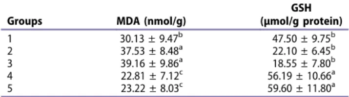
Renal tissue MDA and GSH
The highest renal MDA levels were found in groups 2 and 3 (p < 0.05). Renal MDA levels in groups 4 and 5 were higher than the control levels (p < 0.05), but lower than those for groups 2 and 3 (p < 0.05). The highest renal GSH levels were found in groups 4 and 5 (p < 0.05). Groups 2 and 3 exhibited the lowest GSH levels for erythrocytes (p < 0.05) (Table 2).
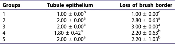
Histology
We found no significant difference in cytoplasmic vacuolization, tubule epithelium or lumen loss among the groups. The greatest brush border loss and cell
Table 1.Plasma MDA and erythrocyte GSH levels.
Groups MDA (nmol/ml) GSH (µmol/g Hb) 1 18.74 ± 3.15b 109.05 ± 16.30b 2 23.22 ± 4.92a 67.30 ± 11.97c 3 23.97 ± 3.95a 69.59 ± 17.63c
4 5.04 ± 3.72d 181.55 ± 25.93a 5 10.21 ± 1.2c 188.73 ± 22.82a Group 1, control; group 2, renal ischemia; group 3, renal ischemia +
reperfusion; group 4, resveratrol + renal ischemia; group 5, resveratrol + renal ischemia + reperfusion. a−dDifferences between group means with different superscript letters in the same column are significant (p < 0.05).
Table 2.Renal MDA and GSH levels.
Groups MDA (nmol/g)
GSH (µmol/g protein) 1 30.13 ± 9.47b 47.50 ± 9.75b 2 37.53 ± 8.48a 22.10 ± 6.45b 3 39.16 ± 9.86a 18.55 ± 7.80b 4 22.81 ± 7.12c 56.19 ± 10.66a 5 23.22 ± 8.03c 59.60 ± 11.80a Group 1, control; group 2, renal ischemia; group 3, renal ischemia +
reperfusion; group 4, resveratrol + renal ischemia; group 5, resveratrol + renal ischemia + reperfusion. a−dDifferences between groups with superscripted letters in the same column are significant (p < 0.001).
necrosis, and the highest Paller score, were found in groups 2 and 3 compared to all other groups (p < 0.05). Values for the same characteristics in groups 4 and 5 were higher than those for group 1, but lower than for groups 2 and 3 compared to other groups (p < 0.00) (Tables 3−5;Figures 1–5).
Discussion
Renal ischemia-reperfusion is a serious clinical complication that can cause tissue damage for which there is no specific treatment. Owing to its effects on cell metabolism, oxidative stress and inflammation, resveratrol has been proposed as a possible treatment for kidney ischemia-reperfusion (Khader et al. 2015). These investigators reported that administration of resveratrol to adult male rats with induced ischemia-reperfusion significantly reduced kidney injury by reducing the levels of interleukin-6 and interleukin-1, nitric oxide synthase and
tumor necrosis factor alpha (TNF-α). Treatment with 5 mg/ kg resveratrol corrected the functional impairment and morphological changes caused by ischemia-reperfusion in renal tissue and reduced elevated MDA levels and increased antioxidant activity (Chander and Chopra2006). Similarly, Saito et al. (2005) reported that resveratrol treatment reversed the loss of renal function that resulted from ischemia-reperfusion injury. Injection of 10 mg/kg/day resveratrol i.p. significantly reduced ROS damage due to renal ischemia-reperfusion in diabetic rats (Xiao et al. 2016). Our findings are consistent with earlier reports that resveratrol supplementation exhibits antioxidant activity. Xiao et al. (2016) administered 10 mg/kg/day resveratrol to diabetic rats for 7 days. Chander and Chopra (2006) administered a single dose of resveratrol and Saito et al. (2005) used resveratrol just before ischemia and 6 days after reperfusion. By contrast to earlier research, we used oral resveratrol supplementation for 3 weeks.
Renal injury causes development of renal fibrosis. It has been suggested that resveratrol administration might protect against both renal injury and the development of fibrosis by inhibiting the effect of transforming growth factor β on matrix metalloproteinase 7 (Xiao et al. 2016).
When the kidney medulla was exposed to extreme oxidative stress due to local hypoxia and hypertonicity, administration of resveratrol prevented tissue injury by inhibiting the expression of inflammatory proteins and activation of nuclear factor-kappa B (Bae et al. 2016). Sener et al. (2006) subjected nephrectomized rats to renal ischemia for 45 min, then reperfused them for 6 h. The rats were injected i.p. with 30 mg/kg resveratrol 30 min before ischemia and immediately following reperfusion. These investigators reported decreased tissue GSH levels and increased MDA levels owing to the antioxidant properties of resveratrol. Resveratrol treatment also ameliorated histopathological changes.
We also found increased MDA and reduced GSH levels as a result of renal ischemia-reperfusion. We found that GSH levels increased and MDA levels decreased after resveratrol administration for both renal ischemia and reperfusion. Clearly, resveratrol prevents tissue injury under renal ischemia-reperfusion conditions by stimulating antioxidant activity. Our findings are consistent with those reported by Sener et al. (2006).
Administration of polydatin, the glucoside form of resveratrol, increased the antioxidant capacity and reduced apoptosis in kidney tissue by ischemia-reperfusion injury (Meng et al.2016). Khader et al. (2015) reported that resveratrol administration after renal ischemia-reperfusion in adult male rats reduced the
Table 3.Histopathology: tubule epithelium and loss of brush border.
Groups Tubule epithelium Loss of brush border 1 1.00 ± 0.00b 1.00 ± 0.00c
2 2.00 ± 0.00a 2.80 ± 0.63a
3 2.00 ± 0.00a 3.00 ± 0.00a 4 1.80 ± 0.42a 2.20 ± 0.63b 5 2.00 ± 0.00a 2.20 ± 1.03b
Group 1, control; group 2, renal ischemia; group 3, renal ischemia + reperfusion; group 4, resveratrol + renal ischemia; group 5, resveratrol + renal ischemia + reperfusion. a−dDifferences between group means with different superscript letters in the same column are significant (p < 0.05).
Table 4.Histopathology: cytoplasmic vacuolization and cell necrosis.
Groups Cytoplasmic vacuolization Cell necrosis
1 1.10 ± 0.31a 1.00 ± 0.31c 2 1.50 ± 0.52a 2.70 ± 0.48a
3 1.60 ± 0.51a 2.70 ± 0.48a
4 1.30 ± 0.48a 1.40 ± 0.00b 5 1.50 ± 0.52a 1.50 ± 0.51b Group 1, control; group 2, renal ischemia; group 3, renal ischemia
+ reperfusion; group 4, resveratrol + renal ischemia; group 5, resveratrol + renal ischemia + reperfusion.a−dDifferences between group means with different letters in the same column are significant (p < 0.05).
Table 5.Histopathology: tubule lumen obstruction and Paller scores.
Groups Tubule lumen obstruction Paller score 1 2.30 ± 0.00a 1.00 ± 0.31c 2 2.90 ± 0.48a 3.60 ± 0.48a
3 2.90 ± 0.31a 3.70 ± 0.48a 4 2.30 ± 0.73a 1.80 ± 0.00b 5 2.50 ± 0.52a 2.10 ± 0.51b Group 1, control; group 2, renal ischemia; group 3, renal ischemia
+ reperfusion; group 4, resveratrol + renal ischemia; group 5, resveratrol + renal ischemia + reperfusion.a−dDifferences between group means with different letters in the same column are significant (p < 0.05).
number of apoptotic cells and ameliorated histopathologic changes after ischemia-reperfusion. Sener et al. (2006) reported that resveratrol treatment prevented histopathological changes that occurred during renal
ischemia-reperfusion. Others have reported that morphological changes resulting from ischemia-reperfusion injury can be corrected by resveratrol administration (Giovannini et al.2001; Khader et al.2015).
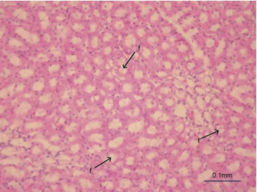
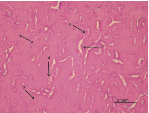
Figure 2.Group 2. Obstruction of renal tubules (o), flattened tubule epithelium (d) and necrotic foci (n) are more obvious and frequent than in groups 4 and 5.
Figure 3.Group 3. Obstruction of tubules (o), flattened tubule epithelium (d) and necrotic foci (n) are more obvious and frequent than in groups 4 and 5.
Figure 4.Group 4. Flattened the tubule epithelium (d), obliterated tubule lumina (o) and cytoplasmic vacuolization (v) are evident.
We found that repeated oral administration of 60 mg/kg resveratrol for 3 weeks may be useful for preventing renal ischemia-reperfusion injury.
Acknowledgments
This study was supported by the Scientific Research Projects Coordinatorship of Selcuk University (SUBAPK; project no. 13202027).
Disclosure statement
No potential conflict of interest was reported by the authors.
Funding
This work was supported by the Scientific Research Projects Coordinatorship of Selcuk University [SUBAPK; project no. 13202027].
References
Alexandre EC, Calmasini FB, de Oliveira MG, Silva FH, Da Silva CP, André DM, Leonardo FC, Delbin MA, Antunes E. 2016. Chronic treatment with resveratrol improves over active bladder in obese mice via antioxidant activity. Eur J Pharmacol. 788:29–36. doi:10.1016/j.ejphar.2016.06.017.
Avundu K MC, Yurdakul T, Erdemli E, Yavuz A. 2003. Prevention of renal damage by alphatocopherol in
ischemia and reperfusion models of rats. Urol Res. 31:280–285. doi:10.1007/s00240-003-0329-y.
Bae EH, Joo SY, Ma SK, Lee J, Kim SW. 2016. Resveratrol attenuates 4-hydroxy-2-hexenal-induced oxidative stress in mouse cortical collecting duct cells. Kor J Physiol Pharmacol. 20:229–236. doi:10.4196/kjpp.2016.20.3.229. Baltaci SB, Mogulkoc R, Baltaci AK. 2016. Resveratrol and
exercise. Biomed Rep. 5:525–530. doi:10.3892/br.2016.777. Chander V, Chopra K.2006. Protective effect of nitric oxide pathway in resveratrol renal ischemia-reperfusion injury in rats. Arch Med Res. 37:19–26. doi:10.1016/j. arcmed.2005.05.018.
Ellman GL. 1959. Tissue sulfhydryl groups. Arch Biochem Biophys. 82:70–77. doi:10.1016/0003-9861(59)90090-6. Ficarra S, Tellone E, Pirolli D, Russo A, Barreca D, Galtieri A,
Giardina B, Gavezzotti P, Riva, De Rosa MC. 2016. Insights into the properties of the two enantiomers of trans-δ-viniferin, a resveratrol derivative: antioxidant activity, biochemical and molecular modeling studies of its interactions with hemoglobin. Mol Biosyst. 12:1276–1286. doi:10.1039/c5mb00897b.
Giovannini L, Migliori M, Longoni BM, Das DK, Bertelli AA, Panichi V, Filippi C, Bertelli A. 2001. Resveratrol a polyphenol found in wine, reduces ischemia reperfusion injury in rat kidneys. J Cardiovasc Pharmacol. 37:262–270.
Khader A, Yang WL, Kuncewitch M, Prince JM, Marambaud P, Nicastro J, Coppa GF, Wang P. 2015. Novel resveratrol analogues attenuate renal ischemic injury in rats. J Surg Res. 193:807–815. doi:10.1016/j. jss.2014.08.015.
Luo G, Li Z, Wang Y, Wang H, Zhang Z, Chen W, Zhang Y, Xiao Y, Li C, Guo Y, Sheng P.2016. Resveratrol protects against titanium particle-induced aseptic loosening through
Figure 5.Group 5. Necrotic foci (n), flattened the tubule epithelium (d), obliteration of the tubule lumina (o) and cytoplasmic vacuolization (v) are evident.
reduction of oxidative stress and inactivation of NF-κB. Inflammation. 39:775–785. doi:10.1007/s10753-016-0306-6. Meng QH, Liu HB, Wang JB. 2016. Polydatin ameliorates
renal ischemia/reperfusion injury by decreasing apoptosis and oxidative stress through activating sonic hedge hog signaling pathway. Food Chem Toxicol. 96:215–225. doi:10.1016/j.fct.2016.07.032.
Mogulkoc R, Baltaci AK, Oztekin E, Aydin L, Sivrikaya A. 2006. Melatonin prevents oxidant damage in various tissues of rats with hyperthyroidism. Life Sci. 79::311–315. doi:10.1016/j.lfs.2006.07.039.
Muhammad MS, Magaji RA, Mohammed A, Isa AS, Magaji MG. 2017. Effect of resveratrol and environmental enrichment on biomarkers of oxidative stress in young healthy mice. Metab Brain Dis. 32:163–170. doi:10.1007/s11011-016-9891-1.
Paul R, Choudhury A, Kumar S, Giri A, Sandhir R, Borah A. 2017. Cholesterol contributes to dopamine-neuronal loss in MPTP mouse model of Parkinson's disease: Involvement of mitochondrial dysfunctions and oxidative stress. Plos One. 12:e0171285.
Reddy KP, Madhu P, Reddy PS. 2016. Protective effects of resveratrol against cisplatin-induced testicular and epididymal toxicity in rats. Food Chem Toxicol. 91:65–72. doi:10.1016/j.fct.2016.02.016.
Saito M, Satoh S, Kojima N, Tada H, Sato M, Suzuki T, Senoo H, Habuchi T. 2005. Effects of a phenolic compound, resveratrol, on the renal function and co-stimulatory adhesion molecule CD86 expression in rat kidneys with ischemia/reperfusion injury. Arch Histol Cytol. 68:41–49.
Sener G, Tuğtepe H, Yüksel M, Cetinel S, Gedik N, Yeğen BC. 2006. Resveratrol improves ischemia/ reperfusion-induced oxidative renal injury in rats. Arch Med Res. 37:822–829. doi:10.1016/j.arcmed.2006.04.003. Tanko Y, Jimoh A, Ahmed A, Mohammed A, Ayo JO.2016.
Resveratrol protects rabbits against cholesterol diet-induced hyperlipidaemia. Niger J Physiol Sci. 31:71–75.
Tartaro Bujak I, Mihaljević B, Ferreri C, Chatgilialoglu C. 2016. The influence of antioxidants in the thiyl radical induced lipid oxidation and geometrical isomerization in micelles of linoleic acid. Free Rad Res. 50:S18–S23. doi:10.1080/10715762.2016.1231401.
Xiao YD, Huang YY, Wang HX, Wu Y, Leng Y, Liu M, Sun Q, Xia ZY. 2016. Thioredoxin-interacting protein mediates NLRP3 inflammasome activation involved in the susceptibility to ischemic acute kidney injury in diabetes. Oxid Med Cell Longev. 2016:2386068. doi:10.1155/2016/2386068.