Hemiarthroplasty For 4-Part Fractures Of The
Proximal Humerus: Is It A Good Treatment
Option?
Müjdat ADAŞ
1*İsmail KALKAR
1Cem Zeki ESENYEL
1Semih DEDEOĞLU
1Yusuf ÖZCAN
1Kürşat BAYRAKTAR
1Murat ÇAKAR
1Ayşın ERSOY
2 AbstractObjective: We examined the clinical, functional and radiological outcomes in patients 65 years and older who underwent primary shoulder hemiarthroplasty for a 4-part acute fracture of the proximal humerus, and evaluated efficacy of treatment, patient satisfaction and our procedural deficiencies.
Patients and Methods: Eighteen patients were treated with primary hemiarthroplasty for acute proximal humerus Neer type-IV fracture. During follow-up, active forward elevation, abduction, internal/external rotation of the shoulder were assessed. The assessments were based on the Constant-Murley Shoulder Score (CMSS), Simple Shoulder Test Score (SSTS), Oxford Shoulder Score (OSS), scales of University of California and Los Angeles (UCLA). Abduction strength was measured by a myometer
1* Sorumlu Yazar, Müjdat ADAŞ, [email protected]
1 Okmeydanı Eğitim ve Araştırma Hastanesi, Ortopedi ve Travmatoloji Kliniği, İstanbul, Türkiye 2 Okmeydanı Eğitim ve Araştırma Hastanesi, Anestezi ve Reanimasyon Kliniği, İstanbul, Türkiye
and VAS scores were measured for pain and disability. Radiographically, Acromiohumeral Distance (AHD) and Humeral Head-Greater Tuberosity Distance (HHGTD) were assessed, examining presence of union and displacement in the tuberosities. Mean follow-up was 32,8 months.
Results: During last follow-up, the mean CMSS was 60,1 (range, 24-77); SSTS, 6,5 (range, 0-10) ; OSS, 25,6 (range, 10-36) and UFSS, 25,2 (range, 16-33). The mean VAS score was 2,6. The mean AHD and HHGTD, on the operated side were 11,7 mm (range, 5– 38 mm) and18,6 mm (range, 8– 29 mm) respectively. Our rate of union of the tuberosity was 89%.
Conclusions: PHA surgery performed according to specific principles in the treatment of Neer type IV proximal humeral fractures particularly of fractures in patients who are older than 65 years of age and cannot undergo osteosynthesis for osteoporosis is a reliable surgical treatment, maintaining the shoulder level and relieving the pain as well as allowing acceptable and adequate range of motion for daily activities.
Keywords: Functional results, Neer classification, Prosthesis, Proximal
humerus fracture.
Level of Evidence: Level III retrospective study.
Proksimal Humerusun 4-Parçalı Kırıklarında Hemiartroplasti: İyi Bir Tedavi Seçeneği mi?
Özet
Amaç: Dört parçalı proksimal humerus kırığı nedeni ile hemiartroplasti uygulanmış 65 yaş ve üstündeki hastaların klinik, fonksiyonel ve radyolojik sonuçlarını muayene etmek ve tedavinin etkinliğini, hastanın memnuniyetini ve prosedürün eksikliklerini değerlendirmektir.
Hastalar ve Metod: Neer tip IV proksimal humerus kırığı nedeni ile 18 hasta primer hemiartroplasti ile tedavi edildi. Takiplerde, omuzun öne aktif elevasyonu, abdüksiyonu, internal/eksternal rotasyonları değerlendirildi. Değerlendirmelerde Constant-Murley Shoulder Score (CMSS), Simple Shoulder Test Score (SSTS), Oxford Shoulder Score
(OSS), University of California ve Los Angeles (UCLA) değerlendirme skalaları kullanıldı. Abdüksiyon kuvveti bir myometer ile ölçüldü ve VAS skoru da ağrı ve sakatlık değerlendirilmesinde kullanıldı. Radyolojik olarak akromiohumeral mesafe (AHD) ve humerus başı daha büyük tuberkülum mesafesi (HHGTD) değerlendirildi, Tuberkülumların kaynamaları ve deplasmanları gözlendi. Ortalama takip süresi 32,8 aydı. Bulgular: Son takiplerde, ortalama CMSS 60,1 (24-77), SSTS 6,5 (0-10), OSS 25,6 (10-36), UFSS 25,2 (16-33) olarak bulundu. Ortalama VAS skoru 2,6 idi. Ameliyat yapılan taraftaki ortalama AHD ve HHGTD değerleri sırasıyla 11,7 (5-38) mm ve 18,6 (8-29) mm idi. Tüberkulumların kaynama oranları % 89 olarak bulundu. Sonuç: Altmışbeş yaş üstünde olan ve mevcut osteoporozu nedeni ile osteosentez uygulanamayan hastalardaki Neer Tip IV proksimal humerus kırıklarının tedavisinde hemiartroplasti uygulanması, ağrının geçmesi ve omuz seviyesinin sağlanmasının yanında günlük aktiviteler için yeterli ve kabul edilebilir omuz hareketlerini sağlamada güvenilir bir cerrahi tedavidir.
Anahtar kelimeler: Fonksiyonel sonuçlar, Neer sınıflaması, Protez,
Proksimal humerus kırıkları.
Proximal humeral fracture (PHF) is the second most common fracture of the upper extremity. It represents approximately 4-5% of all fractures in the emergency service [1]. The prevalence of these fractures increases
as the population ages [2]. Other risk factors which increase the risk of
developing PHF include osteoporosis, female gender and white race [3].
While 4-part PHF accounts for approximately 3% of all humeral fractures, it is considered to be one of the most difficult fractures to treat, and it requires technical skills [1]. In such fractures, circulation to the humeral
head may be disturbed due to pronounced fragmentation, resulting in increased risk of development of pseudoarthrosis and avascular necrosis [4].
While approximately 80% of displaced or minimally displaced PHF can be treated conservatively by-non-surgical methods [5], treatment with
closed reduction becomes more difficult as the degree of displacement and amount of fragmentation increase, and surgical intervention becomes
necessary to prevent painful and non-functional malunions [6]. Surgical
restoration of the proximal humerus anatomy relies on the type of fracture, bone quality, experience of the surgeon and appropriateness of the internal fixation methods [7]. Open reduction and internal fixation (ORIF) is an
approach preferred in younger patients [7] while many treatment alternatives
including conservative therapy, ORIF, and HA are recommended for treatment of such fractures in older patients [8]. However, it has been
reported that number of evidence is limited to support superiority of one treatment method over the other, and there is noconsensus or guidelines for the optimal method of treatment [9,10,11]. Despite advanced fixation
techniques, the risk of failure to achieve an accurate fracture reduction and loss of fixation, malunion, nonunion or development of avascular necrosis is higher in a 4-part PHF [12]. Reconstruction of partial humeral prosthesis
together with residual bone fragments around the prosthesis represents an alternative to osteosynthesis in older patients with 3- and 4-part fractures or fractures and dislocations [7].
The objective of this retrospective study was to evaluate the efficacy of the treatment modality, patient satisfaction and our procedural deficiencies based on the radiographic and physical outcomes in patients with acute 4-part PHF treated by primary HA.
Material and Methods:
We conducted a retrospective review of 18 consecutive patients who were managed with a cemented shoulder hemiarthroplasty for the treatment of 4-part proximal humeral fractures not amenable to open reduction internal fixation between 2010 and 2013 by one of the authors (CZE).The study was approved by the hospital’s institutional review board and patient consent were received.
Global advantage shoulder arthroplasty system prosthesis (J&J) was used in all patients. All procedures were performed through a standard deltopectoral approach.
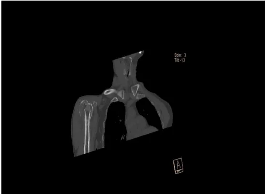
The initial diagnoses were made by direct radiography of the shoulder (Figure 1). In addition to direct radiography, a routine CT analysis (Figure 2) was made in all patients to evaluate dislocation and fragmentation,
classify fractures, and examine vascularization of the humeral head. The fractures were assessed according to the classification system described by Neer [13]. Patients were informed about the treatment method, potential
risks and complications, and their consent was received. The mean time to surgical intervention was 10,4 days (range, 7-15) after the trauma.
Surgical Technique:
All operations were performed by the same surgeon (CZE). A surgical intervention was performed when all patients were under general anaesthesia, with the head fixed to the operation table and the patient seated in the beach chair position. Each patient received prophylactic intravenous systemic cefazolin 2 gr preoperatively. A standard deltopectoral incision was used during the procedure. The cephalic vein in the deltopectoral interval was retracted laterally together with the deltoid muscle. In order to avoid any negative postoperative impact on the shoulder functions, attention was paid not to harm parts of the deltoid muscle attaching to the humerus. The proximal attachment of the pectoralis muscle was cut approximately 1 cm and loosened, allowing for increased external rotation of the shoulder postoperatively. The pectoralis muscle and conjoint tendon were retracted medially. The axillary nerve and musculocutaneous nerve were palpated and preserved. In order to have a clear exposure of the fracture, the long head of the biceps tendon between the greater and lesser tuberosity was reached to expose the tissues up to the superior edge of the glenoid. The bone fragment in the tuberosities was left attached to the rotator cuff. The humeral head was removed, and its diameter was measured. No.5 nonabsorable sutures were placed on the bone-tendon junction of the tuberosity where rotator cuffs were attached. Next, 3 to 4 holes were drilled in the proximal humeral shaft to aid the reconstruction. While the forearm was in neutral rotation and the arm was parallel to the ground, the forearm was externally rotated, a gentle traction (20-30°) was performed through the elbow to determine the length of prosthesis. The head of the prosthesis was turned to face the glenoid, and then retroversion and height of the prosthesis were adjusted. The reference points were marked for accepted height and retroversion. The prosthesis was applied with bone cement, and reduction was achieved using an appropriate modular head. First of all, the greater tuberosity was fixed when the extremity was in neutral rotation. Before tightening the sutures, spongious grafts harvested
from the fractured head were inserted between the tuberosity and the implant. Later on, the lesser tuberosity was fixed with horizontal sutures. Both tuberosities were sutured to each other, and to the prosthesis and to the humerus. The rotator interval was repaired, and a drain was placed. All patients underwent “global advantage shoulder arthroplasty system” (Figure 3). We tested maximum internal rotation and stability of the greater tuberosity, maximum external rotation and stability of the lesser tuberosity. Post-Operative Rehabilitation and Follow Up:
Passive exercises including the pendulum, were initiated one day after the surgery. Then, passive external rotation and elevation were initiated to the tolerable level. The patients were instructed on how to make exercise at home before they were discharged. The objective was to achieve enough range of motion of the shoulder for daily activities. Each patients shoulder was immobilized with a postoperative shoulder sling for two weeks, and passive range of motion exercises were maintained. Active-assisted exercises were initiated after confirmation of the union of the tuberosities by control radiographs.
Patients, at their last control, were evaluated clinically and radiologically. Their x-rays of shoulders (Figure 4), range of motions and functional outcomes were noted (Table 1).
The clinical and functional results were assessed for all patients during follow-up and scored according to CMSS, SSTS, OSS and UFSS (Table 1). Also the operating surgeon evaluated active shoulder flexion, abduction, external/internal rotation. Active internal rotation was evaluated according to spinous processes where the fingers of the involved side extend to the back. A visual analog scale (VAS) was used to assess pain (10 = maximum pain, 0 = no pain).
In radiological evaluation, we evaluated improvement and position of tuberosities as well as presence of nonunion, subluxation, glenoid arthritis, humeral stem osteolysis, migration of the prosthesis, malposition of the greater tuberosity, component and cement fractures, and heterotopic ossification (HO). The most recent available radiography was compared with early postoperative radiography to determine any sign of loosening
in cement. Any radiolucent line more than 2 mm around the stem of the prosthesis and ectopic bone formations around the joint were considered as loosening [14]. Furthermore, we measured AHD and HHGTD on direct
glenohumeral anterior-posterior (AP) radiographs for any changes in distances, and examined the presence of any effects of these changes on the functional outcomes of the shoulder.
Malpositioning of the greater tuberosity was evaluated according to Boileau et al.[14]. When it was not seen on the AP radiograph, but on the
transcapular radiograph, it was considered as malposition. When it was not observed in any plane of the radiograph, tuberosity was considered as resorbed [14]. Proximal migration of the prosthesis was evaluated on the last
AP radiographs. Subluxation was assessed based on the percentage of the failure of humeral head to lie concentrically within the glenoid.
We measured the distance between the most inferior aspect of the acromion and the upper aspect of the humeral head as AHD in the AP radiograph of the glenohumeral joint. The change in AHD was analysed in early postoperative radiograph vs. last control radiograph. A change in AHD less than 7 mm in any of the measurements was considered abnormal, allowing the humeral head ride upwards, and suggesting a rotator cuff failure. All analyses were performed using SPSS II Version 17,0 (SPSS, Inc, Chicago, IL, USA) and Mann-Whitney U test.
Results :
The study group included 8 male and 10 female with a mean age of 69 years (range, 52-80 years) at the time of fracture. Of the patients, 14 (78%) were aged 65 years or more at the time of fracture.The fractures were on the dominant side in 12 patients, involving right extremity in 10, and left extremity in 8 patients.
The cause of fracture was a fall in 15 patients, and a traffic road accident in 3 patients. The patients had no accompanying fracture, open injuries and neuro-vascular problem. None of the patients had peripheral vascular problems or diabetes mellitus.The mean follow-up was 32,8 months (range 24 to 48). All of proximal humerus fractures were Neer type-IV.
This fracture classification was determined by the initial radiographic view when the patients presented to the emergency department. Table 1 and table 2 respectively reports functional, physical examination outcomes and radiographic outcomes.
The mean forward elevation was 87,7° (range, 40°-110°) while abduction was 78,3° (range, 45°-100°) and the mean external rotation was 28,3° (range, 15°-40°). The mean internal rotation was at the level of lumbar 3 vertebrae (Table 1). Among those patients with a mean VAS score of 2,6 (range 1-8) during the last control, 12 (67%) had mild or no pain while 4 (22%) had moderate, and 2 (11%) severe pain. The range of motion was also poor in the patient with severe pain, who also had the lowest CMS score (24 points). Same patient also showed lysis of the greater tuberosity. None of the patients achieved the functional level prior to the injury. Functional assessments showed a mean SSTS of 6,5 (range, 0-10) (good), OSS of 25,6 (range, 10-36) (good), UFSS of 25,2 (range, 16-33) (good) (Table 1). Eighteen patients (85%) were satisfied with the treatment. The mean CMSS was 60,1 (range, 24-77) (Table 1). According to this scoring, the result was good and excellent in 10 shoulders (55,5%), fair in 6 shoulders (33,3%), and poor in 2 shoulders (11,1%).
None of the patients had evidence of prosthetic loosening (osteolysis of humeral stem, migration of the prosthesis, component and cement fractures). While 2 patient (11,1%) had resorbtion of the tuberosity, 16 patients (88,9%) achieved a complete union in the tuberosities. HHGT distance was more than 20 mm in three patients but union was seen. The mean AHD was 13,2 mm (range, 8 – 23 mm) on the intact side, it was 15,1 mm (range, 5 – 28mm) on the operated side in early stage, and 11,7 mm (range, 5– 38 mm) in last controls (Table 2). In two patients (11%) who were older than 75 years, AHD values were smaller than 7 mm in the last control. The functional outcomes were good in these patients. The increase of AHD measurements was not statistically different between intact side and late postoperative (p>0.05). And there was statistically significant difference between early and late postoperative AHD values (p=0.59).
The mean HHGTD on the intact side was 15,2 mm (range, 6 – 20 mm), it was 19,2 mm (range, 8-29 mm) on the operated side in early stage, and 19 mm (range, 8– 29 mm) in last controls (Table 2). The HHGTD was higher than 20 mm in 3 patients (14%). In these patients, the CMS was lower compared to other patients. The increase of HHGTD measurements was not statistically different between intact side and late postoperative (p>0.05). And there was no statistically significant difference between early and late postoperative AHD values (p=0.45).
Discussion:
Treatment of the PHF still remain challenging for an orthopedic surgeon. Higher rate of poor bone quality, tenuous soft tissue and associated medical conditions raise difficulties in the treatment of such fractures. Conservative treatment has been frequently reported to fail in such fractures, particularly in traditional 4-part fractures where each of four segments are dislocated, the segment of the articular surface is deprived of its soft tissue attachments, and damaged blood supply to the humeral head is accompanied with a risk of osteonecrosis [15,16]. For such fractures, open reduction and internal
fixation (ORIF) is recommended as a surgical intervention [17]. Despite
advanced fixation techniques, potential lack of accurate reduction of the fracture and risk of fixation loss, malunion, non-union or development of avascular necrosis are considered higher for internal fixation in patients with such a profile [18]. Therefore, it is important to determine whether the
fracture is suitable for anatomic reduction and stable fixation or not, and whether HA is required or not. Hemiarthroplasty has long been accepted as the standard of care for the treatment of displaced and comminuted proximal humeral fractures [14] and PHA can be a choice of treatment in
Neer type IV PHF in osteoporotic older people. Neer reported that the rate of symptomatic nonunion, malunion of the humeral head, tuberosity failure and osteonecrosis was higher in 4-part fractures, which are considerably dislocated, and indicated that HA produce much better outcomes than ORIF in such patients [19]. Indications include 3- and 4-part fractures,
proximal humeral fracture-dislocations, and headsplitting fractures that involve 40% of the articular surface [14].The best candidates for HA in such
fractures include elderly patients who are older than 65 years with a poor bone quality, patients with a pronounced displacement and/or dislocation of the humeral head, those with a fragmentation of the articular surface,
and patients with a failed osteosynthesis [20]. Accordingly, HA has become
the choice of surgical treatment particulary for fractures in elderly patients with a failed internal fixation or a non-viable humeral head [7]. This
injury leads to high rates of osteonecrosis or posttraumatic arthritis with subsequent pain and loss of function when treated nonoperatively or with internal fixation
Thus, the principal objective of hemiarthroplasty is to decrease pain and produce a functional joint.This treatment may allow us to achieve a painless shoulder with an acceptable range of motion in this group of patients. The treatment in osteoporotic older people was determined based on the mean cortical thickness of the proximal humerus, which is an indicator of bone density, as described by Tingart et al. [21]. A mean index of less
than 4 mm (medial + lateral cortical thickness) has been reported to be an indication for arthroplasty [22]. Although our first choice of treatment
is ORIF in such fractures, we preferred to use HA since the mean cortical thickness was measured as 3 mm (range, 1,8 – 4,2) in our patients.
The time interval between the injury and surgical intervention is controversial. A complete evaluation of the injury, identifying any associated pathology and informing the patient and the family require a period of few days [23,24,25]. The delay in surgery should not be longer than
3 weeks. If there is a waiting period of more than 20 days for a surgical procedure, bony union and resorption will occur, which may complicate tuberosity mobilization, anatomic reduction and fixation [26,27,28]. Any delay
in time from fracture to surgery leads to problems in tuberosity fixation and negative impact on clinical outcomes [15]. In the present study, the patients
underwent surgery within 15 days at latest. We noted that as the surgical intervention was delayed, soft tissue adhesions increased and reduction of fractured fragments became more difficult.
The key factors which have an impact on postoperative success and functional outcomes include adjustments to the actual length of humerus and height of the stem, delivery of a proper prosthesis version and anatomical fixation of tuberosities [25]. A prosthesis positioned too high
prosthesis positioned too low causes shortening of arm length, weakening of the deltoid muscle, and consequently inferior subluxation of the humeral head [27]. Some techniques have been described to adjust the height of the
prosthesis. One of them relies on the intact shoulder. Preoperatively, the distance from the medial humeral cortex is measured on the intact shoulder to evaluate the intraarticular loss at the calcar loss on the involved side. Another reliable criterion for height adjustment is to achieve restoration of the Gothic Arch between the lateral edge of the scapula and the medial edge of the humerus [10]. The mean distance from the superior edge of the
pectoralis major tendon to the top of the humeral head was consistently shown to be 5,5 cm ± 0,5 cm independent of the patient’s height [27,29].
Version adjustment is also important. Anteversion positioning of the prosthesis is associated with anterior instability [30], while excess traction of
the greater tuberosity and posterior instability may occur with neutralization of excess retroversion [25].
We adjusted the prosthetic height and retroversion of the head according to the recommendations of Rockwood during the surgery [31]. We applied
a gentle traction to the forearm while patient’s arm was abducted 45° in parallel to the ground in order to determine the prosthetic height during which the head of the test prosthesis was adjusted to face the glenoid. Upon determination of the prosthetic height, the test prosthesis was fixed. In addition to that, the height adjustment was confirmed by measuring the superior end of the pectoralis major tendon and the superior end of the prosthetic head. In retroversion, the objective was to achieve 30° in relation to the forearm, and 20° in relation to the elbow epicondylar axis. The test prosthesis was placed into the humeral shaft, and the arm was externally rotated 20° while the forearm was hold parallel to the ground, and a gentle traction was applied. Modular humeral head was also inserted, and the head of the test prosthesis was adjusted to face the glenoid for appropriate retroversion. Switching the humeral head to face the glenoid when the arm was in external rotation to 20° allowed us to achieve the intended amount of retroversion of 20°. No instability was observed during follow-up of the patients treated with this prosthetic procedure. After determining the appropriateness of the prosthetic rotation and height by several tests, it was marked with a cautery.
We used bone cement to fix the prosthesis in all patients. However, cement was only used for fixation of the shaft, not for fixation of the tuberosities. It has not been established yet if the humeral stem without cement has any advantage. However, recent trend is to use proximal hydroxyapatite-coated implants without cement in order to enhance proximal consolidation [25].
Malunion of the tuberosities cannot be always tolerated, and its full correction is very difficult (7,32). That is why it has been reported that a failure rate of HA procedures can be as high as 50% even in the hands of experienced shoulder surgeons [10]. During HA, tuberosities must be
anatomically and reliably fixed as tight as possible because anatomic recovery of tuberosities and achievement of a functional rotator cuff are important factors in determining the outcomes for shoulder HA [33,34].
Poorly positioned tuberosities are associated with tension in the rotator cuff muscles and compression syndrome [34]. In order to avoid poor
positioning, we attached the lesser tuberosity to the anterior fin, and the greater tuberosity to the lateral fin of the prosthesis. For a good repair of the tuberosity, we needed to place an autogenous bone graft between the tuberosity and the shaft and under the prosthetic head to enhance the rate of union, use heavy suture material to pass through the bone and tendon, and fix the tuberosities with these sutures to the shaft and the prosthesis in a stable way. We tried to preserve the bone block of the tuberosities attached to the rotator cuffs as safely as possible during the procedure. The belief that successful outcomes for the hemiarthroplasty are related to successful bony union of the greater tuberosity [35]. Overall, all patients
achieved tuberosity union, while only one patient had lysis of bone structure. Follow-up of this patient showed that the patient also had poor functional outcomes. We believe that anatomical union of the tuberosities increased our success rate. During repair of the tuberosities, the HHGTD, i.e., the distance between the top of the humeral head and the superior edge of the greater tuberosity, is reestablished. This distance should be between 5 and 10 mm in order to restore anatomical relationships and improve functional outcomes [7]. Another study on anatomical examples
reported that HHGTD ranged from 3 to 20 mm, and the mean value was 8 mm (± 3 mm) (36). In the present study, the mean HHGTD value was 15.2 mm (range, 6 – 20 mm) on the intact side, and 19 mm (range, 8-29 mm) on the operated side. Our mean HHGTD values are consistent with the literature. A 67 year-old woman had lysis of the greater tuberosity. The
patient had a HHGTD larger than 20 mm as well as the lowest CMS score and functional abilities, and the worst pain scores, and was dissatisfied with the surgical outcome.
We believe that early rehabilitation is one of the important factors which have an impact on the postoperative functional outcomes and range of motion of the joint. Immobilization of the shoulder leads to muscle atrophy and fibrosis, joint capsule and contracture in ligaments. Rehabilitation is relatively more difficult after development of such events, and they may not be improved with rehabilitation [34]. Therefore, all patients who were
given an postoperative arm sling were initiated on passive rehabilitation on postoperative day 1. Gerber et al. [29] reported that during the surgery,
the stability of the tuberosity should be tested following fixation to define a safety arch of the initial passive rehabilitation. We tested the stability of tuberosities during the surgery, and didn’t limit range of motion since we observed no instability. During discharge, patients and their family were instructed on how to do passive exercises. The patients were scheduled for control at weeks 2, 4 and 6. Patients with any sign of union were initiated on active-assisted exercises. For those whose passive range of motion exercises were worse than expected during follow-up, we asked assistance from the physical therapy clinic. Patients were instructed to continue exercise program for18 months.
Recent consensus on the treatment of PHF with HA indicates that long-term results provide satisfactory outcomes with respect to pain, while results related with the range of motion of the shoulder are less satisfactory [10,37].
In older patients, achieving painless and functional outcomes well enough to perform daily activities may be considered satisfactory. These patients usually cannot restore their preoperative shoulder following the surgery. The patients and their relatives should be informed of the potential loss of range of motion. The objective of treating PHF with HA is to return the shoulder to an almost normal level of function to perform usual activities of daily living without any pain in the shortest period.
The limitations of our study include its retrospective design, lack of sufficient number of patients and lack of a closer relationship with patients for rehabilitation. However, in the treatment of 4-part PHFs in older
patients, PHA is a good choice of surgical treatment which provides a reliable and sustained resolution, eliminating the pain in case.
We think of the four-part humeral fractures can be successful in the treatment with partial prosthesis if patient selection and assessment of fracture are appropriately managed; patient and his/her care givers are well informed initially about expectations, potential problems, and details about the shoulder to be reestablished; surgical intervention is performed as early as possible; soft tissue and muscles are preserved; prosthetic height and retroversion are properly adjusted, and tuberosities are anatomically and securely fixated, andappropriate and sufficient rehabilitation is provided. Hemiarthroplasty for acute fractures may achieve the goals of preservation of function and relief of pain in the short-term.
REFERENCES:
[1] Boons HW, Goosen JH, van Grinsven S, van Susante JL, van Loon CJ. Hemiarthroplasty for humeral four-part fractures for patients 65 years and older: a randomized controlled trial. Clin Orthop Relat Res. 2012 Dec; 470 (12): 3483-91.
[2] Palvanen M, Kannus P, Niemi S, Parkkari J. Update in the epidemiology of proximal humeral fractures. Clin Orthop Relat Res. 2006 Jan; 442: 87-92.
[3] Nguyen TV, Center JR, Sambrook PN, Eisman JA. Risk factors for proximal humerus, fore arm and wrist fractures in theelderly men and women: the Dubbo Osteoporosis Epidemiology Study. Am J Epidemiol. 2001 Mar 15;153 (6): 587-95.
[4] Court-Brown CM, Garg A, McQueen MM. The epidemiology of proximal humeral fractures. Acta Orthop Scand. 2001 Aug; 72 (4): 365-71.
[5] Rees J, Hicks J, Ribbans W. Assessment and management of three and four part proximal humeral fractures. Clin Orthop Relat Res. 1998 Aug; (353): 18-29.
[6] Shah N, Iqbal HJ, Brookes-Fazakerley S, Sinopidis C. Shoulder hemiarthroplasty for the treatment of three and four part fractures of the proximal humerus using Comprehensive® Fracture stem. Int Orthop. 2011 Jun; 35(6): 861-7.
[7] Mighell MA, Kolm GP, Collinge CA, et al. Outcomes of
hemiarthroplasty for fractures of the proximal humerus. J Shoulder Elbow Surg. J Shoulder Elbow Surg. 2003 Nov-Dec;12(6):569-77. [8] Gregory TM, Vandenbussche E, Augereau B. Surgical treatment
of three and four-part proximal humeral fractures. OrthopTraumatol Surg Res. 2013 Feb; 99 (1 Suppl): 197-207.
[9] Hawkins RJ, Angelo RL. Displaced proximal humeral fractures. Selecting treatment, avoiding pitfalls. Orthop Clin North Am. 1987 Jul; 18 (3): 421–31.
[10] Boileau P, Krishnan SG, Tinsi L, Walch G, Coste JS, Molé D. Tuberosity malposition and migration: reasons for poor outcomes after hemiarthroplasty for displaced fractures of theproximal humerus. J Shoulder Elbow Surg. 2002 Sep-Oct;11(5): 401-12. [11] Owsley KC, Gorczyca JT. Fracture displacement and screw cut
out after open reduction and locked plate fixation of proximal humeral fractures [corrected]. J Bone Joint Surg Am. 2008 Feb; 90 (2): 233-40.
[12] Lanting B, MacDermid J, Drosdowech D, Faber KJ. Proximal humeral fractures: a systematic review of treatment modalities. J Shoulder Elbow Surg. 2008 Jan-Feb; 17 (1): 42-54.
[13] Iannotti JP, Ramsey ML, Williams GR Jr, Warner JJ. Nonprosthetic management of proximal humeral fractures. Instr Course Lect. 2004; 53: 403–16.
[14] Neer CS 2nd et al: Displaced proximal humeral fractures I. Classification and evaluation. J Bone Joint Surg Am. 1970 Sep; 52 (6): 1077-89.
[15] Spross C, Platz A, Erschbamer M, Lattmann T, Dietrich M. Surgical treatment of Neer Group VI proximal humeral fractures:
retrospective comparison of PHILOS® and hemiarthroplasty. Clin Orthop Relat Res. 2012 Jul; 470 (7): 2035-42.
[16] Edelson G, Safuri H, Salami J, Vigder F, Militianu D. Natural history of complex fractures of the proximal humerus using a three-dimensional classification system. J Shoulder Elbow Surg.
2008 May-Jun; 17 (3): 399-409.
[17] Ozyakın N, Aktuglu K. Treatment of the four-part proxsimal humerus fractures in elderly patients with hemiarthroplasty. Joint Dis Rel Surg. 2008; 19 (3): 101-105.
[18] Leyshon R. Closed treatment of fractures of the proximal humerus. Acta Orthop Scand. 1984 Feb; 55 (1): 48-51.
[19] Hartsock LA, Estes WJ, Murray CA, Friedman RJ. Shoulder hemiarthroplasty for proximal humeral fractures. Orthop Clin North Am. 1998 Jul; 29 (3): 467-75.
[20] Robinson CM, Page RS, Hill RMF, Sanders DL, Court-Brown CM, Wakefield AE. Primary hemiarthroplasty for treatment of proximal humeral fractures. J Bone Joint Surg Am. 2003 Jul; 85-A(7): 1215-23.
[21] Ko JY, Yamamoto R: Surgical treatment of complex fracture of the proximal humerus. Clin Orthop Relat Res. 1996 Jun;
(327): 225-37.
[22] DeFranco MJ, Brems JJ, Williams GR JR, et al: Evaluation and management of valgus impacted four-part proximal humerus fractures. Clin Orthop Relat Res. 1996 Jun; (327): 225-37. [23] Tingart MJ, Apreleva M, vonStechow D, Zurakowski D, Warner
JJ. The cortical thickness of the proximal humeral diaphysis predicts bone mineral density of the proximal humerus. J Bone Joint Surg Br. 2003 May; 85 (4): 611-7.
[24] Nho SJ, Brophy RH, Barker JU, et al: Innovations in the management of displaced proximal humerus fractures. J Am Acad Orthop Surg. 2007 Jan;15 (1): 12-26.
[25] Demirhan M, Kilicoglu O, Altinel L, Eralp L, Akalin Y. Prognostic factors in prosthetic replacement for acute proximal humerus fractures. J Orthop Trauma. 2003 Mar; 17(3): 181-8;
discussion 188-9.
[26] Plausinis D, Kwon YW, Zuckerman JD. Complications of humeral head replacement for proximal humeral fractures. Instr Course Lect. 2005; 54: 371-80.
[27] Sirveaux F, Roche O, Molé D. Shoulder arthroplasty for acute proximal humerus fracture. OrthopTraumatol Surg Res. 2010 Oct; 96 (6): 683-94.
[28] Krishnan SG, Pennington S, Burkhead W. Shoulder arthroplasty for fracture: restoration of the ‘‘gothicarch’’. Techniques Shoulder Elbow Surg. 2005; 6: 57-66.
[29] Gerber A, Warner JJ. Hemiarthroplasty for management of complex proximal humerus fractures: preoperative planning and surgical solution. In: Warner JJ, Iannotti JP, FlatowE, editors. Complex and revision problems in shoulder surgery. 2nd ed. Philadelphia: Lippincott Williams &Wilkins; 2005. p.311—29.
[30] Murachovsky J, Ikemoto RY, Nascimento LG, Fujiki EN, MilaniC, Warner JJ. Pectoralis majör tendon reference (PMT): a new method for accurate restoration of humeral length with
hemiarthroplasty for fracture. J Shoulder Elbow Surg. 2006 Nov-Dec; 15 (6): 675-8.
[31] Greiner SH, Kaab MJ, Kroning I, Scheibel M, Perka C.
Reconstruction of humeral length and centering of the prosthetic head in hemiarthroplasty for proximal humeral fractures. J Shoulder Elbow Surg. 2008 Sep-Oct; 17 (5): 709-14.
[32] Cofield RH. Comminuted fractures of the proximal humerus. Clin Orthop Relat Res. 1988 May; (230): 49-57.
[33] Bigliani LU, Flatow EL, Pollock RG. Fractures of the proximal humerus. In: Rockwood CA, Matsen FA 3rd, editors. The Shoulder. Vol 1, 2nd ed. Philadelphia: W.B.Saunders; 1998. p. 337-91. [34] Kralinger F, Schwaiger R, Wambacher M, Farrell E, Menth-Chiari
W, Lajtai G, Hübner C, Resch H. Outcome after primary hemiarthroplasty for fracture of the head of the humerus. A retrospective multicentre study of 167 patients. J Bone Joint Surg Br. 2004 Mar; 86 (2): 217-9.
[35] Frankle MA, Mighell MA. Techniques and principles of tuberosity fixation for proximal humeral fractures treated with hemiarthroplasty. J Shoulder Elbow Surg. 2004 Mar-Apr; 13 (2): 239-47.
[36] Iannotti JP, Gabriel JP, Schneck SL, Evans BG, Misra S. The normal glenohumeral relationships. An anatomical study of one hundred and forty shoulders. J Bone Joint Surg Am. 1992 Apr; 74 (4): 491-500.
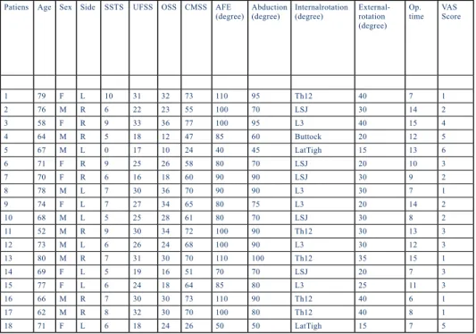
Table 1: It shows the patients’ active forward elevation (AFE), abduction
and internal / external rotation degrees of the shoulder. Also, Simple Shoulder Test Score (SSTS), UCLA Functional Shoulder Score (UFSS), Oxford Shoulder Score (OSS) and Constant –Murley Shoulder Score were evaluated.
Patiens Age Sex Side SSTS UFSS OSS CMSS AFE
(degree) Abduction (degree) Internalrotation (degree) External-rotation (degree)
Op. time VAS Score
1 79 F L 10 31 32 73 110 95 Th12 40 7 1 2 76 M R 6 22 23 55 100 70 LSJ 30 14 2 3 58 F R 9 33 36 77 100 95 L3 40 15 4 4 64 M R 5 18 12 47 85 60 Buttock 20 12 5 5 67 M L 0 17 10 24 40 45 LatTigh 15 13 6 6 71 F R 9 25 26 58 80 70 LSJ 20 10 3 7 70 F R 6 16 18 60 90 90 LSJ 30 9 2 8 78 M L 7 30 36 70 90 90 L3 30 7 1 9 74 F L 7 27 34 65 80 75 L3 20 14 2 10 68 M L 5 25 28 61 80 70 LSJ 30 8 2 11 52 M R 9 30 34 72 100 90 Th12 30 13 3 12 73 M L 6 26 24 68 100 90 L3 30 12 3 13 80 M R 7 31 30 70 110 100 Th12 35 15 1 14 69 F L 5 19 16 51 70 70 LSJ 20 7 3 15 77 F L 6 24 18 64 85 80 L3 25 11 3 16 66 M R 7 30 30 73 110 90 Th12 40 6 1 17 62 M R 8 32 30 70 100 80 Th12 40 8 1 18 71 F L 6 18 24 26 50 50 LatTigh 15 7 5
Table2 : Comparison of the AHD and HHGTD of the injured shoulder and uninjured
shoulder.
Patient
OPERATED SHOULDER UNINJURED SHOULDER EARLY POSTOP LAST CONTROL
AHD (mm) HHGTD (mm) (mm)AHD HHGTD (mm) AHD (mm) HHGTD (mm) CORTICAL THICKNESS (mm) 1 19 20 12 20 11 6 2.7 2 15 19 14 19 13 20 2.8 3 28 22 22 22 15 21 2.4 4 16 21 14 10 15 20 1.8 5 20 8 18 8 20 10 3.9 6 11 24 9 25 8 9 1.4 7 14 26 12 27 14 25 2.5 8 13 19 8 20 14 28 3.4 9 5 17 4 20 9 12 2.7 10 20 29 17 29 23 20 3.1 11 9 29 6 20 6 19 2.4 12 9 10 8 11 8 12 2.1 13 19 20 11 20 16 29 4.2 14 24 27 18 28 22 25 2.8 15 16 18 11 19 18 25 3.6 16 18 22 12 20 12 8 2.2 17 16 20 14 18 14 16 2.6
Figure1: Preoperative AP x-ray of 71-year-old women with right 4-part acute fracture of
Figure 3: Early postoperative AP x-ray view of 71-year-old women with right 4-part