DOI:10.25000/acem.605006 Araştırma makalesi / Research article
Atıf yazım şekli: Şeker M, Çiftçi TT, Akıncı D, Akhan O.Radiologically guided percutaneous nephrostomy: A 6-year single-center experience. Arch Clin Exp Med.
2019;4(3):132-Abstract
Aim: To retrospectively analyze the indications, underlying pathologies, technical success rate, complications and benefit of percutaneous nephrostomies in a single centre.
Materials and Methods: Data of 578 patients who underwent radiologically guided percutaneous nephrostomy between January 1999 and December 2004 were retrospectively reviewed. The mean age of the patients was 42.5 years (range, 6 days–90 years). The indications were urinary obstruction without urinary infection (77.9%), urinary obstruction with urinary infection (13.1%), urinary diversion (6.9%) and diagnostic testing (2.1%). Results: The technical success rate was 99.4%. There was no procedure related mortality. Major hemorrhage or sepsis were not observed in children. Major hemorrhage occurred in 1.55% and sepsis occured in 2.65% of adult patients. Catheter dislodgement was the commonest complication with an overall rate of 11.4%. In 7.2% patients, percutaneous nephrostomy was successful in managing patients without further intervention. 36.5% of patients had surgery and 14.7% had ureteral stenting as definitive treatment.
Conclusion: Radiologically guided percutaneous nephrostomy, can be used effectively, and safely in a wide variety of indications with high technical success and low complications rates.
Keywords: Percutaneous nephrostomy, urinary system obstruction, urinary leakage, urinary fistula, interventional radiology.
Öz
Amaç: Perkütan nefrostomilerin, endikasyonlarını, altta yatan patolojileri, teknik başarı oranını, komplikasyonları ve genel faydalarını retrospektif olarak incelemek.
Materyal ve Metod: Ocak 1999 ile Aralık 2004 tarihleri arasında görüntüleme kılavuzluğunda perkütan nefrostomi yapılan 578 hastanın verileri retrospektif olarak incelenmiştir. Hastaların ortalama yaşı 42,5 yıl idi (6 gün–90 yıl). İşlem endikasyonları üriner enfeksiyon olmadan obstrüksiyon varlığı (% 77,9), üriner enfeksiyon ile beraber obstrüksiyon varlığı (%13,1), üriner diversiyon (%6,9) ve böbrek fonksiyonları değerlendirme (%2,1) idi.
Bulgular: Teknik başarı oranı % 99,4 idi. İşlemle ilişkili mortalite izlenmedi. Çocuklarda major hemoraji ya da sepsis görülmemişti. Erişkin hastalarda major kanama oranı % 1,55, sepsis oranı ise %2,65 idi. Kateter dislokasyonu en sık ortaya çıkan komplikasyondu ve oranı toplamda %11,4 idi. Hastaların %7,2'sinde başka girişim yapılmadan perkütan nefrostomi ile başarılı tedavi sağlandı. Hastaların %36,5'inde cerrahi ile ve %14,7'sinde ise üreteral stent yerleştirilerek kesin tedavi sağlandı.
Sonuç: Radyoloji kılavuzluğunda yapılan perkütan nefrostomi, yüksek teknik başarı ve düşük komplikasyon oranları ile çok çeşitli endikasyonlarda etkili ve güvenli bir şekilde kullanılabilir.
Anahtar kelimeler: Perkütan nefrostomi, üriner sistem obstrüksiyonu, üriner sistem kaçağı, üriner fistül, girişimsel radyoloji.
1 Medipol University, Faculty of Medicine,
Department of Radiology, Bağcılar, Istanbul, Turkey.
2 Hacettepe University, Faculty of Medicine,
Department of Radiology, Çankaya, Ankara, Turkey.
MŞ: 0000-0002-6745-0159 TTÇ: 0000-0002-1284-859X DA: 0000-0002-8189-4688 OA: 0000-0002-6864-0229
Ethics Committee Approval: The study was approved by the local ethical authority (28.04.2005; LUT 05/31).
Etik Kurul Onayı: Çalışma lokal etik komite tarafından onaylanmıştır (28.04.2005; LUT 05/31).
Conflict of Interest: No conflict of interest was declared by the authors.
Çıkar Çatışması: Yazarlar çıkar çatışması bildirmemişlerdir.
Financial Disclosure: The authors declared that this study has received no financial support.
Finansal Destek: Yazarlar bu çalışma için finansal destek almadıklarını beyan etmişlerdir.
Geliş Tarihi / Received: 22.08.2019 Kabul Tarihi / Accepted: 06.11.2019 Yayın Tarihi / Published: 01.12.2019
Sorumlu yazar / Corresponding author: Mehmet Şeker
Adress/Adres: Medipol Mega Hospital, No: 1, Bağcılar, 34214, Istanbul/Turkey.
E-posta: [email protected] Tel/Phone: +90 533 453 38 29 Fax: +90 0212 460 70 70
Copyright © ACEM
Radiologically guided percutaneous nephrostomy: A 6-year
single-center experience
Görüntüleme kılavuzluğunda perkütan nefrostomi: 6 yıllık tek merkez deneyimi
Mehmet Şeker
1P a g e / S a y f a 133
Introduction
Interventional radiology plays a fundamental role in terms of diagnostic and therapeutic support in the field of nephrology and urology. With the accumulation of experience within years, interventional radiologic procedures have been found to be better tolerated than the surgical procedures and they have fully or partially replaced the standard treatment with open abdominal surgery. Percutaneous nephrostomy (PCN) is one of these interventional procedures [1-4].
PCN has conventionally been performed under fluoroscopic guidance and anatomic landmarks or a radiopaque target (eg, stone, opacified collecting system with contrast) are used to localize the collecting system and to select the appropriate entry site. But along with the technical developments in imaging modalities, more procedures were performed with the use of cross-sectional techniques. The use of cross-sectional imaging modalities, alone or in combination with fluoroscopy, to guide the procedure have increased the technical success and reduced the associated complications [2, 3].
Early in its development, PCN was used almost always for temporary or permanent drainage in cases of an obstructed kidney and for evaluation of renal function. Increased success rates and reduced associated complications have significantly broadened the indications. These indications include relieving urinary obstructions without or with infection, urinary diversion for a leak or fistula, and accessing the collecting system for diagnostic and therapeutic procedures [1-4]. Although the advancement of modern endourological techniques has led to a decline in the indications for PCN, it still plays an important role in the treatment of multiple urologic conditions [2-4].
PCN is one of the commonest procedures performed in urologic and interventional radiology units and several reports were published for assessing the indications and outcome of PCN. However, since there is no recently published article on the indications and outcomes of PCN, we conducted this retrospective study to evaluate our experience of PCN. In this retrospective study we aim to analyze the indications, underlying pathologies, technical success rate, complications and overall benefit of PCN.
Material and methods
For this study approval of Ethics Committee of Hacettepe University was obtained (28/April/2005; decision no. LUT 05/31). Informed consent was obtained from all individual participants included in the study. The study was performed in accordance with the ethical standards as laid down in the 1964 Declaration of Helsinki and its later amendments or comparable ethical standards. Written consent could not be taken due to the retrospective design of the study.
Data collection and study population
We retrospectively reviewed the hospital charts and radiology records of all patients whom PCN procedure were performed in non-vascular interventional radiology unit of Hacettepe University between January 1999 and December 2004.
Hemorrhage and sepsis rates were calculated based on the number of patients treated and other complication and technical success rates were reported based on the number of procedures performed according to the Percutaneous Nephrostomy Guideline of Society of Interventional Radiology (SIR) [2, 3].
Preparation before PCN and technique
International normalized ratio (INR) and platelet count of all cases were evaluated and abnormal parameters (INR higher than 1.5 or platelet count less than 80000/mm3) were corrected before procedure. A single dose of prophylactic broad spectrum antibiotic was administered one hour before the procedure to all patients unless the patient was already receiving antibiotics.
The procedures were carried out by an interventional radiologist or by a radiology resident supervised by an interventional radiologist.
Procedures of children were carried out under sedation or general anesthesia with supervision of a specialist an anesthesiologist. In all adult patients, sedation were used.
All procedures, except two, were performed with Seldinger technique under combined use of ultrasonography (US) and fluoroscopy. In the remaining 2 patients, only US was used for guidance.
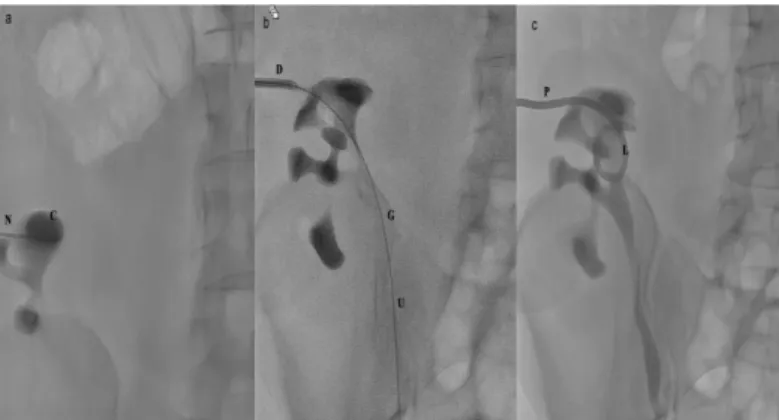
The patients were placed on the fluoroscopy table in prone position. After sterilely cleansed and draped, the skin and fascia were incised with a 11G blade and an 18-21G Chiba needle was inserted through the incision under US guidance and aimed at the direction previously determined. The sonographic view as well as urine confirmed that the needle was at the desired site. A small volume of dilute contrast was injected to opacify the collecting system to verify needle positions. Following succesfull access, needle was removed and a 0.035 or 0.018 inch guidewire (Amplatz Superstiff or 0.018 inch Nitinol guide wire; Boston Scientific, Watertown, MA) was advanced through the needle under fluoroscopic guidance. Nephrostomy tract was dilated with dilators and a PCN catheter (6-12 Fr) was advanced over guidewire. All PCN catheters used in our series had self-retaining mechanisms (650 with and 153 without locking strings) and fixed to the skin. Catheters were placed to a gravity drainage bag. The technique of PCN is shown step by step in figure 1.
The patients were followed in their referring clinics after the procedure.
Statistical analysis
Descriptive statistics, including means and percentages, were used to summarize the data.
Figure 1. Radiographs show the basic technique of percutaneous nephrostomy. (a) With the patient in the prone position, a 21G Chiba needle (N) is passed into the upper pole calyx (C). After urine sample is collected, a small volume of diluted contrast material is injected to opacify the collecting system to verify needle positions. (b) Following succesfull access, needle is removed and a guidewire (G) is advanced through the needle into the ureter (U) under fluoroscopic guidance. Nephrostomy tract is than sequentially dilated with dilators (D). (c) A PCN catheter (P) with self-retaining mechanism is advanced over the wire, and the loop (L) is formed.
Results
Study population
A total of 803 PN procedures were performed in 578 patients (353 male and 225 female). Mean age of these patients was 42.5 years. 160 PCN procedures were performed in 126 patients, aged between 6 days to 15 years (mean age, 4.93 years). The remaining 452 patients were aged between 16 to 90 years (mean age, 52.9 years) and a total of 643 PCN procedures were performed in these patients.
The most common indication was urinary obstruction without urinary infection (77.9%). The other indications were urinary obstruction with urinary infection (13.1%), urinary diversion (6.9%) and diagnostic testing (2.1%). The most common underlying pathologies considered as PCN indication in children and adults were congenital abnormalities (80.1%) and malignities (53.6%), respectively. The indications of PCN procedures and underlying pathologies are shown in tables 1-3.
Table 1: Indications of PCN procedures and underlying pathologies
Indication Malignity Urinary stone disease Congenital pathology+ Other pathology* RPF Total Obstruction without infection 216 (48) 107 (23.8) 94 (20.9) 24 (5.3) 9 (2) 450 (77.9) Obstruction with infection 19 (25) 29 (38.1) 18 (23.7) 10 (13.2) 0 76 (13.1) Urinary diversion 7 (17.5) 3 (7.5) 1 (2.5) 29 (72.5) 0 40 (6.9) Diagnosting testing 3 (25) 4 (33.3) 3 (25) 2 (16.7) 0 12 (2.1) Total 245 (42.4) 143 (24.7) 116 (20.1) 65 (11.2) 9 (1.6) 578 (100)
n (%): numbers of patients and percentage, PCN: percutaneous nephrostomy, RPF; retroperitoneal fibrosis, +: includes ureteropelvic and ureterovesical junction obstruction, vesicoureteral reflux, posterior uretral valve, ureterocele, *: includes urinary, gynecologic and other surgery, trauma, radioterapy.
Table 2. Indications of PCN procedures and underlying pathologies in children.
Indication Malignity
Urinary stone disease
Congenital abnormality Other pathology* Total UP UV VUR Other+ Total
Obstruction without infection 3 (3.2) 11 (11.6) 41 (43.2) 25 (26.3) 8 (8.4) 5 (5.2) 79 (83.1) 2 (2.1) 95 (75.5) Obstruction with infection (0) 2 (9.1) 4 (18.2) 9 (40.9) 3 (13.6) 2 (9.1) 18 (81.8) 2 (9.1) 22 (17.5) Urinary diversion (0) (0) 1 (25) 0 0 0 1 (25) 3 (75) 4 (3.1) Diagnosting testing (0) 1 (20) 3 (60) (0) (0) (0) 3 (60) 1 (20) 5 (3.9) Total 3 (2.4) 14 (11.1) 49 (38.9) 34 (26.9) 11 (8.7) 7 (5.6) 101 (80.1) 8 (6.4) 126 (100)
n (%); numbers of patients and percentage, PCN; percutaneous nephrostomy, UP; ureteropelvic junction obstruction, UV; ureterovesical junction obstruction, VUR, vesicoureteral reflux. +includes posterior uretral valve and ureterocele; *; includes surgery and trauma.
Technical success
The procedure was successfully completed in 798 out of 803 procedures (99.4%). In 80 % of patients with technical failure, no or minimal dilatation were observed in the collecting system.
Complications
There was no procedure related death. In a 10-day-old neonate, methemoglobinemia due to local anesthesia and a subsequent cardiac arrest was observed. The patient survived after succesful resuscitation. Mortality rate within 30 days of PCN was 6.4%. Most of these deaths (91.9%) were as a result of underlying malignancy.
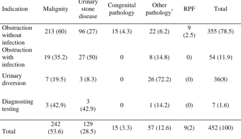
Table 3. Indications of PCN procedures and underlying pathologies in adults. Indication Malignity Urinary stone disease Congenital pathology Other pathology* RPF Total Obstruction without infection 213 (60) 96 (27) 15 (4.3) 22 (6.2) 9 (2.5) 355 (78.5) Obstruction with infection 19 (35.2) 27 (50) 0 8 (14.8) 0) 54 (11.9) Urinary diversion 7 (19.5) 3 (8.3) 0 26 (72.2) (0) 36(8) Diagnosting testing 3 (42.9) 3 (42.9) 0 1 (14.2) (0) 7 (1.6) Total 242 (53.6) 129 (28.5) 15 (3.3) 57 (12.6) 9(2) 452 (100)
PCN; percutaneous nephrostomy, RPF; retroperitoneal fibrosis, numbers in parentheses are raw data, *; includes urinary, gynecologic and other surgery, trauma, radiotherapy.
Complications such as neighboring organ injury, hydro or pneumothorax were not observed in any patient. Major hemorrhage was not observed in children. Major hemorrhage was occurred in 1.55% of adult patients (in 7 of 452 patients). In these patients, stabilization was achieved without any treatment except blood transfusion.
The overall rate of minor hemorrhage was 6.1%. Perirenal hematoma developed in 1.7% of all procedures and all of them resolved spontaneously without any treatment. There was no sepsis in children. In adult patient group, sepsis rate was 2.65%. Of these patients the most common indication was urinary obstruction with urinary infection (91.7%).
The catheter dislodgement rate was 10.6% in children and 11.7% in adults. 52.9% of these children and 41.3% of these adult patients had bilateral PCN catheters. The rate of catheter dislodgement was 10.3% for those with locking strings and 16.4% for those without locking strings.
The major and minor complications are summarized in tables 4 and 5, respectively.
Table 4. Major complications of PCN.
Complication Children Adult Total
Failed PCN+ 0 5/643 (0.78) 5/803 (0.6)
Methemoglobinemi due tolocal
anesthesia* 1/126 (0.79) 0 1/578 (0.17)
Hemorrhage requiring blood
transfusion* 0 7/452 (1.55) 7/578 (1.2)
Sepsis* 0 12/452 (2.65) 12/578 (2.1)
Urinoma requiring percutaneous
intervention+ 1/160 (0.63) 1/643 (0.16) 2/803 (0.25)
Perirenal abscess+ 0 4/643 (0.62) 4/803 (0.49)
PCN catheter dislodgement+ 17/160 (10.6) 75/643 (11.7) 92/803 (11.4)
n (%); numbers of patients and percentage, numbers are raw data, PCN;
percutaneous nephrostomy, +; represents the number of number of PCN
P a g e / S a y f a 135
Table 5. Minor complications of PCN.
Complication Children Adult Total
Hemorrhage not requiring blood
transfusion* 4/126 (3.2) 31/452 (6.9) 35/578 (6.1)
Urinoma not requiring
percutaneous intervention+ 1/160 (0.63) 0 1/803 (0.13)
Urine extravasation not requiring percutaneous intervention+
2/160 (1.25) 8/643(1.24) 10/803 (1.25)
n (%); numbers of patients and percentage, numbers are raw data, PCN; percutaneous nephrostomy, *; Represents the number of patients, +; represents the number of PCN procedures.
Outcomes of the procedure
In 95.1% of patients who underwent PCN due to azotemia, creatinine levels decreased after the procedure and in 42.5% of these patients, the creatinine levels decreased even to normal levels.
Among adult patients who underwent PCN for urinary diversion, successfull treatment was achieved with PCN without further intervention in 19.4% and by with ureteral stenting in 11.1% of patients. 50% children (2 of the 4 children) who underwent PCN for urinary diversion were succesfully treated with percutaneous catheterization alone. Leaks or fistulas were caused by benign causes in 85% of the patients treated with percutaneous treatment and conservative approach. In two patients, there was no urinary leak or fistula on antegrade pyelography. PCN was used for permanent urinary diversion in 13.9% of the patients.
PCN was performed in 12 patients for renal function evaluation. After catheterization, renal function was considered to be sufficient in 25% of these patients.
PCN catheters were placed in transplanted kidneys in 10 patients. Of these patients all except one were adults. The most common indication for PCN procedures performed in this group was urinary obstruction without urinary infection (60%). Of these 6 patients, 83.3% had obstruction in the UVJ and 16.7% had obstruction in the UPJ. 33.3% of these patients were treated succesfully with surgery and 66.7% with PCN, percutaneous ureteral stent placement and-or balloon dilatation without the need for surgery. In the remaining 4 patients the indications were urinary leakage from UVJ and urinary obstruction with infection (30% and 10%, respectively). In one of the 3 patients treated for urinary leakage, no leakage was found after antegrade pyelography. The other 2 patients underwent surgical treatment after catheterization.
The mean duration of PCN drainage was 34.9 days (1 to 480 days) in adults and 28.4 days (2 to 365 days) in children. The mean duration of PCN drainage was longest in patients who were treated for urinary leakage or fistula (46.4 days) and the mean duration of PCN drainage was longer in the procedures performed due to malignant underlying pathology (45.2 days for malignancies, 22 days for urinary tract stones and 21.8 days for retroperitoneal fibrosis). In 14.4% of adult patients, catheters were used for permanent drainage and the most common underlying pathology in these patients was malignancy (83.7%). In 1.6% of children, the catheters were used for long-term drainage and these patients had UPJ obstruction.
In 7.2% patients, PCNs were successful in managing patients without further intervention. 36.5% of patients had surgery and 14.7% had ureteral stenting as definitive treatment. 8.3% of patients underwent nephrectomy. In 4.6% of patients, the catheters were withdrawn as the kidney was nonfunctional. In 11.6% patients, catheters were used for long term drainage and discharged from the hospital with their catheters. 10.5% of patients died. The remaining patients were out of follow-up.
Discussion
The technical success rates of PCN under imaging guidance were reported to reach 98-100% in many reports [1-5]. In our series, technical success rate was 99.4%. Although high success rates have been also reported under sole fluoroscopy guidance, the success rates of combined guidance is higher. The use of US guidance for puncture, reduces the number of puncture attempts. But in some cases it may be compulsory to perform the next steps under fluoroscopy guidance due to the difficulty of monitoring the guidewire and catheter with US [5-7]. In addition the use of US during puncture decreases the risk of complications such as adjacent organ injury and hydro or pneumothorax [7]. In our study, adjacent organ injury and hydro or pneumothorax were not observed.
Dilatation of renal collecting system is an another factor which is thought to be effective on technical success [4, 5]. In recent studies it was reported that in almost all procedures where technical failure was observed, there was no collecting system dilatation and the use of US during puncture increases the success even in the existence of non or minimally dilated collecting systems [2, 5, 7]. In our study, there was no or minimal dilatation in 80% of the cases with technical failure.
The relief of urinary obstruction without or with infection represents the most common indication for PCN, representing 85% to 90% of patients in several large series [1-5]. In our study, this ratio was 91 % in all patients.
The two most common causes of renal obstruction without infection in adults are malignancies and urinary tract stones. In large series, these rates vary between 38.2% to 61% and 26% to 40.6%, respectively [1, 8, 9]. In our study, the rates were similar with the literature and were calculated as 60% and 27%, respectively.
In our series, the most common underlying pathology in adult patients who underwent PCN for obstruction with infection was urinary tract stones (50%). This result is also consistent with the literature [3].
Congenital abnormalities are the most common underlying pathology in children. In the literature [10, 11], common underlying pathologies in patients undergoing PCN for the relief of obstruction with or without infection are UPJ obstruction (49.5%), UVJ obstruction (21%) and urinary tract stones (13.6%) and these are similar with our study (38.5%, 29.1% and 11.1%, respectively).
PCN is often the simplest method for the initial management of obstructive renal failure and meets the need for safe, effective and urgent urinary drainage until the cause of obstruction is eliminated in these cases [1]. Complete recovery of the glomerular filtration rate can be expected with one week of complete obstruction but after 12 weeks, very little recovery may be seen. In addition, relief of obstruction may also contribute to the improvement of renal function [4]. In our series, creatinine levels were improved in almost all patients (95.1%) who underwent PCN due to azotemia. Beyond this, the creatinine levels decreased to normal levels in 42.5% of the these. However there is a dilemma in the therapeutic approach in the presence of malignant obstruction. But PCN or other percutaneous or endourological methods may at least protect the patient from the adverse effects of uremia and save time for selecting and performing the appropriate treatment [12, 13].
On the other hand an infected, obstructed kidney requires emergent drainage. Evacuation of pus and the release of intrarenal pressure with PCN, reduce the risk of aggravation of infection and improve renal perfusion and function [1-4]. Although PCN and retrograde stenting have been shown to have equivalent patient outcomes in some reports, PCN has recently
become the first-line therapy especially in seriously ill patients based on its reduced manipulation of the obstructed, infected ureter [4].
The most common nonobstructive indication for PCN in the literature is urinary diversion [14, 15]. The most common causes of urinary leakage or fistulas, are malignancies, surgical procedures, radiotherapy and trauma [16, 17]. In our study, the most common cause of urinary leakage-fistula was gynecological and urological surgeries (44.4%) in adults and trauma (50%) in children.
Conservative treatment or surgical reconstruction is performed in the presence of urinary fistula or leakage. PCN with or without antegrade ureteric stenting are used effectively both as a primary treatment and as an adjunctive method to surgery. They are also used as a palliative treatment for patients who are not suitable for surgery and may increase the quality of life of these patients [17, 18]. In our series, 32.5% of the patients whom PCN were performed for urinary diversion, were succesfully treated without the need of surgery.
It is thought that the renal function and how much of its function can recover may be evaluated by PCN. Other than diagnostic benefit, functional recovery in kidneys with poor function after PCN has been reported [4]. In our series, after an avarage of 9.8 days follow up, it was decided that 25% of the patients had sufficient renal function and the preprocedurel treatment decision (nephrectomy) was changed.
In our series the indications for PCN in transplanted kidneys were, obstruction without infection, urinary diversion for urinary leakage and obstruction with infection (60%, 30% and 10%, respectively). In the literature urinary leakage are most commonly seen from the ureteroneocystostomy site and obstructions are often observed in the distal ureter proximal to the ureteroneocystostomy [1, 19]. Similar results were observed in our study.
Traditionally surgical treatment is the preferred method in the presence of urinary leakage in transplantated kidneys. However, PCN saves time for patient preparation if surgery is indicated [19]. In addition, PCN can provide diagnostic benefits because in some cases antegrade pyelography is required for definitive diagnosis. In our series, in one of 3 patients who underwent PCN for urinary leakage, no leakage was detected after antegrade pyelography and the catheter was withdrawn. The other two patients underwent surgical treatment after catheterization.
PCN can also contribute to diagnosis and treatment in the presence of obstruction in transplanted kidneys. In the studies performed in transplant kidneys with obstruction, functional improvement was observed in almost all patients by percutaneous intervention in the early postoperative period. However, percutaneous intervention success was low in obstruction diagnosed late in postoperative period [20]. In our series, 33.3% of the patients who underwent PCN for this indication were treated with surgery and 66.7% of the patients were treated successfully with PCN, percutaneous ureteral stent placement and-or balloon dilatation without the need for surgery. The mortality rates associated with PCN are as low as 0.046-0.7% and PCN can be performed safely even in patients with poor general condition [1]. In our series, there was no procedure related mortality.
Minor hemorrhages which are often due to small vessel or venous bleeding are common after PCN [1]. The rate of minor hemorrhage was 6.1% in our series. The rates of major hemorrhage which can take the form of hematuria or retroperitoneal hemorrhage, vary between 1-4% in different series [1-3]. This rate was 1.55% in adults in our series. There was no major hemorrhage in children.
Procedure-related major hemorrhages are usually caused by lacerations in major branches of renal artery and vein. The use of US guidance during puncture reduces the risk of vessel injury by providing an appropriate selection of the entry site. In addition, US guidance allows these hemorrhages to be noticed during the procedure most of the time [4]. Persistent gross hematuria more than 3 to 5 days after PCN or significant drop in hemoglobin levels may indicate arterial injury, and angiographic evaluation may be required in rare cases. Fortunately, these vascular injuries can be managed with endovacular approach without the need for surgery [1, 4]. In our series, in one patient angiography was required. But no pathology was found in angiography and hemorrhage was controlled by catheter upsizing.
Patel et al [21] reported the rate of major hemorrhage as relatively high (7%) in their series compared with others. In this study, PCN procedures were performed in kidneys that had no dilatation in their collecting systems and the authors claimed that the risk of major hemorrhage was higher in the procedures performed in such cases. However, in our series as some other studies, in most of the procedures in which hemorrhage occurred, there were advanced or moderate dilatation in the collecting system and no relationship was found between hemorrhage and collecting system dilatation [8].
There are different reports that coagulation disorders being a risk factor for PCN-related hemorrhage. However, many authors suggest to correct coagulation parameters before the procedure [8, 13]. In our series, 71.5% of patients (5 of 7 patients) with major hemorrhage had coagulopathy. With these findings, we also think that coagulopathies should be corrected before the procedure.
Another important complication of PCN is sepsis and in different series it was reported to be 1-9% [1-3, 8]. In our series sepsis was not observed in children and 2.65% of adult patients developed sepsis despite all were given antibiotic prophylaxis. However in some reports, the prevalence of septic complications were reported to be 25% or higher [22, 23]. This discrepancy may be due a number of factors including differences in the definition of sepsis which are accepted as complications, the differences in the use of prophylactic antibiotics and differences in underlying pathologies.
The use of prophylactic antibiotics is not universally accepted in PCN. Some feel that prophylactic antibiotics is not indicated for routine PCN [21, 24] whereas others give prophylactic antibiotics to all patients undergoing PCN [25]. Some authors defined a group at high risk for development of sepsis including but not limited to patients with positive urine culture or urinalysis, urinary tract stones (especially struvite calculi), a urinary ostomy and those who are immunosuppressed. Their findings showed that administration of antibiotics decreased the frequency of sepsis in the high-risk group and had a beneficial effect in the low-risk group as well [8, 23, 24].
The other factor for this discrepancy may be the underlying pathologies. In the study of Cohran et al [23] in which there was a higher rate of sepsis, 75% of patients who developed sepsis had urinary tract stones. In this study, it was also found that, the probability of a patient with a struvite stone developing signs of sepsis was significantly increased compared with patients with other types of stones. They recommended to use appropriate antibiotics, on the basis of urine test results before PCN and continue for 48-72 hours after the procedure in high-risk group and for 24-48 hours in low-risk group [23, 24].
Urinoma related to PCN procedure is a very rare complication and was reported to be 0.2-0.5% [5]. Small urinomas usually resolve spontaneously but if urinoma is large or infected, percutaneous drainage is required. In our series urinoma
P a g e / S a y f a 137 requiring percutaneous intervention encountered in one child and
one adult patient after the procedure.
Amongst the complications, catheter dislodgement is the commonest problem. We had an overall catheter dislodgement complication rate of 11.4% which was comparable with other studies [1, 9, 26]. Catheter dislodgements are more common with those without self-retaining mechanisms but dislodgements can be seen in any type of catheter. In our series all PCN catheters had self-retaining mechanisms but the rate of catheter dislodgement rate for those without locking strings was higher than those with locking strings (16.4% and 10.3%, respectively). Fixation of the catheter at the skin is also used in order to keep the catheter in place. However, none of these can completely prevent catheter dislodgements. The common opinion is that the most important way to prevent catheter dislodgements is to inform patients and their relatives about catheter care [27].
This was a retrospective study and has limitations. Because the patients were sent back to the referring clinics, a number of complications, especially minor ones, might not have been reported to us.
In conclusion, our technical success and complications rates were within the accepted target ranges proposed by the Society of Interventional Radiology Standards of Practice Committee. Combined US and fluoroscopy guided PCN, can be used effectively, and safely in a wide variety of indications with high techn cal success and low compl cat ons rates.
References
1. Dagli M, Ramchandani P. Percutaneous nephrostomy: technical aspects and indications. Semin Interv Radiol. 2011;28:424–37.
2. Pabon-Ramos WM, Dariushnia SR, Walker TG, d’Othée BJ, Ganguli S, Midia M, et al. Quality improvement guidelines for percutaneous nephrostomy. J Vasc Interv Radiol. 2016;27:410–4.
3. Ramchandani P, Cardella JF, Grassi CJ, Roberts A.C, Sacks D, Schwartzberg M.S, et al. Quality improvement guidelines for percutaneous nephrostomy. J Vasc Interv Radiol. 2003;14:277–81. 4. Young M, Leslie SW. Percutaneous Nephrostomy. [Updated 2019 Jun
17]. In: StatPearls [Internet]. Treasure Island (FL): StatPearls
Publishing; 2019 Jan-. Available from:
https://www.ncbi.nlm.nih.gov/books/NBK493205/
5. Efesoy O, Saylam B, Bozlu M, Çayan S, Akbay E. The results of ultrasound-guided percutaneous nephrostomy tube placement for obstructive uropathy: A single-centre 10-year experience. Turk J Urol 2018; 44: 329-34.
6. Lodh B, Gupta S, Singh AK, Sinam RS. Ultrasound guided direct percutaneous nephrostomy (pcn) tube placement: stepwise report of a new technique with its safety and efficacy evaluation. J Clin Diagn Res 2014;8:84-7.
7. Montvilas P, Solvig J, Johansen TE. Single-centre review of radiologically guided percutaneous nephrostomy using “mixed” technique: success and complication rates. Eur J Radiol. 2011;80:553–8. 8. Farrell TA, Hicks ME. A review of radiologically guided percutaneous
nephrostomies in 303 patients. J Vasc Interv Radiol. 1997;8:769–74. 9. Radecka E, Magnusson A. Complications associated with percutaneous
nephrostomies. A retrospective study. Acta Radiol. 2004;45:184–8. 10. Gupta DK, Chandrasekharam VV, Srinivas M, Bajpai M. Percutaneous
nephrostomy in children with ureteropelvic junction obstruction and poor renal function. Urology. 2001;57:547–50.
11. Hogan M, Coley BD, Jayanthi VR, Shiels WE. Percutaneous Nephrostomy in Children and Adolescents: Outpatient Management. Radiology 2001;218:207–10.
12. Chitale SV, Scott-Barrett S, Ho ET, Burgess NA. The management of ureteric obstruction secondary to malignant pelvic disease. Clin Radiol. 2002;57:1118–21.
13. Meira MDS, Barbosa PNVP, Bitencourt AGV, Almeida MFA, Tyng CJ, Costa MAF et al. Retrospective analysis of computed tomography-guided percutaneous nephrostomies in cancer patients. Radiol Bras. 2019;52:148–54.
14. Millward SF. Percutaneous nephrostomy: a practical approach. J Vasc Interv Radiol 2000;11:955-64.
15. Patel U, Hussain FF. Percutaneous nephrostomy of nondilated renal collecting systems with fluoroscopic guidance: technique and results. Radiology. 2004;233:226-33.
16. Gayer G, Zissin R, Apter S, Garniek A, Ramon J, Kots E, et al. Urinomas caused by ureteral injuries: CT appearance. Abdom Imaging. 2002;27:88–92.
17. Avritscher R, Madoff DC, Ramirez PT, Wallace MJ, Ahrar K, Morello FA, et al. Fistulas of the lower urinary tract: percutaneous approaches for the management of a difficult clinical entity. Radiographics, 2004;24:S217–S36.
18. Titton RL, Gervais DA, Hahn PF, Harisinghani MG, Arellano RS, Mueller PR. Urine leaks and urinomas: diagnosis and imaging-guided intervention. Radiographics 2003;23:1133–47.
19. Hunter DW, Castaneda-Zuniga WR, Coleman CC, Herrera M, Amplatz K. Percutaneous techniques in the management of urological complications in renal transplant patients. Radiology. 1983;148:407-12. 20. Gregory MC, Micklos J, Miller FJ, et al. Percutaneous dilatation and stenting of ureteral stenosis in renal transplantation. Clin Trans. 1988;2:107-9c.
21. Patel U, Hussain FF. Percutaneous nephrostomy of nondilated renal collecting systems with fluoroscopic guidance: technique and results. Radiology. 2004;233:226-33.
22. Ferral H, Stackhouse DJ, Bjarnason H, Hunter DW, Castaneda-Zúñiga WR, et al. Complications of percutaneous nephrostomy tube placement. Semin Intervent Radiol 1994;11:198–206.
23. Cochran ST, Barbaric ZL, Lee JJ, Kashfian P. Nephrostomy tube placement: an outpatient procedure? Radiology 1991;179:843–7. 24. Moon E, Tam MD, Kikano RN, Karuppasamy K. Prophylactic antibiotic
guidelines in modern interventional radiology practice. Semin Intervent Radiol. 2010;27:327–37.
25. Smith TP, Hunter DW, Letourneau JG, Cragg AH, Darcy MD, Castaneda-Zuniga WR, et al. Urine leaks after renal transplantation: Value of percutaneaous pyelography and drainage for diagnosis and treatment. AJR Am J Roentgenol. 1988;151:511–3.
26. Sim LS, Tan BS, Yip SK, Ng CK, Lo RH, Yeong KY, et al. Single centre review of radiologically-guided percutaneous nephrostomies: a report of 273 procedures. Ann Acad Med Singapore 2002;31:76-80. 27. Wah TM, Weston MJ, Irving HC. Percutaneous nephrostomy insertion:
outcome data from a prospective multi-operator study at a UK training centre. Clin Radiol. 2004;59:255–61.