Abstract. Background: Reduced expression of Fas and/or increased expression of FasL is known to exist in some cancer types including lung cancer, so the Fas/FasL system may play a role in the course of cancer. Lack of cell surface Fas expression is one of the main routes of apoptotic resistance in tumor formation and progression. Functional mutations in the Fas gene that impair apoptotic signal transduction are associated with susceptibility to various types of cancer. In this study, we focused on lung cancer. Patients and Methods: The genotypic tendencies that may occur due to a specific point mutation (Fas-1377 G→A) on promoter region for Fas were evaluated. Results: We did not find any relationship between Fas-1377 G→A polymorphism and lung cancer. But there was a significantly higher number of AG patients who smoked than GG ones. Conclusion: There was no relationship between Fas-1377 G→A polymorphism and lung cancer, but it was statistically significant that smoking might increase the possibility of creating lung cancer in AG genotypes more than in other genotypes.
Apoptosis is a form of programmed cell death that is essential for tissue development, repair and also in the pathophysiology of some disorders (1). There are two convergent pathways for apoptosis: the intrinsic, in response to release of mitochondrial products, such as cytochrome, and the extrinsic, occuring in response to activation of specific cell membrane receptors termed "death receptors" (1, 2).
Fas (ApoI=CD95) is a type I membrane protein of 48 kDa, consisting of 335 amino acids, which belongs to one of the subfamilies of death receptors and is part of the TNF-receptor superfamily (3-5). The members of this family are characterized by two to five extracellular cysteine-rich
repeats (5). Fas is especially expressed in cardiac tissues and in liver, spleen, thymus and ovarian tissues (3, 4), in which an intracellular death domain that is responsible for apoptotic signal transduction exists (5). This Fas-associated death domain protein (FADD) was discovered as a protein that interacts with Fas receptor (6). FADD also has two domains: death effector domain and death domain (6). The former interacts with the death effector domain of procaspase-8 forming the death inducing signalling complex (DISC) and the latter interacts with the death domain of Fas receptor (4, 6, 7). All these interactions start apoptotic mechanisms (6).
In basic cancer research, it is now known that genes that control apoptosis have a profound effect on the malignant phenotype (8). Tumour cells develop some defence mechanisms to eliminate apoptotic signals or to induce apoptosis of tumour specific lymphocytes (8, 9). Fas receptor is known to be widely expressed in various tissues, but FasL is especially expressed on cells of the immune system such as activated T-cells and natural killer cells, and also on the cells of immune privileged areas (8, 10, 11). Reduced expression of Fas and/or increased expression of FasL is known to exist in some cancer types (including lung cancer), so the Fas/FasL system may play very important roles in the course of cancer (2, 8, 9). Lack of cell surface Fas expression is one of the main routes of apoptotic resistance in tumorigenesis and tumor progression (12).
It is thought that functional mutations in Fas and FasL genes that impair apoptotic signal transduction are associated with susceptibility to cancer including lung cancer (8). One of the transition mutations in the Fas gene is G→A at position -1377 which is located within an SpI transcriptional factor binding site in the promoter region of the Fas that abolishes the consensus sequence for binding of transcription factor SP-1 (8, 10).
The lung is a complex organ of different types of cells, such as endothelial cells, epithelial cells, fibroblasts and inflammatory cells. In lung tissue, Fas has been found on the surface of alveolar and bronchial epithelial cells, alveolar macrophages, myofibroblasts and Clara cells (13).
663
Correspondence to: Prof. Dr. Turgay Isbir, Institute for Experimental Medical Research, Department of Molecular Medicine, Istanbul University, Capa, Istanbul, Turkey. Tel/Fax: +90 212 6351959, e-mail: [email protected]
Key Words: Fas, polymorphism, lung cancer, Fas/FasL pathway.
in vivo
21: 663-666 (2007)Fas-1377 A/G Polymorphism in Lung Cancer
UZAY GORMUS1,2, ARZU ERGEN1, ILHAN YAYLIM-ERALTAN1, HULYA YILMAZ1, AKIF TURNA3, NILÜFER BOZKURT1and TURGAY ISBIR1
1Istanbul University, Institute for Experimental Medical Research, Department of Molecular Medicine Capa; 2Istanbul Bilim University, Faculty of Medicine, Department of Biochemistry, Esentepe, Istanbul; 3Yedikule Hospital For Chest Disease and Thoracic Surgery, First Surgery Clinic, Istanbul, Turkey
Hence there are two paths for the Fas/FasL system in lung cancer: since FasL on T-lymphocytes can promote apoptosis in Fas-expressing lung cancer cells, this system can play important roles in cell-mediated cytotoxic reactions against cancer; on the other hand, malignant cells of lung cancer can escape from the immune system by down-regulation of Fas and/or by killing the lymphocytes using the expression of FasL (14, 15). Thus it is thought that the balance in the Fas/FasL system is crucial for the progression, metastasis, lymph node involvement and the death of cancerous cells (14, 16, 17). We aimed to evaluate the genotypic tendencies that may occur due to a specific point mutation (Fas-1377 G→A) on the promoter region of the Fas gene. Our aim was to determine the potential for lung cancer because of the polymorphic state of individuals.
Patients and Methods
The study consisted of 94 lung cancer patients that were followed-up at the Yedikule Hospital, Clinics of Chest Surgery and 50 healthy subjects, between 2003 and 2004. Exclusion criteria included existence of previous cancer. Healthy control subjects were chosen from the population that applied to the hospital only for minor complaints. The selection criteria for the control subjects included no individual history of cancer, and they were chosen to be numerically matched to case patients on the basis of gender and age. At recruitment, written informed consent was obtained from each participant.
Polymorphism analysis. A previously described method was used (18). The primers used for PCR for Fas-1377 G/A polymorphism were forward: 5’-TGT GTG CAC AAG GCT GGC GC-3’ and reverse: 5’-TGC ATC TGT CAC TGC ACT TAC CAC CA-3’ from IDT (Integrated DNA Technologies Inc, Iowa, USA). To introduce a restriction endonuclease site, the 3’ end of primer FasIF was changed from CAC to CGC to create a BstU1 site. These fragments were amplified using a 25 ÌL reaction mixture containing approximately 100 ng of template DNA, 1 ÌL of each primer, all four deoxyribonucleoside 5’ triphosphates (each at 0.2 mM), 2 mM MgCl2 and 1 U of Taq polymerase in 1x reaction buffer (MBI Fermentas, Vilnius, Lithuania).
The reaction was carried out with an initial melting step of 2 min at 94ÆC, followed by 35 cycles of 45 sec at 94ÆC, 45 sec at 62ÆC, 60 sec at 72ÆC, and a final elongation step of 7 min at 72ÆC. The restriction endonuclease used was BstU1 to distinguish Fas-1377 G/A polymorphism. The products of this reaction were separated on 3% agarose gels containing ethidium bromide. BstU1 digestion generated the following fragments: -1377G allele, fragments of 104 bp and 18 bp; Fas-1377A allele, a single fragment of 122 bp.
Statistical analysis. Student's t-test was used to determine whether or not significant differences in gender and ages existed between patient and control groups. Pearson's Chi-square analyses were used to examine differences in genotype distribution between cancer patients and control subjects.
Results
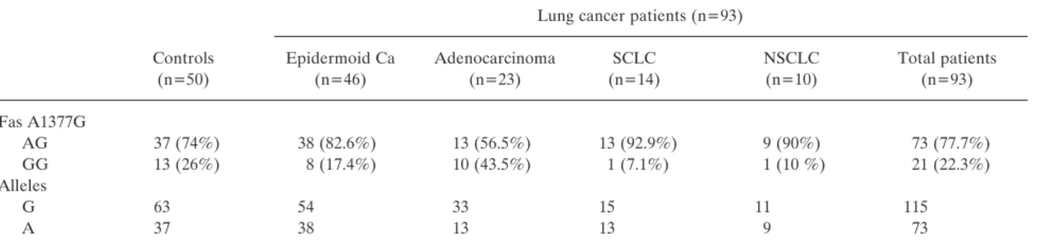
This study included 93 patients with different types of lung cancer and 50 control subjects. The lung cancer patient group contained 46 epidermoid type (49%), 23 adenocarcinoma (24%), 14 small cell lung cancer (SCLC) (15%) and 10 non-small cell lung cancer (NSCLC) of unknown subtype (10%). Of the control group, 14% were women and in the case group there were 11.7% women. The mean age ± standard deviations were 58.62 years ±10.04 years for the control group and 59.94 years ±12.13 years for the case group. Statistically there was no significant differences between case patients and control subjects in terms of age and gender distributions. The genotype and allele distributions of Fas in case and control subjects are shown in Table I.
We observed no homozygotic Fas-1377AA subjects in any of our study groups. The allele frequency of Fas-1377A was 0.37 in the control group and 0.38 in the patient group; the Fas-1377G allele frequency was 0.63 in the control group and 0.61 in the lung cancer patients. The frequencies of Fas-1377GG and Fas-1377GA genotypes among all patients did not differ statistically significantly from those among control subjects (¯2=0.242; p>0.05; df: 1). And frequencies of
Fas-in vivo
21: 663-666 (2007)664
Table I. Prevalence of the Fas A1377G genotypes in patients and control subjects.
Lung cancer patients (n=93)
Controls Epidermoid Ca Adenocarcinoma SCLC NSCLC Total patients
(n=50) (n=46) (n=23) (n=14) (n=10) (n=93) Fas A1377G AG 37 (74%) 38 (82.6%) 13 (56.5%) 13 (92.9%) 9 (90%) 73 (77.7%) GG 13 (26%) 8 (17.4%) 10 (43.5%) 1 (7.1%) 1 (10 %) 21 (22.3%) Alleles G 63 54 33 15 11 115 A 37 38 13 13 9 73
1377A allele and Fas-1377G alleles among all case and control patients did not differ significantly from a statistical point-of-view (¯2: 0.092; p>0.05; df: 1).
There was significant difference between the control group and the total patient group with regard to smoking (¯2: 58.921; p<0.0001; df:1); 30% of the control group, and 91.5% of the patient group were smokers; and there was a significant difference between the smoking AG genotypic patients and GG genotypic patients (¯2: 86.000; p<0.0001; df:1). Hence there might be a direct relationship between the possibility of creating lung cancer in AG genotypic patients, more than GG ones. But there was no relationship between the genotype and the smoking status in any particular type of cancer.
There was significant difference between total non-adenocarcinoma type lung cancer patients of AG genotype and GG genotype (¯2: 8.740; p=0.003; df:1). This may suggest that AG genotype increases the tendency to create lung cancer of a type other than the adenocarcinoma. There were no other significant relationships between any of the lung cancer types and the control group.
Discussion
There are several studies on different kinds of cancer which evaluate the relationship between risk of malignancy development and various genetic polymorphisms. One of the targets for those investigations are the genes playing a role in apoptotic pathways; the Fas/FasL system is one of the most important of these. Since FasL on T-lymphocytes can promote apoptosis in Fas-expressing cancer cells, this system can play important roles in cell-mediated cytotoxic reactions against malignant cells; hence malignant cells can escape from the immune system by down-regulation of Fas and/or by killing the lymphocytes using the expression of FasL (14, 15). Because of these possible dual roles, the Fas/FasL pathway is known to be very important also in therapeutic aspects; there are chemicals and drugs that are known to cure malignant tendencies using this pathway (19, 20). There is also research on the apoptotic pathway that shows the contribution of the Fas/FasL pathway to the proliferative and/or metastatic capacity of cancer. Shimizu et al. mentioned that serum soluble Fas and FasL played important roles in the proliferation and metastasis of SCLC patients (14); and there are many other investigations that support this hypothesis (2, 8).
Zhang et al. suggested that some functional polymorphisms in cell death pathway genes could increase the tendency to develop malignant cells (8, 18). Despite their results, we did not find any significant difference between -1377GA and -1377GG genotypes; nor did we find any statistically significant difference between allele frequencies. But in contrast to our results, there has been much research on the same subject proving the importance of Fas and/or FasL polymorphisms in development of cancerous lesions.
The multiplicative gene-gene interaction between Fas and FasL polymorphisms has been found to increase the risk of lung cancer, suggesting that these two polymorphisms are likely to be active in the same causal pathway (8). It would be better for us to evaluate Fas and FasL genes together in future research to compare the effects and ratios of these different types of polymorphisms. As Fas and FasL work as receptor substrate complexes, it is more logical to evaluate them together.
In SCLC, Fas expression was evaluated by some researchers in relationship with p53 and bcl-2, and as a result it was found that in a large portion of SCLC patients there was a lack of membranous Fas expression. It was thus thought that reduced membranous Fas expression could be considered as one of the basic mechanisms of immune evasion in NSCLC (2, 14). We determined no such supporting result for Fas gene polymorphisms in SCLC patients.
Conclusion
We did not find any relationship between Fas-1377 G→A polymorphism and the development of lung cancer. But there was a significantly higher number of AG patients who smoked than GG ones; hence smoking may increase the possibility of creating lung cancer in AG genotypic patients. This article is the first informative research on this subject in Turkey and leads the way for further research of larger populations. References
1 Martin TR, Hagimoto N, Nakamura M and Matute-Bello G: Apoptosis and epithelial injury in the lungs. Proc Am Thorac Soc 2: 214-220, 2005.
2 Myong NH: Tissue microarray analysis of Fas and FasL expressions in human non-small cell lung cancers; with reference to the p53 and bcl-2 overexpressions. J Korean Med Sci 20: 770-776, 2005.
3 Lee H and Ferguson TA: Biology of FasL. Cytokine and Growth Factor Reviews 14: 325-335, 2003.
4 Poulaki V, Mitsiades CS and Mitsiades N: The role of Fas and FasL as mediators of anticancer chemotherapy. Drug Resistance Updates 4: 233-242, 2001.
5 Walczak H and Krammer PH: CD95 (APO-1/Fas) and TRAIL (APO-2L) apoptosis systems. Exper Cell Res 256(1): 58-66, 2000.
6 Kim KS: Multifunctional role of Fas-Associated death domain protein in apoptosis. J Biochem Mol 35: 1-6, 2002.
7 Bhojani MS, Chen G, Ross BD, Beer DG and Rehemtulla A: Nuclear localized phosphorylated FADD induces cell proliferation and is associated with aggressive lung cancer. Cell Cycle 4(11): 1478-1481, 2005.
8 Zhang X, Miao X, Sun T, Tan W, Qu S, Xiong P, Zhou Y and Lin D: Functional polymorphisms in cell death pathway genes fas and fasl contribute to risk of lung cancer. J Med Genet 42: 479-484, 2005.
Gormus et al: Gene Polymorphisms and Lung Cancer
9 Liu K, Caldwell SA and Abrams SI: Cooperative disengagement of Fas and intercellular adhesion molecule-1 function in neoplastic cells confers enhanced colonization efficiency. Cancer Res 65(3): 1045-1054, 2005.
10 Pinti M, Troiano L, Nasi M, Moretti L, Monterastelli E, Mazzacani A, Mussi C, Ventura P, Olivieri F, Franceschi C, Salvioli G and Cossarizza A: Genetic Polymorphisms of Fas (CD95) and FasL (CD178) in human longevity: studies on centenarians. Cell Death and Differentiation 9: 431-438, 2002. 11 Niederkorn JY: See no evil, hear no evil, do no evil: The lessons
of immune privilege. Nat Immunol 7(4): 354-359, 2006. 12 Commentary on Kim et al: Profiling tumor counter-attack: Do
Fas Ligand-containing microvesicles reduce anticancer immunity? Clin Can Res 11: 968-970, 2005.
13 Lu Q, Harringto EO and Rounds S: Apoptosis and lung injury. Keio J Med 54(4): 184-189, 2005.
14 Shimizu M, Kondo M, Ito Y, Kume H, Suzuki R and Yamaki K: Soluble Fas and Fas Ligand provide new information on metastasis and response to chemotherapy in SCLC patients. Can Det Prev 29: 175-180, 2005.
15 Liu K, Caldwell SA and Abrams SI: Immune selection and emergence of aggresive tumor variants as negative consequences of Fas-mediated cytotoxicity and altered IFN-Á-regulated gene expression. Cancer Res 65(10): 4376-4388, 2005. 16 Tang X, Wu W, Sun S, Wistuba II, Hong WK and Mao L: Hypermethylation of the death-associated protein kinase promoter attenuates the sensitivity to TRAIL-induced apoptosis in human non-small cell lung cancer cells. Mol Cancer Res 2(12): 685-691, 2004.
17 Fokkema E, Timens W, de Vries EGE, de Jong S, Fidler V, Meijer C and Groen HJM: Expression and prognostic implications of apoptosis-related proteins in locally unresectable non-small cell lung cancers. Lung Cancer 52: 241-247, 2006. 18 Sun T, Miao X, Zhang X, Tan W, Xiong P and Lin D:
Polymorphisms of death pathway genes Fas and FasL in esophageal squamous cell cancer. J Nat Can Inst 96(13): 1030-1036, 2004.
19 Liu X, Yue P, Zhou Z, Khuri FR and Sun SY: Death receptor regulation and celecoxib-induced apoptosis in human lung cancer cells. J Nat Can Inst 96(23): 1769-1780, 2004.
20 Du AY, Zhao BX, Miao JY, Yin DL and Zhang SL: Safrole oxide induces apoptosis by up-regulating Fas and FasL instead of integrin ‚4 in A549 human lung cancer cells. Bioorg Med Chem 14: 2438-2445, 2006. Received February 21, 2007 Revised April 12, 2007 Accepted April 23, 2007