Original Article
INTRODUCTION
Idiopathic intracranial hypertension (IIH) is a syndrome in which intracranial pressure increases without hydrocephalus or a space oc-cupying lesion [1-7]. It is frequently seen in young overweight women [8]. The female-male ratio varies between 2:1 and 8:1 [2, 8-10]. The age
distribution is between 3 and 58, with the median age of 30 [11]. The incidence of the disease is 1:100 000 in the United States of America.
This syndrome was first defined by Dandy in 1937. Later, the criteria were modified by Smith in 1985 (modified Dandy criteria), and updated in 2002 by Friedman and Jacobson [12, 13]. Accordingly, they are: 1) symptoms and signs of increased intracranial pressure;
2) normal neurologic exam except 6th cranial nerve abnormalities; 3) increased cerebrospinal fluid (CSF) opening pressure (≥250
mm H2O) but normal CSF constituency; 4) no evidence of hydrocephalus, mass, structural, or vascular lesion on imaging; 5) no other cause of increased ICP identified. The etiology is still obscure. Currently, three major theories are emphasized: 1) increased resis-tance to CSF absorption, 2) increased production of CSF, 3) increased venous sinus pressure [3, 5, 6].
Numerous terms have been used to describe this condition (idiopathic intracranial hypertension, pseudotumor cerebri, benign intracranial hypertension) [12]. Currently, IIH is the most preferred, so the authors will use this term [13].
The symptoms of IIH are; headache, blurred vision, transient or permanent vision disorders, papilledema, diplopia, dizziness, nausea, hearing loss, aural fullness, and tinnitus [11, 12]. It is well known that vision disorders are due to optic nerve compression [13]. Increased
intracranial pressure is transmitted to the optic nerve sheath via the optic canal. Raised pressure blocks the fast and slow axoplasmic transmission in the optic nerve axons and the result is swelling of the optic nerve fibers. With similar mechanisms, it is suggested that audio-vestibular symptoms are the result of vestibulocochlear nerve compression and edema following elevated intracranial pressure
[14]. Giuseffi et al. [15] found that 58% of IIH cases complained of pulsatile tinnitus. Though Rudnick and Sismanis [16] suggested that
hear-ing loss is the result of the maskhear-ing effect of tinnitus; the pathophysiologies of tinnitus and hearhear-ing loss are still unexplained. Another reason for the hearing loss is thought to be the transmission of raised intracranial pressure to the perilymph through the cochlear aqueduct [16]. By this, the tension of the basillar membrane is increased and the mobility of the stapes footplate is hindered, causing
Audio-Vestibular Findings in Increased Intracranial
Hypertension Syndrome
OBJECTIVE: Idiopathic intracranial hypertension (IIH) can be manifested by audiological and vestibular complaints. The aim of the present study
is to determine the audio-vestibular pathologies and their pathophysiologies in this syndrome by performing current audio-vestibular tests.
MATERIALS and METHODS: The study was performed prospectively on 40 individuals (20 IIH patients, 20 healthy volunteers). Pure tone
audiom-etry, tympanomaudiom-etry, vestibular evoked myogenic potentials, and electronystagmography tests were performed in both groups and the results were compared.
RESULTS: The mean age of both groups was found to be 30.2±18.7. There were 11 females and 9 males in each group. The study group patients
had significantly worse hearing levels. Pure tone averages were significantly higher in both ears of the study group (p<0.05). Ten patients (50%) in the study group and one healthy volunteer (5%) had pathologic ENG, possibly related to central pathologies (p=0.008). Eight patients (40%) and one (5%) control had variable abnormal VEMP records (p=0008).
CONCLUSION: Many IIH patients initially visit otolaryngology clinics since cochlear and vestibular systems are frequently affected in this
condi-tion. Our test results suggest inner ear pathologies in these patients. Higher incidence of inferior vestibular nerve and/or saccule dysfunction is detected as a novelty. Increased intracranial pressure may affect the inner ear with similar mechanisms as in hydrops.
KEYWORDS: Idiopathic intracranial hypertension, pseudotumor cerebri syndrome, dizziness, vertigo, hearing loss, tinnitus
Kübra Çoban, Erdinç Aydın, Levent Naci Özlüoğlu
Department of Otorhinolaryngology, Başkent University Alanya Hospital, Antalya, Turkey (KÇ) Department of Otorhinolaryngology, Başkent University Ankara Hospital, Ankara, Turkey (EA, LNÖ)
hearing loss [16]. Otorhinolaryngologists should be familiar with this
syndrome since many patients are presented with audiological and vestibular symptoms. Nevertheless, there are limited studies in the lit-erature, that investigates the audio-vestibular signs in IIH. Our aim is to observe the audio-vestibular effects of IIH and clarify the pathophysi-ology, by using current audio-vestibular tests.
MATERIALS and METHODS
Between September 2008 and June 2010, 20 IIH patients were in-cluded in this study. IIH diagnosis was based on the modified Dandy criteria : 1) symptoms and signs of increased intracranial pressure; 2) normal neurologic exam except 6th cranial nerve abnormalities;
3) increased CSF opening pressure (≥250 mm H2O) but normal CSF constituency; 4) no evidence of hydrocephalus, mass, structural, or vascular lesion on imaging; 5) no other cause of increased ICP iden-tified (12-14). Since this condition is rarely diagnosed, only 20 patients
after exclusions were included in this specific period of time. Twenty healthy individuals constituted the control group. The criteria for es-tablishing the study group patients were as follows:
1) Patients who were diagnosed with IIH,
2) IIH patients, who did not have any other neurotological patholo-gies that may result with hearing loss, tinnitus, or vertigo,
3) IIH patients who were not diagnosed with any other organic cere-bral pathologies,
4) Patients who did not have additional pathologies causing con-ducting-type hearing loss,
5) Patients who could easily accommodate audiometry, tympanom-etry, electronystagmography (ENG), and vestibular evoked myogenic potentials (VEMP) tests.
The control group was selected among age and sex-matched healthy volunteers who did not have audio-vestibular pathologies.
Groups were informed and asked to sign the clinical research volun-teer and informed consent form. All the tests that were practiced in this study were evaluated and approved by the Başkent University Medical and Health Sciences Research Committee (Study no: KA 08/189).
Patients and volunteers had audiometry, tympanometry, electro-nystagmography (ENG) and vestibular evoked myogenic potentials (VEMP) tests [17-19].
Routine otorhinolaryngologic examination was performed initial-ly. Audiologic evaluation was carried out under the International Acoustics Company (IAC) standards with “Clinic Audiometer, AC40”, Interacoustics Co., Assens, Denmark) and “Telephonics TDH 39P” ear-phone (Telephonics Co., Farmingdal, New York, U.S.A). Air and bone-conducting thresholds were determined between 25−8000 Hz. and 250−2000 Hz., respectively. Acoustic impedance analysis was detected using “Impedance Audiometer AZ 26” (Interacoustics Co., Assens, Denmark).
For ENG, a “Micromedical Spectrum ENG”, (Micromedical Technologies, U.S.A) device was used. ENG analyzes central-peripheral vestibular and oculomotor system pathologies using gaze, saccadic pursuit, tracking, optokinetic, positional, Dix-Hallpike, and bithermal caloric tests.
During caloric tests, the patient’s head was elevated approximate-ly 30 degrees, in order to position the lateral semicircular canal in the vertical plane. Cold and warm stimuli were 30°C and 44°C water, respectively. The external ear canal was irrigated for at least 30 seconds. Then, slow phase nystagmus that occurred within 2 minutes was observed and the peak velocity was calculated. Ca-nal paresis or directioCa-nal preponderance was calculated using the Jonkees formula. Results higher than 20% were determined as ca-nal paresis.
If any accompanying pathology was suspected, internal acoustic ca-nal magnetic resonance imaging (IAC-MRI) was considered. Patients with co-existing pathologies were disqualified.
VEMPs were detected by using an “EP 25” (Interacoustics Co., Assens, Denmark) device. The patient was asked to sit straight with the neck rotated towards the opposite side of the ear that was stimulated. The active electrode was placed in the middle one-third of the sternoclei-domastoid muscle (SCM), the reference electrode was placed above the sternoclavicular joint, and the ground electrode was located in the middle forehead. The ipsilateral SCM responses to monoau-ral stimulus were recorded. Electromyographic (EMG) signals were amplified and filtered below 10 Hz and above 3000 Hz. Tone burst stimulus in 500 Hz was used. The stimulation period was 5 msec and stimulus was given for 50 msec with a velocity of 5 Hz via a “Telepho-nics TDH-49P” earphone. The results were recorded by calculating the average of 128 stimuli.
During VEMP analysis, the first positive (p13) and the following neg-ative (p23) wave latencies and inter-peak amplitudes were calculat-ed. The VEMP asymmetry ratio in the study group was calculated using the formula: 100 (An-Ap) / (An+Ap), in which Ap is p13−p23 amplitude in the affected ear and An is the p13−p23 amplitude in the normal ear. In the control group, the formula: 100 (Aright-Aleft) / (Aright+ Aleft) was used [17, 18].
Data were analyzed using the Statistical Package for the Social Sci-ences (SPSS) for Windows (version 15.0; SPSS Inc.; Chicago, IL, USA). The Student’s t-test and the chi-square test were used for compari-sons between the groups (Fisher’s exact test was used if the expected cell count in one or more cells was less than 5). Statistical significance was defined with p values less than 0.05.
RESULTS
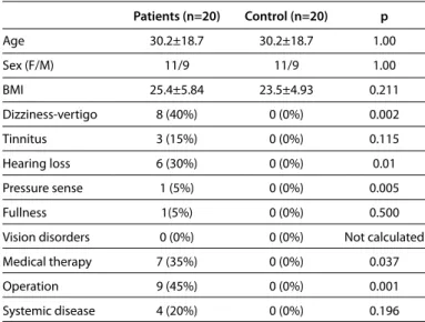
The mean age was 30.2±18.7. There were 11 females and 9 males in each group. The mean BMIs of both the study and control groups were 25.4+5.84 and 23.5+4.93 respectively (p>0.211) (Table 1). The symptoms for IIH were; dizziness (40%, n=8), tinnitus (15%, n=3), hearing loss (30%, n=6), pressure sensation (5%, n=1), and aural fullness (5%, n=1). Among these, the hearing loss was significantly higher (p=0.01). Seven patients (35%) were medically treated [5 with Diazomid (Asetazolamide; Sanofi Aventis, İstanbul, Turkey), 1 with steroids, and 1 with Keppra (Levetiracetam; UCB Pharmaceuticals Inc.; Brussels, Belgium)]. Nine patients (45%) had lumboperitoneal shunt. The control group consisted healthy volunteers, while in 4 study patients, co-morbidities [hypertension (n=2), diabetes mellitus (n=1), and lymphoblastic leukemia (n=2)] were detected (p=0.196) (Table 1).
The study patients had significantly worse hearing levels (in the right ear, at: 250, 500, and 1000 Hz., (p=0.001); in the left ear at: 1000 and 2000 Hz., (p=0.007 and p=0.046). Pure tone averages were signifi-cantly higher in both ears of the study group (p<0.05). The speech discrimination scores were insignificant (p>0.05) (Figure 1).
According to tympanometry results, 3 patients (15%) in the study group had no reflexes while all the healthy subjects had acoustic re-flexes (p>0.05). The median values of impedancemetry in right and left ears of the study group were -20 (-31.5−-13) daPa and -22 (-31−-16) daPa, respectively. In the control group, the results for right and left ears were -20 (-27.25−-12) daPa, and -16 (-27—10.5) daPa, respec-tively (p>0.05).
Ten patients (50%) (saccadic latency prolongation in 4, optokinetic test asymmetry in 5, and tracking test abnormalities in 2 patients) in the study group and one healthy volunteer (5%) (saccadic latency prolongation) had signs in ENG, possibly related to central patholo-gies (p=0.008). Among the subtests, only the optokinetic test results were significant (p=0.024). Three patients (15%) had abnormal posi-tional test results (p>0.05).
Three patients (15%) (2 in the left ear, 1 in the right ear) and 1 (5%) volunteer (left ear) had canal paresis (p>0.05). There was directional preponderance in 1 (5%) patient and in 2 (10%) control individuals (p>0.05). The caloric test results for each ear in each group were sta-tistically insignificant (p>0.05) (Figure 2).
Eight (40%) patients had variable pathological signs in the VEMP [2 (10%) patients with latency prolongation, 1 (5%) patient with increased amplitude, and 1 (5%) patient with decreased ampli-tude in the right ear; 1 (5%) patient with increased ampliampli-tude and 2 (10%) patients with decreased amplitude in the left ear]. Three patients lacked VEMP responses, with one of them bilat-erally). Pathological test results were marked in the right ear of three (15%) and in the left ear of six (30%) patients. Only one (5%) in the control group showed VEMP abnormalities in the left ear (p=0.008). Interaural differences in both groups were insignificant (p=0.478).
DISCUSSION
A significant number of IIH patients attend clinics with audio-vestib-ular symptoms and the audiometry, ENG, and VEMP tests showed significant abnormalities in these patients.
In this study, BMI values were insignificant. The patients were under various treatments during the study including weight loss programs, which as a result might have altered the values.
In 1981, Sismanis [19] analyzed a patient with pulsatile tinnitus, in
whom, bilateral papilledema and increased CSF pressure was ob-served. After lumbar puncture, the patient had a temporary relief from symptoms.
In the study of Saxena et al. [19, 20, 21], including 63 patients with
in-creased intracranial pressure, 38% of patients had hearing loss, 21.3% had tinnitus, and 11% had vertigo complaints In 1943, Fisher and Wolfson [22] observed continuous or intermittent tinnitus, high
fre-quency hearing loss, vertigo, spontaneous nystagmus, and bilateral labyrinth hyper-reaction in their patients.
Kaaber and Zilstorff [23] investigated audio-vestibular signs in 34 IIH
patients in 1978. They observed tinnitus complaints in 4, bilateral hearing loss in 1, and vertigo in 2 patients.
According to our study, similarly, patients attended to our clinic especially with hearing loss, dizziness/vertigo, or pressure sense in Table 1. Characteristics and symptoms of the patient and control groups
Patients (n=20) Control (n=20) p Age 30.2±18.7 30.2±18.7 1.00 Sex (F/M) 11/9 11/9 1.00 BMI 25.4±5.84 23.5±4.93 0.211 Dizziness-vertigo 8 (40%) 0 (0%) 0.002 Tinnitus 3 (15%) 0 (0%) 0.115 Hearing loss 6 (30%) 0 (0%) 0.01 Pressure sense 1 (5%) 0 (0%) 0.005 Fullness 1(5%) 0 (0%) 0.500 Vision disorders 0 (0%) 0 (0%) Not calculated Medical therapy 7 (35%) 0 (0%) 0.037 Operation 9 (45%) 0 (0%) 0.001 Systemic disease 4 (20%) 0 (0%) 0.196
BMI; body mass index
Figure 1. Comparing hearing levels at each frequency in both idiopathic
intra-cranial hypertension (IIH) and control groups. For the right ear, hearing levels at 250, 500, and 1000 Hz; for the left ear hearing, levels at 1000 and 2000 Hz (p<0.05). 25 20 15 10 5 0 R L R L R L R L R L R L 250 Hz 500 Hz 1000 Hz 2000 Hz 4000 Hz 8000Hz Frequencies Hear ing lev el (decibel) IIH Control
Figure 2. The caloric responses of right and left ears in both groups (p>0.05). 35 30 25 20 15 10 5 0
Cal R wam Cal L wam Cal R cold Cal L cold IIH Control
the ears. Lumboperitoneal shunt procedures and medications in use during the otolaryngologic examinations might have relieved the initial audio-vestibular symptoms. Thus the authors believe that symptoms could have been more severe at initial neuro-otologic ex-aminations.
Sismanis [19] studied 20 female IIH patients between the years 1981
and 1985. Pure tone audiometry, tympanometry, and ENG tests were performed. Hearing loss, especially in low frequencies, was observed (250, 500 Hz). After improvement of the intracranial pressure levels, hearing loss in the affected frequencies recovered. These results co-here with our study results.
In our study, hearing loss was observed in 250, 500, and 1000 Hz in the right ear; and in 1000 and 2000 Hz in the left ear. Varying hear-ing levels in each ear and each frequency may be explained with further studies with a greater number of patients. Nevertheless, to our knowledge, the tendency of one side to inner ear pathologies is not mentioned in the English literature. Lower frequency hearing loss suggests a similar mechanism as in the endolymphatic hydrops. This current study differs from the studies of Saxena et al. [20] and
Tan-don et al. [21] because only sensorineural hearing loss was observed.
Saxena et al. [20, 21] studied 65 IIH patients and 81% had hearing loss.
Among them, 77.5% had sensorineural hearing loss, and 22.5% had conductive type hearing loss. Conductive type hearing loss was de-termined as a result of the higher incidence of otosclerosis in the study group. During the study design, we excluded patients with co-morbid ear pathologies causing conductive hearing loss, which may be why we did not observe any such hearing loss.
Speech discrimination scores and tympanometry results were similar in both groups in our study, and the results match the literature [19].
Static compliances were not compared in this study.
Kaaber and Zilstorff [23] evaluated the vestibular functions of 34 IIH
patients with ENG. Affected vestibular functions were seen in 19 pa-tients; 9 patients had a decreased caloric response, 9 patients had directional preponderancy, and hyperreactivity was detected in one patient. Though they could not find any significant relationship be-tween peripheral vestibular functions and intracranial pressure, they observed that, during the follow-up, vestibular functions were im-proved as the pressure was normalized.
We found significantly higher abnormal ENG results in the study group, but there were no significant pathologic signs in positional and caloric tests. ENG showed abnormal responses indicating central dysfunction in our study group. This syndrome primarily affects the central nervous system and neurologic findings are indispensable. Three patients showed abnormal positional tests, raising the possi-bility of co-existing BPPV. All the study group patients were selected from patients that were free of other central or peripheral vestibular pathologies. Increased intracranial pressure, and so inner ear pres-sure alterations, may affect the utricule and saccule, leading to oto-lith dysfunctions nonetheless.
According to our results so far, it is observed that both cochlear and vestibular organs are primarily affected in this syndrome. The
trans-mission of increased cerebrospinal pressure to the cochlear aque-duct and internal acoustic canal, causing compression or increased tension in the cochleovestibular nerve or endolymphatic system, is one of the main pathophysiological explanations [21]. Another
possi-ble mechanism for the vestibular symptoms may be endolymphatic fluid malabsorption as a result of direct compression or decreased vascular supply of the the endolymphatic sac that is wrapped in be-tween both dural leaflets [21].
To our knowledge, no study in the literature considered the inferior vestibular nerve and saccule functions using a VEMP test, in IIH pa-tients. The VEMP responses in our study were significantly abnormal. Abnormal VEMP test results included: increased or decreased ampli-tude, prolonged latency, and absence of VEMP responses. The results suggested that increased intracranial pressure may affect the inferior vestibular nerve and/or saccule. The saccule is in the membranous labyrinth, filled with endolymph, and is located in the bony labyrinth filled with perilymph [24]. The endolymphatic sac responsible for the
absorption of endolymph, is connected to the utricule and the sac-cule via the endolymphatic canal [25]. Pressure alterations in each
lo-cation are directly or indirectly transmitted to others.
The saccule is the second most common site of involvement in en-dolymphatic hydrops [24, 26-28]. It is suggested that increased
endolym-phatic pressure dilates the saccular maculae, which results in delayed VEMP responses [28, 29]. It is known that, especially, decreased
ampli-tude or no response in VEMP may be associated with endolymphat-ic hydrops [29, 30]. Additionally, central pathologies may also change
VEMP responses [30]. Our results in VEMP were: increased amplitude,
decreased amplitude, prolonged latencies, or absence of responses. The results are promising; however, the efficacy, sensitivity, and spec-ificity of VEMP in IIH needs further study. Comprehensive interpreta-tions of the results and their clinical significance need to be clarified with additional studies.
During the time of testing, intracranial hypertension status of many patients was unclear since many had had undergone previous treat-ment for their disorder. However, significant pathological findings were seen in the audiometry, ENG, and VEMP tests, which may sup-port the hypothesis that raised intracranial pressure affects both co-chlear and vestibular end organs and/or the audio-vestibular nerves. Additionally, VEMP results showed that the inferior vestibular nerve may be directly or indirectly (due to saccular dysfunction) affected due to the increased intracranial pressure.
CONCLUSION
Elevated intracranial pressure may induce perilymphatic and endo-lymphatic pressure alterations, causing audio-vestibular complaints. The mechanisms seem to be similar as in the hydrops. The challenge of this study include: having a small study group, being unable to examine some of these patients initially, their medical and surgical therapies, and working on a syndrome demonstrating both central and peripheral vestibular signs. Even so, this clinical research is the first study to search for audio-vestibular signs using all the current audio-vestibular tests including VEMP. Previously, hearing abnormal-ities in IIH were mentioned in the literature; but vestibular dysfunc-tions related with this syndrome comprise a novel research area. In light of IIH, otorhinolaryngologists can consider this syndrome in
pa-tients with audio-vestibular complaints and other departments may consult more frequently with otorhinolaryngology clinics during the diagnostic evaluations. Thus, further studies with an increased number of patients attending as soon as their symptoms begin may be handled. Static compliance differences, and electrocochleograph-ic, VEMP (both cervical and ocular) measurements may be future achievements.
Ethics Committee Approval: Ethics committee approval was received for this
study from the ethics committee of Başkent University Medical and Health Sciences Research and Ethics Committee.
Informed Consent: Written informed consent was obtained from patients
who participated in this study.
Peer-review: Externally peer-reviewed.
Author Contributions: Concept - A.E., O.L., C.K.; Design - A.E., C.K., O.L.;
Super-vision - O.L., A.E.; Materials - C.K., A.E., O.L.; Data Collection and/or Processing - C.K.; Analysis and/or Interpretation - C.K., A.E.; Literature Search - C.K.; Writing Manuscript - C.K.; Critical Review -A.E., O.L.
Conflict of Interest: No conflict of interest was declared by the authors. Financial Disclosure: The authors declared that this study has received no
financial support.
REFERENCES
1. Johnston I, Owler B, Pickard J. The pseudotumor cerebri syndrome. Cam-bridge University Press 2007. [CrossRef]
2. Durcan FJ, Corbett JJ, Wall M. The incidence of pseudotumor cerebri. Population studies in Iowa and Louisiana. Arch Neurol 1988; 45: 875-7.
[CrossRef]
3. Silberstein SD, McKinstry RC 3rd. The death of idiopathic intracranial hy-pertension? Neurology 2003; 60: 1406-7. [CrossRef]
4. Johnston PK, Corbett JJ, Maxner CE. Cerebrospinal fluid protein and opening pressure in idiopathic intracranial hypertension (pseudotumor cerebri). Neurology 1991; 41: 1040-2. [CrossRef]
5. Bono F, Giliberto C, Mastrandrea C, Cristiano D, Lavano A, Fera F et al. Transverse sinus stenoses persist after normalization of the CSF pressure in IIH. Neurology 2005; 65: 1090-3. [CrossRef]
6. Corbett JJ, Digre K. Idiopathic intracranial hypertension: an answer to, “the chicken or the egg?”. Neurology 2002; 58: 5-6. [CrossRef]
7. Johnston I. The historical development of the pseudotumor concept. Neurosurg Focus 2001; 11: E2. [CrossRef]
8. Winn HZ. Non neoplastic disorders mimicking brain tumors. Youmans Neurological Surgery. Philadelphia: Saunders; 2004: 1419-30.
9. Glueck CJ, Goldenberg N, Golnik K, Sieve L, Wang P. Idiopathic intracrani-al hypertension: associations with thrombophilia and hypofibrinolysis in men. Clin Appl Thromb Hemost 2005; 11: 441-8. [CrossRef]
10. Rangwala LM, Liu GT. Pediatric idiopathic intracranial hypertension. Surv Ophthalmol 2007; 52: 597-617. [CrossRef]
11. Brackmann DE, Doherty JK. Facial palsy and fallopian canal expansion as-sociated with idiopathic intracranial hypertension. Otol Neurotol 2007; 28: 715-8. [CrossRef]
12. Andrews LE, Liu GT, Ko MW. Idiopathic intracranial hypertension and obesity. Horm Res Paediatr 2014; 81: 217-25. [CrossRef]
13. Friedman DI, Jacobson DM. Idiopathic intracranial hypertension. J Neu-roophthalmol 2004; 24: 138-45. [CrossRef]
14. Kapoor KG. Etiology of dizziness, tinnitus and nausea in idiopathic intra-cranial hypertension. Med Hypotheses 2008; 71: 310-1. [CrossRef]
15. Giuseffi V, Wall M, Siegel PZ, Rojas PB. Symptoms and disease associa-tions in idiopathic intracranial hypertension (pseudotumor cerebri): a case-control study. Neurology 1991; 41: 239-44. [CrossRef]
16. Rudnick E, Sismanis A. Pulsatile tinnitus and spontaneous cerebrospinal fluid rhinorrhea: indicators of benign intracranial hypertension syn-drome. Otol Neurotol 2005; 26: 166-8. [CrossRef]
17. Akkuzu G, Akkuzu B, Ozluoglu LN. Vestibular evoked myogenic poten-tials in benign paroxysmal positional vertigo and Meniere’s disease. Eur Arch Otorhinolaryngol 2006; 263: 510-7. [CrossRef]
18. Colebatch JG, Halmagyi GM. Vestibular evoked potentials in human neck muscles before and after unilateral vestibular deafferentation. Neurolo-gy 1992; 42: 1635-6. [CrossRef]
19. Sismanis A. Otologic Manifestations of benign intracranial hypertension syndrome: Diagnosis and management. Laryngoscope Supplement 1987; 97: 1-17. [CrossRef]
20. Saxena RK, Tandon PN, Sinha A. Auditory functions in raised intracranial pressure. Acta Otolaryngol 1969; 68: 402-10. [CrossRef]
21. Tandon PN, Sinha A, Kacker SK, Saxena RK, Singh K. Auditory function in raised intracranial pressure. J Neurol Sci 1973; 18: 455-67. [CrossRef]
22. Fisher J, Wolfson LE. In the inner ear. Heineman, London. 1943, p:320. 23. Kaaber EG, Zilstorff K. Vestibular function in benign intracranial
hyper-tension. Clin Otolaryngol 1978; 3:183-8. [CrossRef]
24. Okuno T, Sando I. Localization, frequency, and severity of endolymphatic hydrops and the pathology of the labyrinthine membrane in Meniere’s disease. Ann Otol Rhinol Laryngol 1987; 96: 438-45. [CrossRef]
25. John PY, Kacker SK, Tandon PN. Bekesy audiometry in evaluation of hear-ing in cases of raised intracranial pressure. Acta Otolaryngol 1979; 87: 441-4. [CrossRef]
26. Didier A, Cazals Y. Acoustic responsesrecorded from the saccular bundle on the eighth nevre of the guinea pig. Hear Res 1989; 37: 123-7. [CrossRef]
27. McCue MP, Guinan JJ Jr. Acoustically responsive fibers in the vestibular nevre of the cat. J Neurosci 1994; 14: 6058-70.
28. Young YH, Huang TW, Cheng PW. Vestibular evoked myogenic potentials in delayed endolymphatic hydrops. Laryngoscope 2002; 112: 1623-6.
[CrossRef]
29. Young YH, Huang TW, Cheng PW. Assessing the stage of Meniere’s dis-ease using vestibular evoked myogenic potentials. Arch Otolaryngol Head Neck Surg 2003; 129: 815-8. [CrossRef]
30. Murofushi T. Clinical application of vestibular evoked myogenic poten-tial (VEMP). Auris Nasus Larynx 2016; 43: 367-76. [CrossRef]