Received: 12 October 2016 Revised: 13 March 2017 Accepted: 20 March 2017 https://doi.org/10.1259/bjr.20160803
Cite this article as:
Durur-Subasi I, Durur-Karakaya A, Karaman A, Seker M, Demirci E, Alper F. Is the necrosis/wall ADC ratio useful for the differentiation of benign and malignant breast lesions? Br J Radiol 2017; 90: 20160803.
FULL PAPER
Is the necrosis/wall ADC ratio useful for the differentiation
of benign and malignant breast lesions?
1IRMAK DURUR-SUBASI,MD, PhD,2AFAK DURUR-KARAKAYA,MD,3ADEM KARAMAN,MD,2MEHMET SEKER,MD, 4ELIF DEMIRCI,MDand3FATIH ALPER,MD, PhD
1Department of Radiology, Diskapi Yildirim Beyazit Training and Research Hospital, Ankara, Turkey 2Department of Radiology, Faculty of Medicine, Istanbul Medipol University, Istanbul, Turkey 3Department of Radiology, Faculty of Medicine, Ataturk University, Erzurum, Turkey 4Department of Pathology, Faculty of Medicine, Ataturk University, Erzurum, Turkey
Address correspondence to: Assoc. Prof. Irmak Durur-Subasi E-mail:[email protected]
Objective: To determine whether the necrosis/wall apparent diffusion coefficient (ADC) ratio is useful for the malignant–benign differentiation of necrotic breast lesions.
Methods: Breast MRI was performed using a 3-T system. In this retrospective study, calculation of the necrosis/ wall ADC ratio was based on ADC values measured from the necrosis and from the wall of malignant and benign breast lesions by diffusion-weighted imaging (DWI). By synchronizing post-contrast T1 weighted images, the separate parts of wall and necrosis were maintained. All the diagnoses were pathologically confirmed. Statistical analyses were conducted using an independent sample t-test and receiver operating characteristic analysis. The intraclass and interclass correlations were evaluated. Results: A total of 66 female patients were enrolled, 38 of whom had necrotic breast carcinomas and 28 of whom had breast abscesses. The ADC values were obtained from both the wall and necrosis. The mean necrosis/wall
ADC ratio (6 standard deviation) was 1.61 6 0.51 in carcinomas, and it was 0.656 0.33 in abscesses. The area under the curve values for necrosis ADC, wall ADC and the necrosis/wall ADC ratio were 0.680, 0.068 and 0.942, respectively. A wall/necrosis ADC ratio cut-off value of 1.18 demonstrated a sensitivity of 97%, specificity of 93%, a positive-predictive value of 95%, a negative-predictive value of 96% and an accuracy of 95% in determining the malignant nature of necrotic breast lesions. There was a good intra- and interclass reliability for the ADC values of both necrosis and wall.
Conclusion: The necrosis/wall ADC ratio appears to be a reliable and promising tool for discriminating breast carcinomas from abscesses using DWI.
Advances in knowledge: ADC values of the necrosis obtained by DWI are valuable for malignant-benign differentiation in necrotic breast lesions. The necrosis/ wall ADC ratio appears to be a reliable and promising tool in the breast imaging field.
INTRODUCTION
In the course of interpreting breast MRI, a perplexing condition for breast radiologists may be discriminating an abscess from a necrotic carcinoma. Their similar appear-ance, including irregular borders, peripheral enhancement1 and washout pattern can all be displayed by inflammatory conditions in ways that mimic carcinoma.2 In addition, similar to malignancies, mastitis has been reported to have low apparent diffusion coefficient (ADC) values.1,3
However, in articles concerning breast diffusion-weighted imaging (DWI), generally the ADC of the whole lesion or the enhanced part of the lesion is measured, especially in the case of necrotic lesions.4,5To our knowledge, only one study has evaluated the morphological DWI features of
simultaneously measure the ADC value of the lesions’ necrosis.
In this article, we aimed to measure all parts (necrosis and wall) of the breast lesions to provide an easily referenced criterion reflecting the nature of the entire mass. Therefore, the necrosis/wall ADC ratio was investigated to ascertain whether breast carcinomas and abscesses could be ade-quately discriminated.
METHODS AND MATERIALS Patients
The institutional ethics committee of the Ataturk Univer-sity Faculty of Medicine, Erzurum, Turkey, approved the study protocol, and informed consent was waived due to
Breast MRI data for patients, evaluated between June 2012 and July 2015, with necrotic breast lesions were retrieved from the picture archive and communication system of the Ataturk University Faculty of Medicine Hospital, Erzurum, Turkey. Breast DWI findings were evaluated, with the patients being divided into two groups, according to whether they had malig-nant or benign necrotic breast lesions.
The main indications for breast MRI were (1) to detect multifocal, multicentric or contralateral breast carcinomas; (2) to reveal in-vasive components in ductal carcinomas in situ; (3) to identify occult cancer in patients with metastatic axillary nodes; and (4) to detect unequivocalfindings on conventional imaging. The minor indications were neoadjuvant chemotherapy follow-up (1), eval-uation of breast implants (2), post-surgical evaleval-uation (3).
Breast MRIs
Breast MRIs were obtained by a 3-T system (Skyra; Siemens, Erlangen, Germany) through the use of a breast-dedicated coil in the prone position.
A standard protocol was used: pre-contrast, sagittal, fat-saturated, turbo-spin echo T2 weighted imaging, coronal short-inversion
recovery, transverse turbo-spin echo T1 weighted imaging,
transverse DWI using single-shot echo-planar imaging, and transverse dynamic pre-contrast and post-contrast fat-saturated, fast low-angle shot three-dimensional images were obtained. For all patients, gadolinium chelate was injected intravenously at a dose of 0.1 mmol kg21, followed by a 20-ml saline flush. The injection rate was 2 ml s21, and a power injector was used. Dynamic imaging was started after afixed delay of 30 s following the contrast material injection. Each sequence lasted approxi-mately 65 s. A total offive series were acquired.
DWI was obtained by echo-planar imaging before the dynamic series with the following parameters: time of repetition, 4000 ms; time of echo, 60 ms; slice thickness, 4.0 mm; andfield of view, 380 mm. Diffusion-sensitizing gradients were applied with a diffusion sensitivity of b5 0, 400 and 800 s mm22. Image analyses
The breast MRI data set was evaluated using a standard image interpretation workstation (Syngo.via; Siemens Healthcare, Forchheim, Germany) using breast MRI application software.
All measurements were performed by one radiologist with 10 years of experience (AK), who was blinded to both patient history and histological results. Intraclass reliability was assessed by the same radiologist, with a time interval of 2 weeks, and these data were used to check interclass reliability by a second radiologist with 15 years of experience (FA). Thefinal diagnosis was based on the histopathologicalfindings.
The DWIs were evaluated by drawing free-hand regions of in-terest (ROIs) on lesions on the ADC map images. The ADC values were measured from the longest dimension of the lesion and calculated as the mean of three measurements for both necrosis and the wall of the lesion. For the wall, obvious necrotic or unenhanced areas were excluded by visual correlation with the post-contrast T1 weighted images. A round ROI was
obtained with an area ranging from 2 to 5 mm2. The mean ADC value, not the minimum, of the relevant pixels was used for all calculations. The necrosis/wall ADC ratio was calculated as the ratio of the ADC value of the necrosis to that of the wall. Thefinal patient diagnoses were recorded from the pathological data pool of the Ataturk University Faculty of Medicine.
Statistical analyses
Data were analysed using the Statistical Package for Social Sci-ences program v. 24.0 (IBM Corp., New York, NY; formerly SPSS Inc., Chicago, IL). The mean and standard deviations were calculated for quantitative data. To ensure normal distribution of the data, the one-sample Kolmogorov–Smirnov test was con-ducted. When normal distribution was not ensured, log trans-formation was applied to the data set.
The mean ADC values of necrosis and wall, and the necrosis/ wall ADC ratio of the carcinomas and abscesses, were compared using an independent samples t-test where p, 0.05 was con-sidered statistically significant.
A receiver operating characteristic (ROC) analysis was per-formed to evaluate the diagnostic capability of the ADC values of necrosis and wall, and the necrosis/wall ADC ratio, in terms of malignant–benign differentiation based on the final pathology results. Values of sensitivity, specificity, positive- and negative-predictive values, and accuracy were calculated for the threshold value of the necrosis/wall ADC ratio.
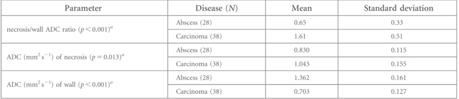
Table 1. For the lesions, mean apparent diffusion coefficient (ADC) values and standard deviation of the necrosis and wall, and the necrosis/wall ADC ratios
Parameter Disease (N) Mean Standard deviation
necrosis/wall ADC ratio (p, 0.001)a Abscess (28) 0.65 0.33
Carcinoma (38) 1.61 0.51 ADC (mm2s21) of necrosis (p5 0.013)a Abscess (28) 0.830 0.115 Carcinoma (38) 1.043 0.155 ADC (mm2s21) of wall (p, 0.001)a Abscess (28) 1.362 0.161 Carcinoma (38) 0.703 0.127
Intraclass correlation was used as a measure of the internal consistency/reliability, and the intraclass correlation coefficient was considered statistically significant if p , 0.001.
RESULTS
38 patients (with a mean age of 416 10 years, ranging from 32 to 65 years) with breast carcinomas and 28 patients (with a mean age of 426 14 years, ranging from 18 to 81 years) with breast abscesses were evaluated. All benign and malignant lesions were pathologically confirmed.
The mean ADC values and standard deviations of the necrosis and wall, and the necrosis/wall ADC ratios, are given inTable 1. Between the carcinomas and abscesses, there was a statistically significant difference between the ADCs of necrosis and wall (p5 0.013 and ,0.001, respectively). The mean necrosis/wall ADC ratios for the carcinomas and abscesses were 1.616 0.51 and 0.656 0.33, respectively, which show a statistically signifi-cant difference (p, 0.001).
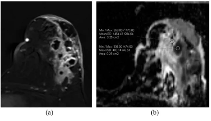
For carcinomas, the wall of the lesion showed a lower mean ADC than the necrosis (0.7033 1023mm2s21 and 1.0433 1023mm2s21, respectively). Benign necrotic lesions (abscesses) exhibited lower mean ADC values in necrosis compared with wall (0.8303 1023mm2s21 and 1.3623 1023mm2s21, respectively) (Figures 1 and 2). Figure 3 represents more diffusion-restricted areas of lesions as dark-grey regions. Box plots show ADC values for both necrosis and wall, and the necrosis/ wall ADC ratio (Figures 4and5).
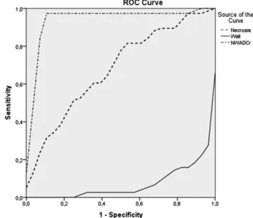
The area under the curve values for necrosis and wall ADC values and the necrosis/wall ADC ratio were 0.680, 0.068 and 0.942, respectively (Figure 6). A wall/necrosis ADC ratio cut-off value of 1.18 demonstrated a sensitivity of 97%, specificity of 93%, positive-predictive value of 95%, negative-predictive value of 96% and accuracy of 95% in determining malignant necrotic breast lesions.Table 2shows the ROC analysis results for ADC of both necrosis and wall, and the necrosis/wall ADC ratio.Table 2also shows the sensitivity and accompanying specificity rates. There was a good intra- and interobserver reliability for the wall [intraclass correlation 0.97 (0.96–0.98) and 0.92 (0.88–0.95) for intra- and interobserver, respectively] and necrosis [intraclass correlation 0.98 (0.95–0.99) and 0.92 (0.86–0.95) for intra- and interobserver, respectively].
DISCUSSION
Our study revealed that the necrosis/wall ADC ratio might be more useful for malignant–benign discrimination in necrotic breast lesions than for measuring ADC only from the wall. For the determined threshold value, the calculated specificity, sen-sitivity, positive- and negative-predictive values, and accuracy indicated the necrosis/wall ADC ratio to be a reliable tool for the differential diagnosis of necrotic breast lesions.
Recently, breast MRI has gained considerable clinical importance.7,8 For the detection of carcinomas, breast MRI has a superior sensitivity of 94–100%.9,10Nevertheless, the specificity of breast
MRI remains below these values due to the overlapping char-acteristics of benign and malignant lesions.2,11,12 Both mor-phologic and kinetic features on breast MRI have to be evaluated during the analysis of benign and malignant lesions,11 and several authors have also reported the importance of DWI to aid differential diagnosis.4,5,13 The Almeida et al14 study, which in-vestigated whether the use of DWI improves diagnostic perfor-mance of breast imaging reporting and data systems categorization on MRI, concluded that ADC measurements should be applied for better diagnostic performance.
Figure 1. (a, b) Breast carcinoma. Subtraction image (a) shows enhanced mass with irregular borders. The ADC values obtained from necrosis and wall (b) of a carcinoma. Max, maximum; min, minimum; SD, standard deviation.
Figure 2. (a, b) Breast abscess. Subtraction image (a) shows a peripherally enhanced lesion. The ADC values obtained from necrosis and wall (b) of the abscess. Max, maximum; min, minimum; SD, standard deviation.
Figure 3. (a, b) Representative figures show greater or lesser diffusion-restricted areas in the carcinoma (a) and abscess (b). Dark grey and light areas show greater and lesser restriction, respectively.
DWI principally measures Brownian motion, referred to as diffusibility. Diffusion occurring in individual voxels can be calculated from images with different diffusion weightings, resulting in a quantitative ADC map. DWI has been reported as being able to increase the specificity and diagnostic accuracy of breast MRI.3,15,16 DWI with quantitative ADC measurements has been reported to be a reliable tool for differentiation of malignant and benign breast lesions.4
Generally, ADC measurements have been performed from contrast-enhanced solid parts of the lesions.4 A number of
studies have discovered major differences in the ADC values of breast lesions.2,15,17,18Greater ADC values are seen in benign lesions pertaining to their lower cellularity and greater mo-lecular independence. In addition, the tissue-surrounding necrosis exhibits vascular fibroblastic proliferation and in-flammation for abscess. Necrosis of the abscess contains cel-lular debris and high viscosity. All these features may contribute to the diffusion appearance of the abscess. On the other hand, lower ADC values are seen in malignant lesions. For carcinomas, the wall of the lesion show high cellular proliferation rate, cells with large size of nucleus, intracellular macromolecules, high nucleus–cytoplasm rate and limited size of the extracellular matrix.19,20Those all may contribute to the diffusion restriction. Moreover, ADC values are affected by multiple factors, and comparisons between different reports are difficult owing to non-standardization and the use of dif-ferent techniques.21
A study on the value of ADC in differentiating malignant and benign breast lesions conducted by a 1.5 Tesla system concluded that the mean ADC values were significantly different in malignant vs benign lesions (1.0460.29 31023vs 1.6160.50 31023cm2s21 for the malignant and benign lesions, respectively, p5 0.03).22 A cut-off value of 1.303 1023mm2s21for ADC detected with ROC analysis yielded 89.1% sensitivity and 100% specificity for the differentiation between benign and malignant lesions. In that study, the ROI was placed manually within the lesions, avoiding the cystic–necrotic and hemorrhagic components.22
Some other studies23,24 have found similar results. However, such applications by ignoring necrosis might not resemble all internal characteristics of the whole lesion, especially necrotic ones. Additionally in our study, all the mean ADC values for both carcinoma and abscess were under these cut-off values. Therefore, it could not be enough to be a malignant-benign discrimination criterion.
Figure 4. Box plot showing apparent diffusion coefficient (ADC) values obtained from necrosis (centre) and periphery (wall) of abscesses and carcinomas (Circles show excessive values).
Figure 5. Box plot showing necrosis/wall apparent diffu-sion coefficient ratio values obtained from abscesses and carcinomas (Stars show excessive values).
Figure 6. Receiver operating characteristic (ROC) analyses show diagnostic performance of apparent diffusion coefficient (ADC) of necrosis and wall and necrosis/wall ADC ratio of the lesions.
A parameter such as the necrosis/wall ADC ratio may be helpful to standardize measurements and provide a more objective di-agnosis. In addition, the necrosis/wall ADC ratio reflects the internal characteristics of the lesions. For example, the periph-eral portions of abscesses can show a moderate diffusion re-striction, which mimics a malignant lesion. However, the use of the necrosis/wall ADC ratio of the lesion corrects this illusion because the central portion is far more diffusion restricting for benign lesions.25 Moreover, in the centre of a carcinoma, the ADC values can be higher. Such a ratio may provide a visual perspective as well. Someone may think that if the restriction is much more in the necrosis, it could be an abscess, and if in wall, it could be a carcinoma.
There are some limitations with our study. First is the ret-rospective design of the study. The second limitation is the potential population bias because all patients with necrotic
breast lesions—especially benign tumours—are not routinely referred for breast MRI. The third limitation is the limited number of patients enrolled. The fourth limitation is that single-shot echo-planar DWI suffers from blurring due to T2*
decay, and it is particularly vulnerable to off-resonance due to the narrow effective bandwidth in the phase-encoding di-rection.21For this imaging technique, artefacts and distortion can be encountered due to magnetic field inhomogeneities, susceptibility effects, eddy currents and chemical shift.21 In addition, DWI is motion sensitive.
In this study, we evaluated the necrosis/wall ADC ratio for dis-criminating breast carcinomas from abscesses. We found that the necrosis/wall ADC ratio may serve as an easy and reliable tool, and this parameter can be used as a way of succinctly expressing both the peripheral and internal characteristics of a lesion using MRI, thereby providing a holistic approach to identify breast lesions.
REFERENCES
1. Neubauer H, Platzer I, Mueller VR, Meyer T, Liese J, Koestler H, et al. Diffusion-weighted MRI of abscess formations in children and young adults. World J Pediatr 2012;8: 229–34. doi:https://doi.org/10.1007/s12519-012-0362-4
2. Kul S, Cansu A, Alhan E, Dinc H, Gunes G, Reis A. Contribution of diffusion-weighted imaging to dynamic contrast-enhanced MRI in the characterization of breast tumors. AJR Am J Roentgenol 2011;196: 210–17. doi:
https://doi.org/10.2214/AJR.10.4258
3. Caivano R, Villonio A, D’Antuono F, Gioioso M, Rabasco P, Iannelli G, et al. Diffusion weighted imaging and apparent diffusion coefficient in 3 tesla magnetic resonance imaging of breast lesions. Cancer Invest 2015; 33: 159–64. doi:https://doi.org/10.3109/ 07357907.2015.1019674
4. Altay C, Balci P, Altay S, Karasu S, Saydam S, Canda T, et al. Diffusion-weighted MR imaging: role in the differential diagnosis of breast lesions. JBR-BTR 2014;97: 211–16.
5. Ramirez-Galvan YA, Cardona-Huerta S, Ibarra-Fombona E, Elizondo-Riojas G. Apparent diffusion coefficient (ADC) value to evaluate BI-RADS 4 breast lesions: correlation with pathologicalfindings. Clin Imaging 2015; 39: 51–5.
6. Kang BJ, Lipson JA, Planey KR, Zackrisson S, Ikeda DM, Kao J, et al. Rim sign in breast lesions on diffusion-weighted magnetic res-onance imaging: diagnostic accuracy and clinical usefulness. J Magn Reson Imaging 2015;41: 616–23. doi:https://doi.org/ 10.1002/jmri.24617
7. Durur-Karakaya A, Durur-Subasi I, Karaman A, Akcay MN, Palabiyik SS, Erdemci B, et al. The use of breast magnetic resonance imaging parameters to identify possible signaling path-ways of a serum biomarker, HE4. J Comput Assist Tomogr 2016;40: 436–41.
8. Durur-Subasi I, Durur-Karakaya A, Alper F, Karaman A, Kılıc RM, Sipal S, et al. Breast lesions with high signal intensity on T1-weighted
MR images. Jpn J Radiol 2013;31: 653–61. doi:
https://doi.org/10.1007/s11604-013-0239-z
9. Warren R, Pointan L, Thompson D. Reading protocol for dynamic contrast-enhanced MR images of the breast: sensitivity and speci fic-ity analysis. Radiology 2005;236: 779–88. doi:
https://doi.org/10.1148/radiol.2363040735
10. Mahoney MC, Gatsonis C, Hanna L, DeMartini WB, Lehman C. Positive pre-dictive value of BI-RADS MR imaging. Radiology 2012;264: 51–8. doi:https://doi. org/10.1148/radiol.12110619
11. Bluemke DA, Gatsonis CA, Chen MH, DeAngelis GA, DeBruhl N, Harms S, et al. Magnetic resonance imaging of the breast prior to biopsy. JAMA 2004; 292: 2735–42. doi:https://doi.org/10.1001/ jama.292.22.2735
12. Wiener JI, Schilling KJ, Adami C, Obuchowski NA. Assessment of suspected breast cancer by MRI: a prospective clinical trial using a combined kinetic and
Table 2. Receiver operating characteristic analysis results
Sensitivity (%) Threshold value (accompanying specificity, %)
ADC (mm2s21) of necrosisa ADC (mm2s21) of wallb Necrosis/wall ADC ratioa
100 0.402 (4) 0.128 (0) 0.37 (11)
90 0.608 (18) 0.413 (0) 1.25 (93)
80 0.737 (50) 0.433 (0) 1.31 (93)
70 0.813 (54) 0.515 (0) 1.35 (97)
60 0.871 (68) 0.555 (0) 1.46 (97)
ADC, apparent diffusion coefficient.
aHigher than these thresholds indicate malignancy. bLower than these thresholds indicate malignancy.
morphologic analysis. AJR Am J Roentgenol 2005;184: 878–86.
13. Cabuk G, Nass Duce M, Ozgur A, Apaydin FD, Polat A, Orekici G. The diagnostic value of diffusion-weighted imaging and the apparent diffusion coefficient values in the differentiation of benign and malignant breast lesions. J Med Imaging Radiat Oncol 2015;59: 141–8. doi:
https://doi.org/10.1111/1754-9485.12273
14. Maltez de Almeida JR, Gomes AB, Barros TP, Fahel PE, de Seixas Rocha M. Subcategori-zation of suspicious breast lesions (BI-RADS Category 4) according to MRI criteria: role of dynamic contrast-enhanced and diffusion-weighted imaging. AJR Am J Roentgenol 2015; 205: 222–31. doi:https://doi.org/10.2214/ ajr.14.13834
15. Partridge SC, Rahbar H, Murthy R, Chai X, Kurland BF, De Martini WB, et al. Improved diagnostic accuracy of breast MRI through combined apparent diffusion coefficients and dynamic contrast-enhanced kinetics. Magn Reson Med 2011;65: 1759–67. doi:https:// doi.org/10.1002/mrm.22762
16. Durur-Subasi I, Durur-Karakaya A, Karaman A, Demirci E, Alper F, Yılmazel-Ucar E, et al. Value of MRI sequences for prediction of invasive breast carcinoma size. J Med Imaging Radiat Oncol 2014;58: 565–8.
17. Woodhams R, Ramadan S, Stanwell P, Sakamoto S, Hata H, Ozaki M, et al. Diffusion-weighted imaging of the breast: principles and clinical applications. Radio-graphics 2011;31: 1059–84.
18. Hatakenaka M, Soeda H, Yabuuchi H, Matsuo Y, Kamitani T, Oda Y, et al. Apparent diffusion coefficients of breast tumors: clin-ical application. Magn Reson Med Sci 2008; 7: 23–9.
19. Henzler T, Schmid-Bindert G, Schoenberg SO, Fink C. Diffusion and perfusion MRI of the lung and mediastinum. Eur J Radiol 2010; 76: 329–36. doi:https://doi.org/10.1016/ j.ejrad.2010.05.005
20. Lyng H, Haraldseth O, Rofstad EK. Mea-surement of cell density and necrotic fraction in human melanoma xenografts by diffusion weighted magnetic resonance imaging. Magn Reson Med 2000;43: 828–36. doi:https://doi. org/10.1002/1522-2594(200006)43:6,828:: aid-mrm8.3.3.co;2-g
21. Barentsz MW, Taviani V, Chang JM, Ikeda DM, Miyake KK, Banerjee S, et al. Assess-ment of tumor morphology on diffusion-weighted (DWI) breast MRI: diagnostic value of reducedfield of view DWI. J Magn Reson Imaging 2015;42: 1656–65. doi:https://doi. org/10.1002/jmri.24929
22. Bozkurt Bostan T, Koç G, Sezgin G, Altay C, Fazıl Gelal M, Oyar O. Value of apparent diffusion coefficient values in differentiating malignant and benign breast lesions. Balkan Med J 2016;33: 294–300. doi:https://doi.org/10.5152/ balkanmedj.2016.141007
23. Durando M, Gennaro L, Cho GY, Giri DD, Gnanasigamani MM, Patil S, et al. Quanti-tative apparent diffusion coefficient mea-surement obtained by 3.0 Tesla MRI as a potential noninvasive marker of tumor aggressiveness in breast cancer. Eur J Radiol 2016;85: 1651–8.
24. Bougias H, Ghiatas A, Priovolos D, Veliou K, Christou A. Whole-lesion apparent diffusion coefficient (ADC) metrics as a marker of breast tumour characterization-comparison between ADC value and ADC entropy. Br J Radiol 2016;89: 20160304.
25. Karaman A, Durur-Subasi I, Alper F, Durur-Karakaya A, Subasi M, Akgun M. Is it better to include necrosis in apparent diffu-sion coefficient (ADC) measurements? The necrosis/wall ADC ratio to differentiate malignant and benign necrotic lung lesions: preliminary results. J Magn Reson Imaging. 2 February 2017. [Epub ahead of print]. doi: