Original papers
DOI: 10.11152/mu.2013.2066.173.khuAbstract
Aims: The aim of this study is to assess the applicability of shear wave elastography (SWE) in the diagnosis of chronic
autoimmun thyroiditis (CAT) patients. Material and methods: The study group consisted of 50 patients with first-diagnosed CAT and 40 control subjects (CS). In all patients with CAT and CS, sonoelastographic measurements were made in both thyroid lobes. Optimal cut-off values were chosen to maximize the sum of sensitivity and specificity. Positive predictive value (PPV), negative predictive value (NPV), and accuracy values were also calculated. Results: Quantitative elastographic analysis evaluated by SWE in CAT patients (2.56 ± 0.30 m/s) was significantly higher compared with CS (1.63 ± 0.12 m/s) (p<0.001). The optimal cut-off value was 2.42 m/s. SWE had 77% sensitivity, 71% specificity, 92% PPV, 81% NPV, and 87% accuracy for the presence of CAT. Conclusions: Our data indicate that SWE correctly defines the elasticty of thyroid paren-chyma, and this technique may assist in the diagnosis and treatment monitoring of CAT.
Keywords: shear wave elastography, ultrasonography, chronic autoimmun thyroiditis,
The role of shear wave elastography in the diagnosis of chronic
autoimmune thyroiditis
Koray Hekimoglu
1, Fuldem Yildirim Donmez
1, Serdar Arslan
1, Adnan Ozdemir
1,
Canan Demir
2, Canan Yazici
31Department of Radiology, 2Department of Endocrinology, 3Department of Biostatistics, Baskent University, School
of Medicine, Ankara, Turkey
Received 01.02.2015 Accepted 04.05.2015 Med Ultrason
2015, Vol. 17, No 3, 322-326
Corresponding author: Koray Hekimoglu Assoc Prof MD Baskent Universitesi Hastanesi,
Radyoloji ABD, Fevzi Cakmak Cad. 10. sok. o: 45, Bahcelievler, Ankara 06490 Turkey. Phone: 90-312 212 68 68/1439
Fax : 90-312-223 73 33
E-mail: [email protected]
Introduction
Due to the impact of enviromental factors, the in-cidence of chronic thyroiditis and thyroid cancer has increased markedly. In industrial populations, chronic autoimmune (Hashimoto’s) thyroiditis (CAT) is charac-terized by variable degrees of lymphocytic inflammatory infiltration and fibrosis of thyroid tissue [1]. CAT is a severe, diffuse thyroid pathology, presenting with hy-pothyroidism, increased antithyroid autoantibodies, and diffusely decreased echogenicity of the thyroid paren-chyma on conventional ultrasonography (US). Primarily, CAT can be diagnosed with thyroid functional tests and elavated antithyroid autoantibodies. Clinically it is silent
and typically is not characterized by painful thyroid en-largement or biochemical signs of inflammation. Though elavated antithyroid autoantibodies are highly indicative of CAT, these do not exclude the diagnosis of other types of thyroiditis [2] and correct diagnosis may be difficult in some cases without biopsy. US examination is a non-invasive and commonly used imaging modality for CAT diagnosis [3]. However, it reveals limited findings such as heterogenous-decreased echogenity, lobulated con-tour, and hyperemia on color Doppler US at initial phase of the disease. As a consequence of the pathology, thy-roid parenchyma becomes stiff comparediwith the healty thyroid tissue due to progression of fibrosis and this find-ing can be evaluated by elastography [4,5].
Strain elastography is a US based dynamic tool to evaluate tissue elasticity by measuring the degree of dis-tortion under the application of an external force. The sys-tem allows the instant evaluation of tissue elasticity and, as in color Doppler imaging, it superimposes the infor-mation in color on the grayscale images. However, being a qualitative imaging technology it can not reveal quan-titative data. Thus, elastography with freehand compres-sion has several limitations being highly dependent on the
compressibility limits or on the extent of tissue compres-sion; also it is an operator dependent technique [5].
Recently, a new elastography method has been de-veloped that uses tracking of shear-wave propagation through tissue to obtain the elastic modulus [6]. In this technology, the target tissue generates small (1-10 µm) lo-calized displacements when it is mechanically “pushed” by short-time acoustic radiation force transmitted from the probe. The displacement of the target tissue gener-ates the “shear wave” which is detected by sonographic detection pulses. This shear-wave elastography (SWE) is operator-independent, reproducible and point quantifica-tion capability [7]. SWE is proporquantifica-tional to the square root of tissue elasticity as a quantitative technique that allows local elasticity estimation of tissues [8].
Acoustic Radiation Force Impulse (ARFI) quantifi-cation is a type of SWE [9,10]. ARFI quantifiquantifi-cation es-timates the elasticity of the US wave by Virtual Touch Tissue Quantification (VTQ) technique. VTQ application is used to create local tissue displacements using lateral shear wave propagation of US beam in SWE. VTQ gen-erates objective and reproducible numeric data. In this mode, numeric values –meters per second- (m/s) of SWE were depicted from ROI evaluation area [11].
The aim of the present study was to evaluate the diag-nostic value of SWE by ARFI in CAT patients by creat-ing a cut-off value for diagnosis.
Materials and methods
This prospective nonrandomized study was per-formed in accordance with the ethical guidelines of the Declaration of Helsinki and was approved by the Eth-ics Committee of University. All of the participants were informed of the research and gave their written informed consent. From August 2012 to November 2014, 50 con-secutive patients diagnosed with CAT (42 male and 8 female) were included. Forty consecutive subjects with no thyroid pathology (23 male and 17 female) were se-lected from the normal population as the control group. The diagnosis of CAT was confirmed by histopathologic examination obtained by fine needle aspiration (FNA) or biopsy. The patients were first evaluated by endocrinolo-gists. Patients with suspected CAT pathology (clinical examination and specific markers) were referred for US-elastography examination and FNA biopsy. Exclusion criteria were prior thyroid surgery or fine needle biopsy in the previous 6 months for possible alterations in the parenchyma, the presence of nodule, calcification, or cystic lesions over 5 mm in thyroid lobes.
All patients underwent a conventional US examina-tion of the thyroid gland, including color Doppler US,
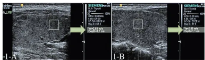
followed by real-time SWE imaging. An Acuson S3000 diagnostic US system (Siemens Medical Solutions, 2013, Erlangen, Germany) was used for SWE imaging with ARFI technique using 4-9 MHz broadband frequency 9L4 lineer probe. The technique was performed with the patient in a supine decubitus position on the exami-nation couch and the neck in hyperextension, eased by positioning of a pillow behind the neck. The examina-tions were conducted by two radiologists with more than 3 years of experience in elastography (S.A., A.O.), who were blinded to the cytology. The probe was placed on the gel gently and, slight compression was maintained during the SWE study. Ten successful measurements per patient were performed, with the ROI (5x6 mm) placed in five different points in per thyroid lobe. While the ve-locities were obtained, the patients held their breath to avoid respiratory movement artifacts. The average SWE examination time was 6±3 minutes (range 3-9 minutes). The measurement values revealed as meter/second (m/s) from ROI area depending on the VTQ calculation (fig 1). Serum assays were performed at the same day by a classical method. Assessment of hormone concentration was performed using Hitachi Cobas e601 chemilumi-nescent analyser and included measurement of serum thyroid stimulating hormone (TSH) and free thyroxine (FT4). Thyroid autoantibody concentrations of anthyroid peroxidase antibody (TPO-Ab) and anthyroglobulin an-tibody (Tg-Ab) were assessed by radioimmunological method using commercially available kits and scintilla-tion gamma counter (LKB Wallac CliniGamma 1272, USA).
The US, SWE, and FNA biopsy results were collect-ed by the statistical coordinator of the study (A.C.Y.) and the values were prospectively entered into a computer database.
Statistical analysis
Statistical analysis was performed with SPSS version 17.0 software (SPSS Inc, Chicago, IL). From the collect-ed variable numeric ARFI measurements of CAT patients and control groups, descriptive statistics were calculated. Reliability of the ten successful measurements per patient was analyzed by intraclass correlation coefficient (ICC).
Fig 1. A) 47 year-old woman with CAT. Diffuse heterogeneous
parenchyma and increased velocities of SWE with 2.83 m/s; B) SWE sonoelastographic evaluation of a 50 year-old male CAT patient. Measurements of ROI area showsn as2.99 m/s.
Normality of distribution of the variables was ana-lyzed using the Shapiro-Wilk test, and the Levene test was used to assess the homogeneity of variances in the groups. Parametric test assumptions were not available; thus comparison between the group of patients with CAT and the control group were performed with the Mann-Whitney U test. P values smaller than 0.05 were consid-ered statistically significant. Diagnostic performance of ARFI elastography was assessed using the receiver op-erating characteristic curve (ROC) that was constructed from sensitivity and specificity values due to a reference line. Optimal cut-off value was chosen to maximize the sum of sensitivity and specificity. Positive predictive val-ue (PPV), negative predictive valval-ue (NPV), and accuracy values were also calculated.
Results
A total of 90 thyroid US-SWE examinations were performed successfully (50 patients and 40 healthy con-trol group subjects).
Conventional US revealed diffuse hypoechogenicity and heterogeneous parenchyma in all CAT patients. The mean SWE value was 2.564±0.30 m/s in CAT patients (range 2.098- 3.164 m/s) (fig 1-2) and in the control group 1.630±012 m/s (range 1.322-1.838). The mean SWE ve-locity result and serum assays of CAT patients were sig-nificantly higher than the mean value of the normal con-trol group (p<0.001) (table I). ICC for right and left lobe were 0.968 (p<0.001) and 0.963 (p<0.001) respectively.
The optimal cut-off value for CAT prediction was 2.42 m/s (77% sensitivity, 71% specificity, 92% PPV, 81% NPV, and 87% accuracy). Area under the curve was 0.849. The 95% confidence interval was 0.741 to 0.958.
Discussions
In the recent years, elastography has been introduced into clinical practice enabling the determination of tissue elasticity using ultrasound devices. Preliminary studies have been performed as a qualitative method real-time elastography in thyroid gland. Usefullness of this meth-od in thyroiditis and differentiating benign - malignant nodules has been widely described. Studies concerning about thyroid nodules concluded that most of the benign nodules were significantly softer comparing with malig-nant nodules and SWE imaging had a good diagnostic quality with high sensitivity and specificity in evaluating this pathology [5,12-16]. Thyroiditis was another major scope of the real-time elastography. First studies empha-sized that, SWE correctly defines the elasticity changes in thyroiditis. Although SWE may provide some useful information in differentiating thyroiditis from the multi-nodular goiter, it gives misleading results in the differen-tial diagnosis of thyroiditis and thyroid cancer [17-19]. Ruchala et al emphasized that, CAT was associated with only a minimal increase in the stiffness of the thyroid parenchyma, and that remains unchanged during thera-py. Thus, SWE had a restricted value for diagnosing the types of thyroiditis and the required clinical assistance [17]. Magri et al concluded that the presence of CAT does not affect the ability of SWE to correctly define the elasticity of thyroid nodules [18]. In the study of Fried-rich-Rust et al the median velocity of SWE was 1.98 m/s in the healthy thyroid gland, a value very similar to our results (1.63 ± 0.13) [14]. According to the other related articles published in the literature, the SWE velocity in thyroid tissue ranged between 0.5 and 4.9 m/s with the highest values reported for malignant nodules ranging
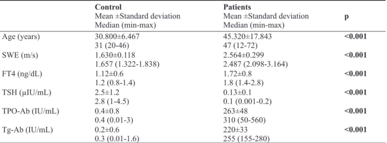
Table I. Mean Age, SWE values and Serum assays of Patients and Control Group.
Control
Mean ±Standard deviation Median (min-max)
Patients
Mean ±Standard deviation
Median (min-max) p Age (years) 30.800±6.467 31 (20-46) 45.320±17.84347 (12-72) <0.001 SWE (m/s) 1.630±0.118 1.657 (1.322-1.838) 2.564±0.2992.487 (2.098-3.164) <0.001 FT4 (ng/dL) 1.12±0.6 1.2 (0.8-1.4) 1.72±0.8 1.8 (1.4-2.8) <0.001 TSH (µIU/mL) 2.5±1.2 2.8 (1-4.5) 0.13±0.1 0.1 (0.001-0.2) <0.001 TPO-Ab (IU/mL) 0.4±0.8 0.4 (0.01-3) 263±48 310 (50-560) <0.001 Tg-Ab (IU/mL) 0.2±0.6 0.3 (0.01-1.6) 220±33 255 (155-280) <0.001 FT4: Free thyroxine; TSH:Thyroid-stimulating hormone; TPO-Ab: Antithyroid peroxidase antibody; Tg-Ab:Antithyroglobulin antibody
from 2.09 to 4.9 m/s. The cut-off point was found to be 2.75 m/s for SWE for distinguishing between malignant and benign thyroid nodules [13,15,16,19-21].
In a recently published study by Sporea et al acoustic radiation force impulse-imaging was used for the evalu-ation of 37 patients with CAT pathologies [22]. Median values in healthy subjects were calculated significantly lower (2 ±0.40 m/s) than in CAT patients (2.43 ±0.58 m/s). They found an optimal cut-off value of 2.36 m/s for diffuse thyroid pathology with a relatively lower sensitiv-ity (62.5% versus 77%). In comparison to this research, we calculated a little higher cut-off value (2.42 m/s) with similar sensitivity and specificity rates. However, the values obtained by Sporea et al could be affected from their heterogeneous patient groups which include Base-dow-Graves or diffuse thyroid goiter patients.
There were some limitations in the present study that need to be addressed. First, the research had a relatively small group. Secondly, the main objective of this study was to show the SWE quantitative evaluation of CAT pa-tients. Hence, detailed description of elastographic im-age variation for each of the particular conditions was not included.
Conclusions
In clinical practice, SWE may assist in the diagno-sis of CAT patients. SWE could be used easily after per-forming routine US examination of thyroid gland. For predicting the presence of autoimmune diffuse thyroid pathology with high sensitivity, the cut-off value should be selected as 2.42 m/s or more in SWE imaging. Fur-ther investigations are required to compare these findings with various thyroid pathologies.
Acknowledgments: We appreciate very much the
help of Assoc.Prof. Canan Yazici for the advanced sta-tistical analysis.
Conflict of interest: none References
1. Weetman AP. Chronic autoimmun thyroiditis. In: Braver-man LE, Utiger RD (eds.) Werner and Ingbar’s The Thy-roid. Philadelphia, Lippincot Williams and Wilkins 2000: 721-732.
2. Espinoza PG, Guendelman CL, Quvedo Limon LN, Fer-nandez RJ. A comparison between two imaging techniques for the diagnosis of subacute thyroiditis (de Quarvain thy-roiditis): brief communication. Clin Nucl Med 2010; 35: 862-864.
3. Mazziotti G, Sorvillo F, Iorio S, et al. Grey-scale analysis allows a quantitative evaluation of thyroid echogenicity in the patients with Hashimoto’s thyroiditis. Clin Endocrinol 2003; 59: 223-229.
4. Asteria C, Giovanardi A, Pizzocaro A, et al. US-elastogra-phy in the differantial diagnosis of benign and malign thy-roid nodules. Thythy-roid 2008; 18: 523-531.
5. Kim H, Kim JA, Son EJ, Youk JH. Quantitative assessment of shear-wave ultrasound elastography in thyroid nodules: diagnostic performance for predicting malignancy. Eur Ra-diol 2013; 23: 2532-2537.
6. Bercoff J, Tanter M, Fink M. Supersonic shear imaging: a new technique for soft tissue elasticity mapping. IEEE Trans Ultrason Ferroelectr Freq Control 2004; 51: 396-409. 7. Palmeri ML, Wang MH, Dahl JJ, Frinkley KD, Nightin-gale KR. Quantifying hepatic shear modulus in vivo using acoustic radiation force. Ultrasound Med Biol 2008; 34: 546-558.
8. Rouze NC, Wang MH, Palmeri ML, Nightingale KR. Pa-rameters affecting the resolution and accuracy of 2D quan-titative shear wave images. IEEE Trans Ultrason Ferroelec-tr Freq ConFerroelec-trol 2012; 59: 1729-1740.
9. Dudea SM, Botar-Jid C. Ultrasound elastography in thyroid disease. Med Ultrason 2015; 17: 74-96.
10. Ophir J, Cespedes I, Ponnekanti H, Yazdi Y, Li X. Elas-tography: a quantitative method for imaging the elasticity of biological tissues. Ultrason Imaging 1991; 13: 111-134. 11. Zhai L, Palmeri ML, Bouchard RR, Nightingale RW,
Nightingale KR. An integrated indenter-ARFI imaging system for tissue stiffness quantification. Ultrason Imaging 2008; 30: 95-111.
12. Bojunga J, Herrmann E, Meyer G, Weber S, Zeuzem S, Friedrich-Rust M. Real-time elastography for the differen-tiation of benign and malignant thyroid nodules: a meta-analysis. Thyroid 2010; 20: 1145-1150.
13. Gu J, Du, L, Bai M, et al. Preliminary study on the diag-nostic value of acoustic radiation force impulse technology for differentiating between benign and malignant thyroid nodules. J Ultrasound Med 2012; 31: 763-771.
14. Friedrich-Rust M, Romenski O, Meyer G, et al. Acoustic radiation force impulse-imaging for the evaluation of the thyroid gland: a limited patient feasibility study. Ultrason-ics 2012; 52: 69-74.
15. Zhan J, Diao XH, Chai QL, Chen Y. Comparative study of acoustic radiation force impulse imaging with real-time elastography in differential diagnosis of thyroid nodules. Ultrasound in Med and Biol 2013; 39: 2217-2225.
16. Zhang FJ, Han RL. The value of acoustic radiation force impulse (ARFI) in the differential diagnosis of thyroid nod-ules. Eur J Radiol 2013; 82: e686-e690.
17. Ruchala M, Szczepanek-Parulska E, Zybek A, et al. The role of sonoelastography in acute, subacute and chronic thyroiditis: a novel application of the method. Eur J Endo-crinol 2012; 166: 425-432.
18. Xie P, Xiao Y, Liu F. Real-time ultrasound elastography in the diagnosis and differential diagnosis of subacute thyroid-itis. J Clin Ultrasound 2011; 39: 435-440.
19. Magri F, Chytiris S, Capelli V, et al. Shear wave elastog-raphy in the diagnosis of thyroid nodules: feasibility in the case of coexistent chronic autoimmune Hashimoto’s thy-roiditis. Clin Endocrinol 2012; 76: 137-141.
20. Zhan J, Diao XH, Chai QL, Chen Y. Comparative study of acoustic radiation force impulse imaging with real-time elastography in differential diagnosis of thyroid nodules. Ultrasound Med Biol 2013; 39: 2217-2225.
21. Han R, Li F, Wang Y, Ying Z, Zhang Y. Virtual touch tissue quantification (VTQ) in the diagnosis of thyroid nodules with coexistent chronic autoimmune Hashimoto’s thyroidi-tis. Eur J Radiol 2015; 84: 327-331.
22. Sporea I, Sirli R, Bota S, Vlad M, Popescu A, Zosin I. ARFI elastography for the evaluation of diffuse thyroid gland pathology: Preliminiary results. World J Radiol 2012; 28: 174-178.