© 2014 by the Texas Heart ®
Institute, Houston
Evaluation of Coronary
Artery–Saphenous Vein
Composite Grafts:
The Aortic No-Touch Technique
We retrospectively compared the results of conventional coronary artery bypass grafting (CABG) performed on patients who showed no preoperative evidence of serious athero-sclerosis of the ascending aorta with the results of the aortic no-touch technique (using coronary artery–saphenous vein composite grafts) on CABG patients who did show such evidence.
From 2003 through 2012, 3,152 consecutive patients underwent isolated primary CABG at our hospital. We chose 360 for the current study. The study group (n=120) com-prised patients who had undergone operation via the aortic no-touch technique. Propen-sity-score-matching (1:2) was used to select the control group of 240 patients who had undergone conventional CABG. Early and late survival rates, reintervention-free survival rates, and freedom from cardiac death were compared.
Early and late mortality rates were similar in the study and control groups (P=0.19 vs
P=0.29, respectively), as were cardiac-related death (2.5% vs 2.1%, respectively; P=0.53)
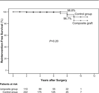
and overall death (8.3% vs 7.9%, respectively; P=0.51). Overall survival rates were 91.7% vs 92.1% and freedom-from-cardiac-death rates were 97.4% vs 97.5% (P=0.71 vs P=0.78, respectively; mean follow-up period, 5.27 ± 2.51 yr). Reintervention-free survival rates were also similar (96.7% vs 98.8%, respectively; P=0.2).
As a result of the similar rates of early and late survival, reintervention-free survival, and freedom from cardiac death, we conclude that the aortic no-touch technique with com-posite grafts might be a reasonable option in patients who have atherosclerotic ascending aorta that cannot be clamped. (Tex Heart Inst J 2014;41(1):26-32)
P
orcelain aorta or disseminated atherosclerotic involvement of the entire ascend-ing aorta presents major surgical limitations durascend-ing coronary artery bypass grafting (CABG). Chief among these is the need to avoid aortic cross-clamp-ing, in order to circumvent neurologic and other sequelae.1 One alternative tocross-clamping is to bypass the aorta with a composite of one or more saphenous vein (SV) and radial artery grafts anastomosed to the left internal mammary artery (LIMA), which previously has been anastomosed to the left anterior descending coronary artery. These grafts are also known as Y or T composite grafts.
Although the safety and efficacy of arterial composite grafts for total arterial revas-cularization have been proved,2,3 the safety and efficacy of venous composite grafts are
still widely debated. Hwang and colleagues4 have reported that the SV composite graft
can be used as an alternative to the arterial composite graft. However, Gaudino and associates5 have maintained that the patency of SV composite grafts is suboptimal.
The objectives of our study were to compare SV-composite-graft results with con-ventional coronary-artery-graft results in particular regard to reintervention-free sur-vival, freedom from cardiac death, and early and late mortality rates.
Patients and Methods
Retrospectively, we collected the data of 3,152 consecutive patients who had under-gone either conventional CABG or “aortic no-touch” CABG (with the aid of coronary artery–SV composite grafting) from January 2003 through January 2012. Of those 3,152 patients at our institution, 360 were included in this current study. Patients with ascending aortic disease (porcelain aorta or disseminated atherosclerotic involvement of the entire ascending aorta) had undergone “aortic no-touch” CABG with use of an
Clinical
Investigation
Isa Coskun, MD Yucel Colkesen, MD Orhan Saim Demirturk, MD Huseyin Ali Tunel, MD Riza Turkoz, MD Oner Gulcan, MD
Key words: Aortic dis-eases/complications; aortic no-touch technique; calcino-sis/complications; coronary artery bypass; graft occlu-sion, vascular; myocardial revascularization/methods; porcelain aorta; retrospec-tive studies; saphenous vein/transplantation; saphe-nous vein composite grafts; treatment outcome From: Departments of Car-diovascular Surgery (Drs. Coskun, Demirturk, Gulkan, Tunel, and Turkoz) and Cardiology (Dr. Colkesen), Baskent University, 01250 Adana, Turkey Dr. Turkoz is now at the De-partment of Cardiovascular Surgery, Baskent University, 34662 Istanbul, Turkey. Address for reprints: Isa Coskun, MD, Depart-ment of Cardiovascular Surgery, Baskent University Faculty of Medicine, Dadal-oglu mh. 39. Sk. No. 6, 01250 Adana, Turkey E-mail:
SV composite graft; these patients (n=120) comprised the study group (Table I). The control group (n=240) was drawn (using 1:2 propensity-score-matching; see Tables II and III) from the 3,032 patients who had un-dergone standard CABG.
Preoperative Evaluation
All patients scheduled for CABG underwent preopera-tive evaluation of atherosclerosis of the ascending aorta, the aortic arch, and the descending aorta. In patients older than 70 years, this screening was performed via multidetector computed tomography (CT). In those younger than 70 years, CT was performed only if there
was carotid bruit; a history of stroke or transient isch-emic attack (TIA); the presence of diabetes mellitus, peripheral vascular disease, or left main coronary artery disease; or the detection of calcification in the aortic arch on chest radiography.
For this study, all CT scans were obtained using a Siemens Somatom® Sensation 4 CT scanner (Siemens
Medical Solutions; Forchheim, Germany). The scan settings were 3-mm slice thickness, 2.5-mm collima-tion, and 5-mm table increment. The area scanned extended from the thoracic inlet to the level of the aortic root, 3 cm distal from the origin of the coronary artery.
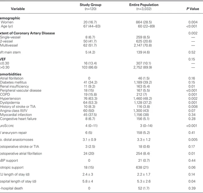
TABLE I. Preoperative and Postoperative Baseline Characteristics of the Study Cohort and the Entire Patient Population
Study Group Entire Population
Variable (n=120) (n=3,032) P Value
Demographic
Women 20 (16.7) 864 (28.5) 0.004
Age (yr) 67 (44–83) 60 (22–89) <0.001
Extent of Coronary Artery Disease 0.002
Single-vessel 8 (6.7) 259 (8.5) —
2-vessel 50 (41.7) 625 (20.6) —
Multivessel 62 (51.7) 2,147 (70.8) —
Left main stem 5 (4.2) 139 (4.6) 0.52
LVEF 0.15 ≤0.30 16 (13.4) 307 (10.1) — >0.30 103 (86.6) 2,752 (89.9) — Comorbidities Atrial fibrillation 0 46 (1.5) 0.16 Diabetes mellitus 41 (34.2) 1,189 (39.2) 0.15 Renal insufficiency 11 (9.2) 163 (5.4) 0.01
Peripheral vascular disease 18 (15) 167 (5.5) <0.001
COPD 19 (15.8) 212 (7) 0.001
Hypertension 76 (63.3) 1,460 (48.2) 0.002
Dyslipidemia 64 (53.3) 1,128 (37.2) 0.001
History of stroke or TIA 10 (8.3) 116 (3.8) 0.008
Angina class III/IV 60 (50) 1,300 (43) 0.07
Myocardial infarction 45 (37.5) 1,156 (38) 0.34
Congestive heart failure 8 (6.7) 156 (5.1) 0.28
EuroScore 4 (0–11) 3 (0–14) <0.001
LV aneurysm repair 6 (5) 158 (5.2) 0.41
No. distal anastomoses 3.1 ± 0.9 3.3 ± 1.2 0.005
Postoperative stroke or TIA 3 (2.5) 18 (0.6) 0.17
Postoperative atrial fibrillation 24 (20) 254 (8.4) 0.01
IABP support 0 21 (0.7) 0.44
Inotropic support 18 (15) 638 (21) 0.06
ICU length of stay (d) 2.4 ± 3 2.2 ± 1.7 0.14
Hospital length of stay (d) 5.8 ± 4 5.3 ± 2.6 0.04
In-hospital death 0 52 (1.7) 0.39
COPD = chronic obstructive pulmonary disease; EuroScore = European System for Cardiac Operative Risk Evaluation; IABP =
intra-aortic balloon pump; ICU = intensive care unit; LVEF = left ventricular ejection fraction; TIA = transient ischemic attack
Data are presented as mean ± SD, median and interquartile range, or number and percentage. P <0.05 was considered statistically significant.
Disease of the ascending aorta was determined to be untouchable if the vessel exhibited extensive intimal thickening, atheroma >3 mm in diameter, protruding atheroma, ulcerated atheroma, or one or more calcifica-tions of any size.
Carotid artery Doppler ultrasonography was per-formed in all patients older than 60 years of age; in patients with carotid bruit; in those with a history of stroke or TIA; in those with diabetes mellitus, pe-ripheral vascular disease, or left main coronary artery disease; and in those with aortic calcification on chest radiography.
Surgical Technique
Median sternotomy was performed in all 360 patients. The LIMA and SV were harvested by means of stan-dard techniques. The decision in regard to whether a patient would undergo the procedure on-pump or off-pump was made by the same surgeons (RT and OG), in consideration of the absence or presence of ascending aortic disease.
The patients with ascending aortic disease (unclamp-able ascending aorta) underwent surgery with the no-touch technique.1,2,4,5 Off-pump coronary surgery was
preferred whenever possible (70/120). On-pump pro-cedures were used in patients whose circumflex systems were considered unable to tolerate off-pump myocardial revascularization (50/120).
In each of the 3,152 subjects, the ascending aorta was routinely evaluated by intraoperative aortic palpation (the method described by Mills and Everson6). The site
of aortic cannulation in the study-group patients was determined after the patients’ CT scans were studied preoperatively. Patients with Mills and Everson mod-erate-grade atherosclerosis were cannulated through the soft site in the aorta identified upon palpation (n=35/50).6 Patients with Mills and Everson
severe-grade atherosclerosis underwent femoral artery cannula-tion (n=15/50).6
An Optima XP® membrane oxygenator (Cobe
Car-diovascular, Inc., a subsidiary of Sorin S.p.A.; Arvada, Colo) was used in conventional CABG. Tepid
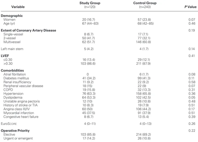
hypo-TABLE II. Baseline Characteristics of the Study and Control Groups after Propensity-Score-Matching
Study Group Control Group
Variable (n=120) (n=240) P Value
Demographic
Women 20 (16.7) 57 (23.8) 0.07
Age (yr) 67 (44–83) 68 (42–85) 0.46
Extent of Coronary Artery Disease 0.19
Single-vessel 8 (6.7) 17 (7.1)
2-vessel 50 (41.7) 77 (32.1)
Multivessel 62 (51.7) 146 (60.8)
Left main stem 5 (4.2) 4 (1.7) 0.14
LVEF 0.41 ≤0.30 16 (13.4) 29 (12.1) >0.30 103 (86.6) 211 (87.9) Comorbidities Atrial fibrillation 0 6 (1.7) 0.08 Diabetes mellitus 41 (34.2) 99 (41.3) 0.11 Renal insufficiency 11 (9.2) 22 (9.2) 0.58
Peripheral vascular disease 18 (15) 22 (9) 0.07
COPD 19 (15.8) 32 (13.3) 0.31
Hypertension 76 (63.3) 158 (65.8) 0.36
Dyslipidemia 64 (53.3) 102 (42.5) 0.05
Unstable angina pectoris 12 (10) 26 (10.8) 0.48
History of stroke or TIA 10 (8.3) 19 (7.9) 0.51
Angina class III/IV 60 (50) 106 (44.2) 0.17
Myocardial infarction 45 (37.5) 91 (37.9) 0.51
Congestive heart failure 8 (6.7) 13 (5.4) 0.39
EuroScore 4 (0–11) 4 (0–13) 0.26
Operative Priority 0.22
Elective 103 (85.8) 214 (89.2)
Urgent or emergent 17 (14.2) 26 (10.8)
COPD = chronic obstructive pulmonary disease; EuroScore = European System for Cardiac Operative Risk Evaluation; LVEF = left
ventricular ejection fraction; TIA = transient ischemic attack
Data are presented as mean ± SD, median and interquartile range, or number and percentage. P <0.05 was considered statistically significant.
thermia (33 °C) was maintained during cardiopulmo-nary bypass.
In the study group, an Octopus® 2 tissue stabilizer
(Medtronic, Inc.; Minneapolis, Minn) or an epicardial retracting stabilizer (Chase Medical; Richardson, Tex) was used to stabilize the target coronary vessel. The tar-get vessel was occluded proximally by using a double loop with a Surg-I-Loop® Plus Occlusive Loop (Scanlan
International, Inc.; St. Paul, Minn) or an 8-mm bulldog clamp. The distal portion of the vessel was occluded when excessive backflow began to obstruct the surgeon’s view. A carbon dioxide blower was used routinely. In the present study, SV composite grafting with the in situ LIMA was the preferred method of revascu-larization. For proximal anastomosis, the target vessel was anastomosed to the LIMA. The inflow was the LIMA in all patients. In patients who required distal anastomosis of the circumflex system, both venae cavae were released from the pericardium and the pleura was opened on the right side, enabling the heart to rotate to
the right. Deep pericardial traction sutures were used to elevate and rotate the heart. A suction-based Starfish®
Heart Positioner (Medtronic) maintained hemodynam-ic stability during lateral-wall anastomosis.
Evaluation of Long-Term Clinical Outcomes
Patients underwent regular postoperative follow-up monitoring through the outpatient clinic at 3-, 6-, and 12-month intervals, and were contacted by telephone in the event that the last clinic visit was not conducted at the scheduled time. Follow-up monitoring was com-pleted for all surviving patients on 1 September 2012. Cardiac death was defined as any death that arose from cardiac events during the follow-up period.
Statistical Analysis
Statistical analysis was performed with the use of SPSS software version 17.0 (IBM Corporation; Armonk, NY). All numerical data were expressed as mean ± SD, median and interquartile range, or proportions. The
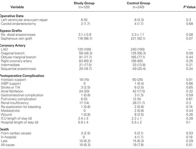
TABLE III. Surgical Outcomes after Propensity-Score Matching
Study Group Control Group
Variable (n=120) (n=240) P Value
Operative Data
Left ventricular aneurysm repair 6 (5) 8 (3.3) 0.3
Carotid endarterectomy 2 (1.7) 4 (1.7) 0.68
Bypass Grafts
No. distal anastomoses 3.1 ± 0.9 3.3 ± 1.1 0.08
Saphenous vein graft 116 (96.7) 221 (92.1) 0.07
Coronary Artery
LAD 120 (100) 240 (100) —
Diagonal branch 58 (48.3) 135 (56.3) 0.09
Obtuse marginal branch 91 (71.7) 185 (77.1) 0.44
Right coronary artery 83 (69.2) 156 (65) 0.25
Intermediate 21 (17.5) 33 (13.8) 0.21 Sequential anastomosis 20 (16.7) 49 (20.4) 0.24 Postoperative Complication Inotropic support 18 (15) 60 (25) 0.01 IABP support 0 1 (0.4) 0.66 Stroke or TIA 3 (2.5) 6 (2.5) 0.65 Atrial fibrillation 24 (20) 42 (17.5) 0.32 Gastrointestinal complication 1 (0.8) 3 (1.3) 0.59 Pulmonary complication 6 (5) 12 (5) 0.61 Renal insufficiency 17 (14) 28 (11.7) 0.3
Re-exploration for bleeding 1 (0.8) 2 (0.8) 0.74
Mediastinitis 0 2 (0.8) 0.44
Wound 1 (0.8) 6 (2.5) 0.26
ICU length of stay (d) 2.4 ± 3 2.2 ± 1 0.28
Hospital length of stay (d) 5.8 ± 4 5.5 ± 2 0.1
Death
From cardiac causes 3 (2.5) 5 (2.1) 0.53
In-hospital 0 4 (1.7) 0.19
Late 10 (8.3) 15 (6.3) 0.29
All-cause 10 (8.3) 19 (7.9) 0.51
IABP = intra-aortic balloon pump; ICU = intensive care unit; LAD = left anterior descending coronary artery; TIA = transient ischemic attack
Kolmogorov-Smirnov test was used to evaluate the nor-mality of data. Continuous variables were compared by the use of Student’s t test or the Mann-Whitney U test, depending on parametric and nonparametric distribu-tion, respectively. The categorical variables between the groups were analyzed by using the c2 test or the Fisher
exact test. In order to determine which patients were to be included in our control group, we performed a 1:2 matching of study group–control group patients in regard to age, sex, coronary artery disease, myocardial infarction, left ventricular dysfunction (expressed by ejection fraction), congestive heart failure (CHF), dia-betes mellitus, smoking, renal insufficiency, peripheral vascular disease, hypertension, chronic obstructive pul-monary disease (COPD), dyslipidemia, and emergency operation. Propensity-score-matching analysis was per-formed to correct the effects of nonrandomization in this retrospective study and of selection bias. Propensity scores were calculated with the aid of Stata® version 10
software (StataCorp LP; College Station, Texas). Sur-vival rates were estimated by the Kaplan-Meier method, and comparisons between the 2 groups were performed with use of the log-rank test. The level for statistical significance was predetermined at P <0.05.
Results
Table I shows the baseline clinical characteristics of the study cohort and the entire patient population. The pa-tients in the study group were older than the patient group as a whole (P <0.001), they had more peripheral vascular disease (P <0.001), and they had higher Euro-Score values (P <0.001). The study group had
statisti-cally significantly higher rates of COPD, dyslipidemia, and history of stroke or TIA.
Table II shows the baseline clinical characteristics of the study and control groups after propensity-score-matching. The mean age of the study group was 67 ± 7 years, and 20% were women. Most patients in both groups had multivessel disease (P=0.19). Mean left ven-tricular ejection fractions were similar (0.46 ± 0.09 vs 0.48 ± 0.11; P=0.08) between the study group and the entire patient population.
Patients with composite grafts had similar preopera-tive comorbidities when compared with control sub-jects. Of no statistical significance were differences between the 2 groups in EuroScore or in their histories
of diabetes, peripheral vascular disease, dyslipidemia, renal insufficiency, COPD, CHF, angina class III/IV, stroke or TIA, or unstable angina pectoris.
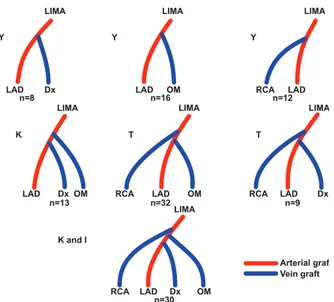
In 30% of patients (n=36), LIMA–SV graft Y anas-tomoses were performed. As necessary, K anasanas-tomoses were done in 13 patients (10.8%), T in 41 (34.1%), and K and I in 30 (25%) (Figs. 1 and 2). The percentages of sequential anastomoses were similar (16.7% vs 20.4%, respectively; P=0.24) in the study and control groups.
Table III shows the surgical outcomes of the study and control groups after propensity-score-matching. Postoperative inotropic support, atrial fibrillation, pul-monary complications, and renal insufficiency were similar in the 2 groups.
Postoperative stroke and TIA were similar in the 2 groups (P=0.65). In the study group, 3 patients (2.5%) experienced neurologic complications. In the control
Fig. 2 Coronary angiogram performed at 9-year follow-up in a 72-year-old male patient shows a left internal mammary artery– saphenous vein “T” composite graft: the left internal mammary artery was anastomosed to the left anterior descending coronary artery, and the saphenous vein grafts were anastomosed to the diagonal and obtuse marginal branches.
Fig. 1 Diagrams show the construction of composite grafts. Dx = diagonal branch; LAD = left anterior descending coronary artery; LIMA = left internal mammary artery; OM = obtuse marginal branch; RCA = right coronary artery
LIMA LAD Y K K and I T T Y Y Dx Dx LAD LAD LAD Dx Dx n=8 n=13 n=32 n=30 n=9 n=16 n=12 LAD OM OM OM OM LAD RCA RCA RCA LAD RCA LIMA LIMA Arterial graft Vein graft LIMA LIMA LIMA LIMA
group, 6 patients (2.5%) had neurologic complications. Only one patient had perioperative embolic stroke in the study group, whereas 4 patients experienced peri-operative embolic stroke in the control group.
The mean follow-up time was 5.27 ± 2.51 years for the study group and 5.28 ± 2.53 years for the control group (P=0.97). Overall follow-up was 5.27 ± 2.52 years. During this period, there were 29 deaths: 10 (8.3%) among study patients (cardiac-related deaths in 3 patients, stroke in 3, cancer in 2, and sudden death in 2) and 19 among control patients (cardiac-related deaths in 5 patients, stroke in 4, cancer in 3, gastroin-testinal bleeding in 1, and unknown in 6).
Hospital mortality rates (0 vs 1.7%, respectively;
P=0.19) and late mortality rates (8.3% vs 6.3%,
re-spectively; P=0.29) were similar. Cardiac-related deaths (2.5% vs 2.1%, respectively; P=0.53), and overall mor-tality rates (8.3% vs 7.9%, respectively; P=0.51) were similar between groups (Table III).
In 2 individuals in the study group, revasculariza-tion was incomplete because of technical difficulties in attaining access to the right coronary artery (posterior descending branch) system. In these cases, hybrid treat-ment (with percutaneous transluminal coronary angio-plasty) was used to deal with the right coronary lesion. The overall survival rates at a mean follow-up time of 5.27 ± 2.52 years were 91.7% for the study group and 92.1% for the control group; freedom-from-car-diac-death rates were 97.5% and 97.9%, respectively. No significant difference was found in freedom-from-cardiac-death rates between the 2 groups (P=0.71 vs
P=0.78, respectively) (Figs. 3 and 4).
Freedom-from-reintervention rates at a mean follow-up time of 5.27 ± 2.52 years were 96.7% and 98.8%, in the study and control groups, respectively (P=0.2) (Fig. 5).
Discussion
The incidence of porcelain or diffusely atherosclerotic aorta ranges between 4% to 29% of all cases referred for myocardial revascularization procedures.1,7 Because
of its potential to radically modify the surgical ap-proach, the preoperative recognition of diseased aorta is of major importance. Regrettably, no reliable diag-nostic criteria have been established to date, and the discovery of diseased aorta remains an intraoperative surprise in the great majority of patients in whom no preoperative evaluation of ascending aortic calcifica-tion is performed.1
Some previous studies have shown significantly lower patency rates of SV grafts, when compared with the pa-tency rates of radial artery grafts.8,9 On the other hand,
a prominent study5 reported the results of CABG in
28 patients in whom the LIMA–SV Y-composite graft had been used as the composite graft: the vein-graft patency rate was 96% at 2.5 postoperative years.
Simi-Fig. 3 Graph shows the Kaplan-Meier overall survival curve.
Years after Surgery
12 10 8 6 4 2 0 Survival Rate (% ) 100 80 60 40 20 0 Patients at risk Composite graft Control group 91.7% 92.1% P=0.71 Composite group Control group 110231 89167 5581 2227 101
Fig. 4 Graph shows the comparison of freedom from cardiac death in the 2 groups.
Years after Surgery
12 10 8 6 4 2 0
Freedom from Cardiac Death
(% ) 100 80 60 40 20 0 Control group Composite graft P=0.78 97.9 % 97.5 % Patients at risk Composite group Control group 110222 89175 55125 2245 17
Fig. 5 Graph shows the reintervention-free-survival curve in the 2 groups.
Years after Surgery
12 10 8 6 4 2 0 Reintervention-Free Survival (% ) Control group Composite graft 98.8% 96.7% P=0.20 Patients at risk Composite group Control group 110222 89175 55125 2245 17 100 80 60 40 20 0
larly, a study by Hwang and colleagues4 revealed that
the patency rate of SV grafts at 1 year was 92.1%, which was similar to that of arterial composite grafts (91%). Patency rates of venous grafts in accordance with their target coronary artery territories were also similar to those of arterial composite grafts.
Our present study revealed 2 main findings. First, the early and long-term mortality rates of patients who experienced the no-touch technique with a composite graft anastomosed to the in situ LIMA were similar to those of patients who underwent conventional CABG. Second, there were no significant differences in early and long-term clinical outcomes, including freedom from cardiac death, reintervention-free survival, and overall survival rates between the groups when both techniques were used.
In the present study, freedom from cardiac death (97.5%), freedom from reintervention (96.7%), and overall survival rates (91.7%) between composite graft with no-touch aorta technique and matched groups of conventional CABG were similar. Our clinical results are parallel with and corroborated by studies in the lit-erature.4
The present study showed that the hospital mortality and postoperative morbidity rates associated with SV composite grafts, as well as the late clinical outcomes, are comparable to those of conventional CABG per-formed in patients with nonatherosclerotic aorta. In the past, the no-touch technique has been shown to reduce the risk of cerebrovascular accident and has been associ-ated with low rates of morbidity, stroke, and mortality during hospital stay and at mid-term follow-up.10 Our
study has also found low neurologic complication rates. A study comparing radial artery and SV composite grafts revealed early clinical outcomes similar to ours in hospital deaths and postoperative morbidity, as well as in 1-year follow-up CT angiographic patency rates between the 2 groups.4 Presumably, an SV graft
anas-tomosed to the LIMA might be exposed to less pressure trauma or shear stress than a graft anastomosed to the ascending aorta. Mean and diastolic graft pressures of an SV graft anastomosed to the LIMA have been shown to be lower than those of an SV graft anastomosed to the ascending aorta.3 Hypothetically, the quality of the
SV graft is identical to that of the radial artery, because the radial artery is equally prone to the development of intimal hyperplasia or fibrous or calcified plaques. Minimal manipulation and tension (for example, one should avoid the use of a pressure syringe for dilation), together with the use of relatively short lengths of com-posite vein graft, might improve the early and 1-year patency rates of SV grafts.4
Limitations of the Study
Our study has all the inherent weaknesses of retrospec-tive studies. Also, the number of patients was relaretrospec-tively
small and follow-up patency tests (CT and angiography) were not performed in all patients, so the sample size might not be sufficient to enable a definite conclusion.
Conclusion
We compared the results of conventional CABG as per-formed on patients who showed no preoperative evi-dence of serious atherosclerosis of the ascending aorta with the results of the aortic no-touch technique (using SV composite grafts) on CABG patients who did show such evidence. In comparing the conventional CABG and SV-composite-graft groups, our study showed simi-lar early and late survival rates, simisimi-lar reintervention-free survival rates, and similar reintervention-freedom from cardiac death. As a result, we conclude that the aortic no-touch technique with composite grafts might be a reasonable option in patients who have an atherosclerotic ascend-ing aorta that cannot be clamped.
References
1. Gaudino M, Glieca F, Alessandrini F, Luciani N, Cellini C, Pragliola C, Possati G. The unclampable ascending aorta in coronary artery bypass patients: a surgical challenge of in-creasing frequency. Circulation 2000;102(13):1497-502. 2. Muneretto C, Negri A, Manfredi J, Terrini A, Rodella G,
Elqarra S, Bisleri G. Safety and usefulness of composite grafts for total arterial myocardial revascularization: a prospective randomized evaluation. J Thorac Cardiovasc Surg 2003;125 (4):826-35.
3. Tedoriya T, Kawasuji M, Sakakibara N, Ueyama K, Wata-nabe Y. Pressure characteristics in arterial grafts for coronary bypass surgery. Cardiovasc Surg 1995;3(4):381-5.
4. Hwang HY, Kim JS, Kim KB. Angiographic equivalency of off-pump saphenous vein and arterial composite grafts at one year. Ann Thorac Surg 2010;90(2):516-21.
5. Gaudino M, Alessandrini F, Pragliola C, Luciani N, Trani C, Burzotta F, et al. Composite Y internal thoracic artery-saphe-nous vein grafts: short-term angiographic results and vasoreac-tive profile. J Thorac Cardiovasc Surg 2004;127(4):1139-44. 6. Mills NL, Everson CT. Atherosclerosis of the ascending aorta
and coronary artery bypass. Pathology, clinical correlates, and operative management. J Thorac Cardiovasc Surg 1991;102 (4):546-53.
7. Leyh RG, Bartels C, Notzold A, Sievers HH. Management of porcelain aorta during coronary artery bypass grafting. Ann Thorac Surg 1999;67(4):986-8.
8. Kim KB, Lee C, Chae IH, Oh BH, Lee MM, Park YB. Off-pump coronary artery bypass may decrease the patency of saphenous vein grafts. Ann Thorac Surg 2001;72(3):1033-7. 9. Athanasiou T, Saso S, Rao C, Vecht J, Grapsa J, Dunning J,
et al. Radial artery versus saphenous vein conduits for coro-nary artery bypass surgery: forty years of competition--which conduit offers better patency? A systematic review and meta-analysis. Eur J Cardiothorac Surg 2011;40(1):208-20. 10. Gulcan O, Turkoz R, Demirturk OS, Oguzkurt L, Turkoz A.
Extending the boundaries of no-touch aorta technique usage for coronary artery bypass grafting in patients with diseased ascending aorta. J Cardiovasc Surg (Torino) 2008;49(3):351-7.