The article was published by ACG Publications
http://www.acgpubs.org/journal/organic-communications © October-December 2020 EISSN:1307-6175 DOI: http://doi.org/10.25135/acg.oc.86.20.10.1853
Org. Commun. 13:4 (2020) 175-183
Facile microwave synthesis of a novel phenothiazine derivative
and its cytotoxic activity
Cenk A. Andac
1,2*1Department of Medicinal Chemistry, School of Pharmacy, Istinye University, 34010, Istanbul, Turkiye
2Department of Medical Pharmacology, School of Medicine, Ankara Yıldırım Beyazıt University, 06800, Ankara, Turkiye
(Received October 20, 2020; Revised November 27, 2020; Accepted November 30, 2020) Abstract: Herein, a facile procedure for microwave synthesis and NMR characterization of phenothiazine derivatives
10-(3-hydroxypropyl)-2-(methylsulphanyl)-10H-phenothiazine (3) (25% yield) and 1-[3-(2-methylsulfanyl-10H-phenothiazine-10-yl)-propyl]pyrimidine-2,4-(1H,3H)-dione (6) (67% yield) is described. After successful microwave synthesis steps, MTT cytotoxicity experiments gave rise to greater cytotoxic effect of compound 6 in MCF-7 human breast adenocarcinoma cell line (IC50= 1.35 µM) as compared to literature values for tamoxifen (IC50= 8.3 µM) and doxorubicin (IC50= 27 µM).
Keywords: Microwave synthesis; phenothiazine derivatives;
1-(3-(2-methylsulfanyl-10H-phenothiazine-10-yl)-prop-1-yl)pyrimidine-2,4(1H,3H)-dione; NMR; IC50. ©2020 ACG Publication. All right reserved.
1. Introduction
Phenothiazine derivatives with a propyl bridge are known to be antagonists of D2-type (D2 and
D3) dopaminergic receptors, generating both antipsychotic1 and anticancer effects.2 The anticancer effect
of phenothiazine derivatives such as thioridazine2 is through the inhibition of D2-type dopaminergic
receptors in certain cancer stem cells.2
Phenothiazine type antipsychotic drugs contain a phenothiazine ring (2 in Scheme 1), a basic heterocyclic ring with an amine group, and a propyl group that bridges the basic heterocyclic group to the phenothiazine ring at position 10, such as 6 and 7 in Scheme 1. In drug discovery, an electronegative substituent like -S-CH3, -CF3, -Cl, or -CN at position 2 of the phenothiazine ring (2 in Scheme 1)
increases the neuroleptic effect of the corresponding psychotic drug.1 Furthermore, any reduction or
increase in the carbon chain of the propyl bridge will significantly decrease the neuroleptic effect of phenothiazine type antipsychotic drugs.1 For instance; promethazine with a two-carbon bridge possesses
antihistaminic effect3 rather than neuroleptic effect.
In general, the synthesis of phenothiazine derivatives with a propyl bridge involves N-alkylation reactions in toluene using a very strong base such as NaNH2 (sodium amide).4,5 Although reaction
product yields for the N-alkylation of the phenothiazine ring using NaNH2 is reported to vary between
80-90%,5 some alkene by products are likely to form by the reaction of NaNH
2 (as a very strong base)
with alkyl halides and also the import of NaNH2 is banned at the customs of some countries (including
Turkey) as NaNH2 is used in making explosives. Alternatively, N-propionyl conjugation of
3-chloropropionyl chloride on phenothiazine followed by reduction of the N-propionyl group with borane (BH3) was reported in literature.6 However, the latter reactions increase the number of reaction steps as
well as the cost to produce phenothiazine derivatives.7
N-alkylation reactions of phenothiazine derivatives are the most critical steps in terms of cost, time and yield. Aside from amine alkylation reactions under basic conditions, modern green chemistry methods utilizing microwave (MW) irradiation for rapid synthesis of alkylated amines in solvent7,8 and
solvent-free9 systems have attracted much attention. For instance, step-wise alkylation of secondary
amines in basic water solution by MW irradiation was reported to give rise to tertiary amines only.7
However, MW reactions in solvent systems commonly face some problems due to solubility (of the solute) and volatility (of the solvent) issues as well as temperature related limitations arising from the boiling point of the solvent used. Interestingly, Sarmiento et al.8 synthesized (in ~60% yield) a
N-propionyl derivative of phenothiazine in DMF catalyzed by MW irradiation, which could then be reduced by diborane (B2H6) to yield a N-propyl linked phenothiazine derivative. However, Sarmiento et
al. work requires additional steps and work-up to obtain the final phenothiazine product with a propyl linker, which may be costly and time consuming. Using solvent-free conditions, a microwave irradiation procedure for the N-alkylation of an amine compound (isatin) using alkyl halides and Na2CO3 (as base)
was previously published by Shmidt et al.9
Herein, this paper presents a facile microwave irradiation method under solvent-free conditions to synthesize a novel phenothiazine derivative with a propyl linker, 1-(3-(2-methylsulfanyl-10H-phenothiazine-10-yl)-prop-1-yl)pyrimidine-2,4-(1H,3H)-dione, which was initially designed in silico as an antagonist of human D2 dopaminergic receptor.
2. Experimental
2.1. Chemical Material and Instrumentation
Glassware were dried at 120 °C over 24 hours before using for chemical reactions. For reactions, 5 mL, 25 mL, 50 mL round-bottom flasks were used as needed. Reaction systems were sealed with rubber-septums. All reactions were performed under N2 atmosphere. NaH used in the reaction was
washed with CH2Cl2 four times before use. Fluorescent silicagel on Al Foils (F: 254 nm)
(Sigma-Aldrich, Milwaukee, WI, USA) was used for thin layer chromatography. Silicagel with a pore size of 60 Å was used in column chromatography as the solid phase. The required solvents were all chemical grade and used without purification as purchased from companies. 2-(methylsulphanyl)-10H-phenothiazine was purchased from Sigma-Aldrich (Milwaukee, WI, USA).
1D and 2D NMR spectra were recorded on a 400 MHz Varian NMR instrument at the Advanced Technology and Research Institute at Konya Selcuk University, Turkey. Deuterated chloroform (CDCl3,
δ=7.23 ppm) was used as the solvent in NMR experiments. NMR Fourier Induction Decay (FID) data were processed and converted into spectra using VNMRJ4.2 Software. 1H,1H-ROESY NMR FID data
were collected between -1 ppm and 14 ppm with 1024 data points in F1 dimension and 1024 data points in F2 dimension. 1H,1H-ROESY NMR FID data were zero-filled and linear-predicted to 2K data points
in both dimensions. The FID data were then apodized in both dimensions using 90o Sine and 0.07 Hz
Gaussian functions in both dimensions. Apodized ROESY FID data were Fourier transformed in inverse and quadrature modes along t2 dimension and in inverse and quadrature modes using the hypercomplex protocol in t1 dimension. ROESY spectra were displayed and analyzed in phase sensitive mode.
Perkin Elmer Spectrum 100 Infrared Spectrometer (Waltham, MA, USA) was used to collect FT-IR spectra. After the samples were pelleted with KBr, FT-IR spectra were recorded in the spectral range 650-3800 cm-1. Mass spectroscopy experiments were implemented in ESI+ mode by ESI+-LCMS
(m/z %) on Waters Micromass ZQ Mass Spectrometer (Milford, MA, USA). Microwave synthesis of compounds 3 and 6 were carried by MARS 5 Microwave Accelerated Reaction System (CEM Corporation, North Carolina, USA).
2.2. Biological Materials and Apparatus
Human breast ductal adenocarcinoma cell line [MCF-7 (ER+/PR+)] was purchased from ATCC
(American Type Culture Collection, Gaithersburg, Maryland, USA). These cells were cultured in flasks (at 37 °C and under 5% CO2 atmosphere) using RPMI medium (Biochrome, Germany) containing 10%
FBS (Fetal Bovine Serum) (Biochrome, Germany) and 1% penicillin/streptomycin (100 U/mL penicillin and 100 μg/mL streptomycin) (Biochrome, Germany). After cells filled out 80% of the culture flasks, they were then washed out with 1xPBS (Gibco, USA) and detached with treatment of 0.25% of
Trypsin-EDTA (Biochrome, Germany) enzymatic solution. Required amount of detached cells were used for IC50 studies, while the rest was treated with 10% DMSO in RPMI medium and stored at -80 °C for later
use.
2.3. Chemistry
2.3.1. Microwave Synthesis of Compound 3
Into an explosion proof 15 mL teflon chamber, 1750 mg 2-(methylsulphanyl)-10H-phenothiazine (7.13 mmol), 907 mg Na2CO3 (8.55 mmol), 715 μL 3-chloropropanol (d: 1.131 g/ mL;
8.55 mmol) and 15 μL of dry N,N-dimethylformamide (DMF) was added and mixed gently by a thin spatula. The reaction vessel was then sealed tightly and mounted into MARS 5 Microwave Accelerated Reaction System (CEM Corporation, North Carolina, USA). 800 watts of microwave energy was applied for 15 minutes followed by monitoring by TLC (Rf = 0.4, Hexane/Ethylacetate, 7:3). Fifteen
minutes of microwave irradiation of the reaction was repeated 3 more times (a total of 1 hour) to confirm the completion of the reaction by TLC. The desired compound was purified by silicagel column chromatography (Hexane/Ethylacetate, 7:3). Yield: 542 mg (25%, 1.78 mmol). ESI+-LCMS (m/z %)
MW: 303.29; computed MW: 304 Da. 1H NMR (400 MHz, CDCl
3) δ (ppm) = 6.80-7.20 (7H, m), 4.78
(1H, s), 4.02 (1H, t, J = 6.40 Hz and 6.00 Hz), 3.78 (1H, t, J = 5.60 Hz), 2.04 (1H, m), 1.60 (3H, s). FT-IR (KBr, cm-1): 3328, 2944, 2878, 1566, 1458, 1300, 1248, 1034, 932, 748.
2.3.2. One-pot Microwave Synthesis of Compound 6
100 mg of 3 (0.32 mmol, 1 eq) previously dissolved in dichloromethane (CH2Cl2, 4 mL) was
transferred via a dry syringe into a dry 25 mL round-bottom flask sealed with a rubber septum under N2
atmosphere. Then, 67 μL of triethylamine (48.6 mg, 48 mmol, 1.5 eq) and 35 μL of PBr3 (0.35 mmol,
1.2 eq) were micropipetted into the reaction vessel, respectively. The reaction was stirred under N2
atmosphere for 2 hours. The completion of the synthesis of the intermediary product, 10-(3-bromoprop-1-yl)-2-(methylsulfanyl)-10H-phenothiazine (4 in Scheme 1), was monitored by TLC (Rf = 0.38-0.85,
Hexane/Ethylacetate, 9:1). Then, solvent of the mixture was evaporated under vacuum and the residue of 4 was transferred into an explosion proof 15 mL teflon chamber for microwave synthesis. Into the teflon chamber, Na2CO3 (407 mg, 3.84 mmol, 1.2 eq), pyrimidine-2,4-(1H,3H)-dione (uracil, 51 mg,
0.45 mmol, 1.4 eq) and 15 μL of dry N,N-dimethylformamide (DMF) were added respectively, and then mixed gently by a thin spatula. The reaction vessel was then sealed tightly and mounted into MARS 5 Microwave Accelerated Reaction System (CEM Corporation, North Carolina, USA). 800 watts of microwave irradiation energy was applied for one hour to complete the reaction, which was confirmed by TLC (Rf = 0.57, Hexane/Ethylacetate, 1:1). Compound 6 was isolated by silicagel column
chromatography (Hexane/Ethylacetate, 1:1). Off-white solid (83 mg, 0.21 mmol, 67% isolated yield). ES+-LCMS (m/z %): 397.87 Da (calculated M.W. 397.51 Da). 1H NMR (400 MHz, CDCl 3, ppm): δ 8.76 (1H, s), 7.22-6.76 (7H, m), 7.05 (1H, d, 3J HH 7.20 Hz), 5.29 (1H, d, 3JHH 5.60 Hz), 3.91 (1H, t, 3JHH 5.60 Hz), 3.79 (1H, t, 3J HH 6.40 Hz), 2.23 (1H, p), 1.25 (1H, s). 2.4. Biological Assay 2.4.1. MTT Citotoxicity Assay
MCF-7 cancer cell inhibition assay (IC50) was determined using the MTT
[3-(4,5-dimethylthiazol-2-yl)-2,5-diphenyltetrazoliumbromide] kit by Promega (Madison, WI, USA).10-12 A
certain number of cells were transferred into the inner wells of 96-well plates and incubated for 24 hours. Plate wells were added a fixed concentration of MTT solution and incubated for 2.5 hours at 37 °C under 5% CO2 atmosphere. 200 μL of solution from each well was transferred to the inner wells of 96-well
plate for reading absorbances at 490 nm by Elisa Plate Reader. Increasing concentrations of compound 6 were added into the wells (three determinations for each concentration scale) and incubated for 72 hours at 37 °C under 5% CO2 atmosphere. Cells in the wells were then treated with MTT solution for
reading absorbance values at 490 nm by Elisa Plate Reader. Percent inhibition was then computed as follows:
Inhibition % = [1 – (avg-OD490
compound+MCF-7 / avg-OD490MCF-7) ] × 100 EQ.1
where avg-OD490
compound+MCF-7 is the average of absorbance values at 490 nm for MCF-7 cells treated
with compound 6 and avg-OD490
MCF-7 is the average of absorbance values at 490 nm for intact MCF-7
cells.
2.5. Quantum Mechanics Computational Studies
3D structures of compounds 6 and 7 were determined by GAMESS US quantum mechanics software v.2010.R2.13 The computations were implemented in the gas phase using the density functional
theory (DFT) at the B3LYP/6-31+G(d,p) basis set. Starting structure coordinates and geometry optimization input parameters (gradient convergence tolerance=10−4, density convergence = 10−5, linear dependence treshhold = 10−6, self-consistent field = RHF) for GAMESS US were prepared and visualized by Avogadro Software v.1.2.0.14
3. Results and Discussion
3.1. Chemistry
A synthetic route to synthesize compound 3 and 6 by microwave irradiation method is given in Scheme 1. Starting from 2-methylthio-10H-phenothiazine, 3-(2-methylthio-10H-phenothiazine-10-yl-propyl)-pyrimidine-2,4-(1H,3H)-dione (6 in Scheme 1) was synthesized in two steps by microwave irradiation and characterized by NMR, FT-IR and mass spectroscopy techniques.
For solvent-free synthesis of N-alkylation reactions by MW irradiation, Shmidt et al.9 reported
that optimal conditions for the N-alkylation reaction was obtained in presence of Na2CO3 and a few
drops of DMF. In similar to Shmidt et al.9 work, Na
2CO3 was initially added to carry out microwave
synthesis of reactions i and iii (Scheme 1) and was found out that the addition of Na2CO3 was not
sufficient to reach high yields in N-alkylation. After several more trials, it was found that addition of a tiny amount of DMF (N,N-dimethylformamide) along with Na2CO3 into reactions i and iii significantly
improved the yields for the synthesis of compounds 3 and 6 (Scheme 1), respectively, which was later noticed to be consistent with Shmidt et al. work.9
Microwave irradiation of reaction i (Scheme 1), which uses 2-methylthio-10H-phenothiazine and 3-chloropropanol, gave rise to compound 3 [ESI+-LCMS (m/z %): 303.29; calculated MW: 304 Da]
(Figure S1) with relatively an affordable yield, 25%. Although several other MW reaction conditions (i.e.; doubling Na2CO3 concentration, increasing DMF concentration upto 200 µL) were tried, the
reaction yield of compound 3 could not be optimized for an unknown reason. 1H-NMR (Figures S2 and
S3) and FT-IR (Figure S4) spectra confirmed compound 3 in high purity. As seen in Figure S2, multiplet signals around 6.8-7.2 ppm are assigned to the phenothiazine protons, a broad peak at 4.78 ppm belongs to the –OH proton. The triplet signals at H 4.02 and 3.78 and the multiplet signal at H 2.04 belong to
the methylene (-CH2-) protons (Figure S3). Also the singlet signal at H 1.60 matches –S-CH3 methyl
protons (Figure S3).
FT-IR spectrum in Figure S4 reveals a broad peak at 3328 cm-1 for -OH stretching.
Characteristic vibrations pertaining to the aliphatic C-H stretching of the –CH2- groups were observed
at 2944-2878 cm-1 in FT-IR spectrum of compound 3 (Figure S4). Characteristic aromatic -C=C
stretching were observed at 1566 and 1458 cm-1. Aromatic and aliphatic -C-N- stretching were observed
at 1300 and 1248 cm-1, respectively. Moreover aromatic -C, aliphatic -C-C(OH), and aliphatic
-S-C(H3) stretchings were observed at 1034 cm-1, 932 cm-1 and 748 cm-1, respectively.
In general, preparation of phenothiazine derivatives with a propyl bridge involves N-alkylation reactions in toluene using a very strong base such as NaNH2 (sodium amide).3 Instead of NaNH2,
synthesis of compound 3 was tried using several other bases including NaH, 4-dimetilaminopyridine (DMAP), Na2CO3, and triethylamine to carry out reaction i in Scheme 1 at varying high temperatures.
failed. Therefore, these laboratory results indicate that N-alkylation reactions by microwave irradiation is a good choice of method as a replacement of using NaNH2 in the preparation of phenothiazine
derivatives.
Scheme 1. Microwave synthesis of a derivative of 2-methylsulfanyl-10H-phenothiazine. Reaction conditions, i: Na2CO3/DMF/microwave (800 watts)/1 h; ii: PBr3/TEA/CH2Cl2/25 oC/1 h; iii:
Na2CO3/DMF/microwave (800 watt)/1 h. (DMF: Dimethylformamide; TEA: triethylamine).
In an attempt to synthesize compound 3 using NaH, five different reactions were carried at various temperatures including 25 oC, 50 oC, 70 oC, 100 oC, and 120 oC. As a general reaction procedure,
to a solution of 515 mg of 2-(methylsulfanyl)-10H-phenothiazine (2 mmol) in 13 mL of xylene and 7 mL of CH2Cl2 in a 50 mL round-bottom flask was added 120 mg of 60% NaH (3 mmol) and 208 mg of
3-chloropropanol (2.2 mmol) and stirred under N2 atmosphere for 4 days at the aforementioned
temperatures. The course of the reactions were monitored many times by TLC (Hexane:Ethylacetate 75:25). The reactions were terminated after four days by addition of 20 mL of ethylacetate into the reactions vessels, which were then transferred into separate extraction funnels. HCl formed in the reactions was neutralized by addition of 10 mL of 10% NaHCO3 solutions into the extraction funnels.
After extractions, the upper organic phase was separated and dried over anhydrous Na2SO4. In all cases,
the reactions did not give rise to high yields for a possible new product as determined by TLC (Hexane:Ethylacetate 75:25). The best TLC result was obtained for the reaction setup at 110 oC, which
gave rise to two new weakly spots at Rf=0.26 (spot-1) and Rf=0.15 (spot-2) for the organic phase.
Silicagel column chromatography purification (Hexane:Ethylacetate 75:25) of the organic extraction phase led to very low yields for the new products, ~25 mg (spot-1) and ~15 mg (spot-2), respectively. Unfortunately, 1H-NMR spectra for the unknown products did not match the 1H-NMR spectrum of 3
obtained by microwave synthesis (Figure S5), proving that using NaH as a base did not lead to the synthesis of compound 3.
Using the same procedure above, the base NaH was replaced with Na2CO3 (2.2 mmol), DMAP
(4-dimethylaminopyridine, 2.2 mmol, pKa = 9.6), and TEA (triethylamine, 2.2 mmol, pKa = 10.75), respectively. Unfortunately, the last three bases did not give rise to new products at all as determined by TLC. Therefore, the microwave synthesis of compound 3 is currently the best method in the laboratory scale as compared to the synthetic procedure described above using NaH, Na2CO3, DMAP, and TEA as
bases.
Compound 6 was obtained after a two-step reaction carried out in one pot. In the first reaction step 10-(3-bromoprop-1-yl)-2-(methylsulfanyl)-10H-phenothiazine (compound 4) was synthesized by reacting compound 3 with PBr3. In the second reaction step, compound 6 was synthesized by reacting
compound 4 with uracil in presence of Na2CO3 and tiny amount of DMF under microwave irradiation.
After work-up and purification by silicagel chromatography (Hexane/Ethylacetate, 1:1), compound 6 was obtained in high yield (67%). As seen in Scheme 1, compounds 6 [1’-(2-methylthio-10H-phenothiazine-10-yl-propyl)-pyrimidine-2’,4’-(1’H,3’H)-dione] and 7 [3’-(2-methylthio-10H-phenothiazine-10-yl-propyl)-pyrimidine-2’,4’-(1’H,3’H)-dione] are isomers with the same molecular weight (calculated M.W. 397.87 Da). Therefore, compound 6 was first confirmed by ES+-LCMS (m/z
%: 397.87 Da; calculated MW. 397.51 Da) (Figure S6) and then 1H-NMR (Figure S7) and ROESY
NMR data (Figure S8).
Assignment of the methylene proton peaks belonging to the propyl bridge requires more attention. The N-CH2– proton peaks appear as triplets at 3.91 ppm (3JHH 5.60 Hz) and 3.79 ppm (3JHH
6.40 Hz) due to their neighboring –CH2– methylene protons which appears as a pentate peak at 2.23
ppm (Figure S8). NOE cross-peak intensities were correlated to proton-proton distances in three ranges: 1.8-2.8 Å for strong NOEs, 2.8-3.3 Å for medium NOEs and 3.3-5.0 Å for weak NOEs.15 Density
functional theory QM computations revealed that a reference distance between the C5’H-C6’H protons of the pyrimidine-2,4-dione (uracil) ring is about 2.45 Å for compound 6 (Figure 1), which leads to a strong ROESY NOE cross-peak at 5.29 ppm (set to -1000 integral units for negative NOE), see Figure S8. A strong negative NOE cross-peak (with -993 integral units) that appears at 3.79 ppm spatially connects the N-CH2- methylene protons of the propyl bridge to the C6’H proton of the uracil ring (Figure
S8). According to the Integrated Spin-Pair Approximation (ISPA) method16 formulated as r
ij = rref (aref /
aij)1/6 (where rref=2.45 Å, aref= -1000 iu, and aij= -993 iu) to measure through-space proton-proton
distances, the rij distance (for the C5’H-C6’H proton-proton system) is found to be roughly 2.50 Å,
which is highly comparable to the C5’H-C6’H proton-proton distance (2.55 Å) of compound 6 as determined by quantum mechanics computations (Figure 1). Such a strong NOE connectivity is not expected for compound 7 as C5’H-C6’H proton-proton distance was computed to be more than 5 Å, which is way far from the limits of NOE detection by NMR (< 5 Å). Thus, the ROESY spectrum shown in Figure S8 strongly proves that the desired product is compound 6.
One-pot synthesis of compound 6 was also tried by using NaNH2 (sodium amide) as the base in
the last step of the reaction pathway (Scheme 1). After the bromination reaction of 3 was complete to synthesize 4 as described in section 2.3.2, the solvent (CH2Cl2) of the reaction mixture was evaporated
under N2 atmosphere. To the mixture of 4 was then added 46 mg of 5 (uracil, 0.41 mmol, 1.4 eq) and 18
mg of NaNH2 (0.46 mmol, 1.6 eq) followed by magnetically stirring under N2 atmosphere at room
temperature for 16 hours. Completion of the reaction was confirmed by TLC (Hexane:Ethylacetate, 1:1). The reaction product 6 was purified by silicagel column chromatography (Hexane/Ethylacetate, 1:1). Off-white solid (76 mg, 0.19 mmol, 59% isolated yield). Comparison of 1H-NMR spectrum with that of 6 obtained by microwave synthesis reaction confirms product 6 (Figure S9).
Figure 1. Quantum mechanics structures of compounds 6 and 7.
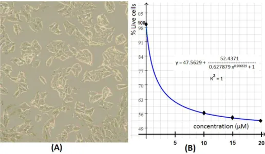
Figure 2
. (A) Cultured MCF-7 human breast adenocarcinoma cells,3.2. MTT Cytotoxicity Assays
Cytotoxic activity of 3-(2-methylthio-10H-phenothiazine-10-yl-propyl)-pyrimidine-2,4-(1H,3H)-dione (6) was determined by the MTT cytotoxicity assay against the MCF-7 cancer cell line (Figure 2A). Percent inhibition determined by EQ.1 in section 2.4.1 was converted to percent live cells by % live-cells = 100 - % inhibition, which were plotted against concentration as seen in Figure 2B. The graph was then line-fitted to determine an IC50 value of 1.35 µM at 76% live cells, which is the average
of upper and lower bounds for % live cells.
Indeed, the IC50 value of compound 6 is considerably better as compared to prescribed
chemotherapy agents applied for the treatment of breast cancer, i.e.; doxorubicin (IC50 = 27 µM in
MCF-7 cells )17 and tamoxifen (IC
50 = 8.3 µM in MCF-7 cells).18 4.
Conclusion
Herein, compound 6 was successfully synthesized in two steps by microwave irradiation method, purified and characterized by NMR, FT-IR and MS techniques. Our laboratory results indicate that the solvent-free microwave synthesis of compound 6 is about 5-10 % more efficient than that of the reaction catalyzed by NaNH2. In addition, the MW reactions applied in this work are less costly as the
reaction is carried out under solvent-free conditions. Compound 6 was found to possess greater cytotoxic activity (IC50= 1.35 µM) as compared to some other prescribed chemotherapy agents (doxorubicin and
tamoxifen) used in the treatment of breast cancer.
To the best of our knowledge, solvent-free microwave synthesis of phenothiazine type neuroleptics has not been reported before. Therefore, the solvent-free microwave synthesis of compounds 3 and 6 is likely to pave the ways to establish a general procedure for the synthesis of other phenothiazine derivatives possessing a propyl bridge.
Acknowledgements
For its novelty and great cytotoxic activity, a Turkish patent was received for 3-(2-methylthio-10H-phenothiazine-10-yl-propyl)-pyrimidine-2,4-(1H,3H)-dione (Turkish Patent #: 2013/139199).
Supporting Information
Supporting information accompanies this paper on http://www.acgpubs.org/journal/organic-communications
ORCID
Cenk A Andac: 0000-0002-9359-4295
References
[1] Jaszczyszyn, A.; Gsiorowski, K.; Oewitek, P.; Malinka, W.; Cieoelik-Boczula, K.; Petrus, J.; Czarnik-Matusewicz, B. Chemical structure of phenothiazines and their biological activity. Pharmacol. Rep. 2012,
64, 16-23.
[2] Sachlos, E.; Risueno, R. M.; Laronde, S.; Shapovalova, Z.; Lee, J-H.; Russel, J. L.; Malig, M.; McNicol, J. D.; Fiebig-Comyn, A.; Graham, M.; Levadoux-Martin, M.; Lee, J. B.; Giacomelli, A. O.; Hassell, J. A.; Fischer-Russell, D.; Trus, M. R.; Foley, R.; Leber, B.; Xenocostas, A.; Brown, E. D.; Collins, T. J.; Bhatia, M. Identification of drugs including a dopamine receptor antagonist that selectively target cancer stem cells.
Cell 2012, 149, 1-14.
[3] Adam, K.; Oswald, I. The hypnotic effects of an antihistamine: promethazine. Br. J. Clin. Pharmacol. 1986,
22(6), 715–717.
[4] Horclois, R. J. Phenothiazine derivatives and processes for their preparation. U.S. Patent 1959, 2,902,484. https://patents.google.com/patent/US2902484A/en
[5] Bourquin, J.P.; Schwarb, G.; Gamboni, G.; Fischer, R.; Ruesch, L.; Guldimann, S.; Theus, V.; Schenker, E.; Renz, E. Syntheses in the phenothiazine family. II. N-Substituted phenothiazinethiol derivatives. Helv.
Chim. Acta 1958, 41, 1072-1108.
[6] Sarmiento, G. P.; Vitale, R. G.; Afeltra, J.; Moltrasio, G. Y.; Moglioni, A. G. Synthesis and antifungal activity of some substituted phenothiazines and related compounds. Eur. J. Med. Chem. 2011, 46, 101-105. [7] Ju, Y.; Varma, R.S. Aqueous N-alkylation of amines using alkyl halides: direct generation of tertiary amines
under microwave irradiation. Green Chem. 2004, 6, 219–221.
[8] Sarmiento, G.P.; Moltrasio, G.Y.; Moglionib, A.G. An alternative synthetic route to the neuroleptic compound Pipothiazine. Arkivoc 2009, 7, 33-41.
[9] Shmidt, M. S.; Reverdito, A. M.; Kremenchuzky, L.; Perillo, I. A.; Blanco, M. M. Simple and efficient microwave assisted N-alkylation of isatin. Molecules 2008, 13, 831-840.
[10] Doğan, İ.S.; Sellitepe, H.E.;, Kayıkçı, N.; Sipahi, H.; Reis, R.; Yaylı, N. Synthesis and anticancer (MCF-7, PC-3) activities of new 2-hydroxy-2,2-bis(4-N'-[(1E)-(3/4- substitutedphenyl)-methylene]-acetohydrazides.Org.Commun. 2018, 11,142-148.
[11] Bader, A.; Abdallah, Q.M.A.; Abdelhady, M.I.S.; De Tommasi, N.; Malafronte, N.; Shaheen, U.; Bkhaitan, M.M.; Cotugno, R. Cytotoxicity of some plants of the Asteraceae family: Antiproliferative activity of
Psiadia punctulata root sesquiterpene. Rec.Nat.Prod. 2019, 13,307-315.
[12] Emerce, E.; Gürbüz, P.; Doğan, S.D.; Kadioglu E.; Süntar, İ. Cytotoxic activity-guided isolation studies on Fumana procumbens (Dunal) Gren. & Godr.Rec.Nat.Prod. 2019, 13,189-198.
[13] Barca, G. M. J.; Bertoni, C.; Carrington, L.; Datta, D.; Nuwan, De S., Deustua, J. E.; Fedorov, D.G.; Gour, J.R.; Gunina, A.O.; Guidez, E.; Harville, T.; Irle, S.; Ivanic, J.; Kowalski, K.; Leang, S.S.;Li, H.; Li, W.; Lutz, J.J.; Magoulas, I.; Mato, J.; Mironov, V.; Nakata, H.; Pha;m, B.Q.; Piecuch, P.; Pooel, D.; Pruitt, S.R.; Rendell, A.P., Roskop, L.B.; Ruedenberg, K.; Sattasathuchana T.; Schmidt, M.W.; Shen, J.; Slipchenko, L.; Sosonkina, M.; Sundriyal, V.; Tiwari, A.; Vallejo, J.L.G.; Westheimer, B.; Włoch, M., Xu, P.; Zhariev, F.; Gordon, M.S.Recent developments in the general atomic and molecular electronic structure system. J.
Chem. Phys. 2020, 152, 154102.
[14] Hanwell, M.D.; Curtis, D.E.; Lonie, D.C.; Vandermeersch, T.; Zurek, E.; Hutchison, G.R. “Avogadro: An advanced semantic chemical editor, visualization, and analysis platform” J. Cheminformatics 2012, 4, 17. [15] Andrews, P.R.; Chang, C.H.; Cooke, R.M.; Craik, D.J.; Edwards, A.J.; Feeney, J.; Gerik, J.T.; Hodge, C.N.; Jaroszewski, J.W.; Kessler, H.; King, G.F.; Konat, R.; Mackay, J.; Nicholson, L.K.; Pavlopoulos, S.; Reid, D.G.; Searle, M.S.; Schmitt, W.; Sweeney, P.J.; Wickham, G. NMR in drug design. CRC Press Inc: USA,
1996.
[16] Borgias, B.A.; James, T.L. Two-dimensional nuclear Overhauser effect: Complete relaxation matrix analysis. Method. Enzymolog. 1989, 176, 169-189.
[17] Oncul, S.; Ercan, A. Discrimination of the effects of doxorubicin on two different breast cancer cell lines on account of multidrug resistance and apoptosis. Ind. J. Pharm. Sci. 2017, 79(4), 599-607.
[18] Seeger, H.; Huober, J.; Wallwiener, D.; Mueck, A.O. Inhibition of human breast cancer cell proliferation with estradiol metabolites is as effective as with tamoxifen. Horm. Metab. Res. 2004, 36(5), 277-80.