©Copyright 2019 by Turkish Society of Physical Medicine and Rehabilitation - Available online at www.turkishjournalpmr.com Original Article
The effect of magnetic field therapy and electric stimulation on
experimental burn healing
Received: April 19, 2018 Accepted: September 24, 2018 Published online: November 22, 2019
Corresponding author: Yaşar Keskin, MD. Bezmi Alem Vakıf Üniversitesi Tıp Fakültesi Fiziksel Tıp ve Rehabilitasyon Anabilim Dalı, 34093 Fatih, İstanbul, Türkiye. e-mail: [email protected]
Cite this article as:
Keskin Y, Taştekin N, Kanter M, Top H, Özdemir F, Erboğa M, et al. The effect of magnetic field therapy and electric stimulation on experimental burn healing. Turk J Phys Med Rehab 2019;65(4):352-360. Presented at the 24th National Physical Medicine and Rehabilitation Congress March 27-31, 2013, Antalya, Turkey.
ABSTRACT
Objectives: : In this study, we aimed to compare morphological and histological differences between magnetic field and electric stimulation therapies in an experimental burn injury model in rats.
Materials and methods: Between February 2011 and July 2011, a total of 21 Sprague-Dawley female rats were used in this study. Second-degree burns were induced on the back areas of the rats. All rats were equally divided into three groups including seven in each: the first burn group was treated with antibacterial pomade (Group 1, control group); the second group was treated with both antibacterial pomade and pulsed electromagnetic field therapy (Group 2); and the third group was treated with antibacterial pomade and electric stimulation for 14 days (Group 3).
Results: Earlier re-epithelialization, wound area contraction, reduction of edema, and hyperaemia were observed on gross examination in the pulsed electromagnetic fields and electric stimulation therapy groups compared to the control group. Neovascularization, collagen density, granulation tissue formation, cell proliferation, and inflammatory cell response of the pulsed electromagnetic fields and electric stimulation group increased, compared to the control group, in the histopathological evaluation (p<0.05).
Conclusion: Our study results showed the positive healing effects of electric stimulation and pulsed electromagnetic fields on burn injury. Pulsed electromagnetic fields therapy produced more positive signs of healing than the electric stimulation group.
Keywords: Burn healing, electrical stimulation, pulsed electromagnetic field therapy.
Burn caused by the disruption of tissue integrity related with high heat, strike of electric/lightning, contact with chemical substances, and effect of radioactive rays is a serious health problem due to both difficulty and cost burden in treatment and long-lasting rehabilitation after treatment, still leaving
serious sequels despite all these efforts.[1]
Although there have been significant improvements in burn care in recent years, the cure is still not fully understood for scar formation and contractures after burn. Physical treatment agents are considered to
accelerate and facilitate healing and increase the scar
quality.[2-4] Partial-thickness burns with skin loss can
heal spontaneously with minimal or no scarring. However, efficacy of physical treatment agents including electrical stimulation (ES), magnetic field therapy (MFT), laser, and ultrasound on burn injury with advocated effects of accelerating burn injury
healing have been investigated.[5-7]
Magnetic field therapy and ES are the first physical treatment modalities used in this area. The MFT is a non-interventional treatment modality based on
Yaşar Keskin1, Nurettin Taştekin2, Mehmet Kanter3, Hüsamettin Top4, Ferda Özdemir2, Mustafa Erboğa3,
Özgür Taşpınar5, Necdet Süt6
1Department of Physical Medicine and Rehabilitation, Bezmialem Vakıf University Medical School, Istanbul, Turkey 2Department of Physical Medicine and Rehabilitation, Trakya University Medical School, Edirne, Turkey 3Department of Histology and Embryology, Trakya University Medical School, Edirne, Turkey 4Department of Plastic and Reconstructive Surgery, Trakya University Medical School, Edirne, Turkey 5Department of Physical Medicine and Rehabilitation, Istinye University Medical School, Istanbul, Turkey 6Department of Biostatistics and Medical Informatics, Trakya University Medical School, Edirne, Turkey
magnetic field interaction which is a natural treatment option. Pulsed electromagnetic field stimulates the release of insulin-like growth factor (IGF) and transforming growth factor-beta (TGF-β) by increasing the capillary vessel formation and endothelial cell proliferation and accelerates fracture healing by
playing a role in all phases of fracture healing.[8] It
has been shown that it stimulates lysosome, ribosome, and mitochondria and increases the enzymatic activity. It also increases partial oxygen pressure, while helping for the removal of metabolic toxins from the tissue. Electrical stimulation is the stimulation of neuromuscular system by low-voltage electric current. It has been scientifically proven that ES has a bio-stimulation property, inhibits infection, has an analgesic effect, activates immune system, increases lymph circulation, and regulates blood circulation and
metabolism through neovascularization.[9-13]
In the present study, we aimed to compare morphological and histological differences between MFT and ES in an experimental burn injury model in rats.
MATERIALS AND METHODS
This study was conducted at Animal Experiments Laboratory of Trakya University Medical School between February 2011 and July 2011. The study protocol was approved by the local Ethics Committee for Animal Experiments and the study was carried out in accordance with the National Institutes of Health Guide for the Care and Use of Laboratory Animals (NIH Publications No. 8023, revised 1978).
Characteristics of animals and care conditions
The study was conducted with eight to 10-month, healthy, 21 female Sprague-Dawley rats with a mean weight of 200 g (range, 190 to 210 g). The rats were accommodated in laboratory conditions with 21±1°C temperature, 40 to 60% humidity, 12-hour night/day cycle, and surveillance by a veterinarian. All rats were fed with freely available water and pellet feed containing 21% protein. Each experiment animal was numbered.
Burn procedure
Before the burn procedure, general anesthesia was provided by intraperitoneally administration of 50 to 100 mg/kg dose of ketamine hydrochloride (Ketas 500 mg/10 mL flacon, Pfizer, Turkey). Under general anesthesia, hairs on the right and left area skin to 0.5 cm ventrolateral of processus spinosuses (Fossa paralumbalis) of lumbar vertebras were depilated by
the scalpel and, then, disinfected using povidone-iodine. A burn was formed on the depilated area of the rats fixed on the evaluation board. The burns were located on 4 to 5-cm far from the caudal area, 16 to 14-cm far from the cranial area, and 0.5 to 1-cm ventral of processus spinosuses of the lumbar vertebras. Burn injuries compatible for the study were
formed by a single surgeon using a 1¥1.5-cm (1.5 cm2)
aluminum plaque heated at 110°C in incubator on specified areas for 10 sec. All rats were equally divided into three groups including seven in each:
- Group 1 (Control group) (n=7): Control group
with an injury area receiving only topical antibacterial nitrofurazone (Furacin 0.2% pomade, Zentiva, Kirklareli, Turkey) Group 2 (MFT group) (n=7): Treatment group receiving open medical dressing, topical antibacterial pomade, and MFT. Group 3 (ES group) (n=7): Treatment group receiving open medical dressing, topical antibacterial pomade, and ES.
Treatment protocol
Group 1: Open medical dressing and topical
antibacterial pomade was applied on burn areas for 14 days under the same conditions. In the following days, each treatment area was washed with saline before treatment. Neither ES nor MFT was performed.
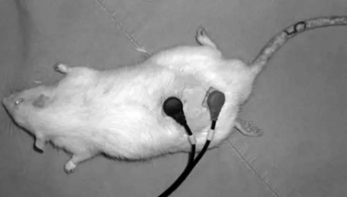
Group 2: The BLT-09 model (BTL, Benesov, Czech
Republic, AC input 230 v/50-60 Hz, 2¥ Fuse T6.3A, input power: 600VA) magnetic field device was used. Pulsed electromagnetic field therapy (PEMFT) was applied at a dose of 15 mT for 30 min once daily at the same time for 14 days with a total of 14 sessions (Figure 1).
Group 3: Two channels as one for right side and
the other for left side application with the Compex vitality (Compex Médical SA, Ecublens, Switzerland)
Figure 1. Power supply and solenoids of pulsed electromagnetic
ES device were used. An active electrode in the same channel was placed on the proximal of injuries, while a passive electrode was placed on distal of injuries. Current intensity ranging between 10 mA and 60 mA, depending on contraction condition of the skin was applied. Frequency was set between 30 Hz and 100 Hz. Duration of contraction was eight sec, rest was 16 sec, rise was 1.5 sec, and fall was 0.75 sec. Consecutive, biphasic, symmetrically synchronized type waves with 450 µS wavelength were used (Figure 2).
Treatment Evaluation Criteria
Gross examination
Gross examination of healing burn injury in rats was done according to the injury size, edema, hyperemia, and epithelialization.
Evaluation of burn area
Evaluation was started on Day 3, as the depth and boundaries of necrosis of burn injury were not fully shaped during the first days. Starting from Day 3, photographs of both burn injuries in rats were taken by a single researcher every other day. Then, burn injury areas transferred to the Image Tool software (UTHSCSA Image Tool for Windows version 3.00, The University of Texas Health Science Center, San Antonio, Texas, USA) were measured (Figure 3).
Evaluation of edema, hyperemia, and epithelialization
Gross examination of edema, hyperemia, and epithelialization were performed by a single researcher using a scoring system based on 0: not present,
1: minimal, 2: moderate, and 3: maximum.[6,14]
Histopathological evaluation
All rats were sacrificed on Day 14 and histopathological examination of biopsies of the burn area was done.
The sections obtained to identify overall characteristics of the skin tissue were stained using the Masson's trichrome stain and examined under light microscopy. On histopathological examination, formation of vascularization, collagenization, and granulation tissue and inflammatory cell response (i.e., neutrophils, monocytes, and lymphocytes) were evaluated. Evaluations were performed based on the following scoring system: 0: not present, 1: minimal,
2: moderate, and 3: maximum.[6,14]
Immunohistochemical examination
Immunohistochemical examination was carried out according to the method described by Hsu et
al.[15] Skin tissue sections of 6 µm in thickness were
obtained for examination and the sections were placed into water following de-paraffinization procedure. The sections placed into water were boiled in antigen retrieval in microwave oven for 20 min. After waiting for cooling at room temperature for 20 min, the sections were washed with phosphate buffer solution (PBS). Subsequently, the sections were treated with
3% hydrogen peroxide (H2O2) prepared in methanol
(Riedel-de Häen 24229) for 20 min to eliminate hydrogen peroxidase activity. The sections were, then, washed in PBS (pH 7.6) by shaking in distilled water. The sections were applied with 1% pre-immune rabbit serum (Ultra V Block, LabVision, TA-015-UB) to block non-specific antibody binding. Then, the sections were incubated in humid chambers within 1/100 diluted primary antibody for one hour. The used antibodies, rabbit polyclonal anti-proliferating cell nuclear antigen (PCNA) antibody (ABCAM (2426), USA), and mouse monoclonal keratin antibody were incubated using the Pan Ab-1 (AE1/AE3, Thermo LabVision, USA). After washing the sections with PBS three times, they were incubated in a second antibody solution (Biotinylated Goat Anti-Mouse, LabVision, TM-015-BN) for 20 min. The sections washed with PBS for three times were treated with streptavidin peroxidase solution (Streptavidin Peroxidase, LabVision, TS-015-HR) for
20 min. After washing with PBS for three times, the sections were treated with 3-amino 6-ethyl carbazole (AEC) chromogen solution (LabVision, TA-002-HAC) for 10 min. Following washing the sections with distilled water, contrast staining was performed using the Mayer’s hematoxylin for five min. The sections which were washed under running water for five min were covered with lamella by adding the covering solution (Mounting Medium, LabVision, TA-060-UG) and the sections were evaluated under light microscope. Nuclear staining was considered positive in cells of tissue sections marked by PCNA antibody. Cell proliferation was scored by cell count in the area with maximum staining. The cells with positive and negative staining were counted under large magnification area (¥400). The number of positively staining cells was detected as the PCNA index, counting 100 cells in each
preparation.[16]
The epithelialization rate was examined by evaluating this staining in all groups, since cytokeratin immune staining occurred only in the epithelial cells. Evaluation was graded as follows: 0: No epithelialization, 1: epithelialization, focal, 2: epithelialization, thin and on the all surface, and
3: epithelialization, thick and on the all surface.[6,14]
Statistical analysis
Statistical analysis was performed using the SPSS for Windows version 16.0 software (SPSS Inc., Chicago, IL, USA). The chi-square test was used to compare edema, hyperemia, and epithelialization rates among the groups. The Kruskal-Wallis test was
used to compare the injury area and histopathological findings among the groups and the Bonferroni corrected Mann-Whitney U test was applied as post-hoc for comparisons among the groups with significant differences. A p value of <0.05 was considered statistically significant.
RESULTS
Gross examination findings
1- Injury area: Burn area in all injuries were
smaller in Group 2 compared to Group 1, indicating statistical significance only on Day 13 (p<0.05). During measurements of burn area every other day, the injury area was becoming much smaller in Group 3, compared to Group 1 at all time points; however, this difference was not statistically significant (Figure 4).
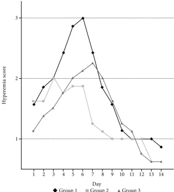
2- Hyperemia: In all groups, hyperemia was
observed on Day 1. Hyperemia was still present on Day 14 in all groups. There was less hyperemia in Group 2 and 3, compared to Group 1. Hyperemia rates were statistically significantly different among the groups on Days 5, 6, 7, 8, and 9 (p<0.05) (Figure 5).
3- Edema: In all groups, edema occurred as of
Day 2 and reached its maximum on Day 6 in Group 1 and 3 and on Day 4 in Group 2. In all groups, edema disappeared on Day 13. At all time points, there was
330 300 270 240 210 180 150 120 90 60 30 0 3 5 7 Day In ju ry a re a ( m m 2) 9 11 13
Group 1 Group 2 Group 3
Figure 4. Change of injury area size of groups according
to days. Day H yp ere m ia sc ore
Group 1 Group 2 Group 3
1 1 2 3 5 10 3 7 12 2 4 6 8 9 11 13 14
Figure 5. Changes of the macroscopic hyperemia scores of
less edema in Group 2 and 3, compared to Group 1. There was a statistically significant difference in the edema rate among the groups on Day 3, 6, 7, 11, and 12 (p<0.05) (Figure 6).
4- Epithelialization: Epithelialization started on
Day 6 in Group 1 and on Day 5 in Group 2 and 3. Epithelialization was completed on Day 13 in Group 2. Epithelialization still continued on Day 14 in Group 1 and 3. There was a statistically significant difference in
the epithelialization rate among the groups on Day 5, 6, 9, 10,11,12,13, and 14 (p<0.05) (Figure 7).
Histopathological examination findings
A) Light microscopic findings
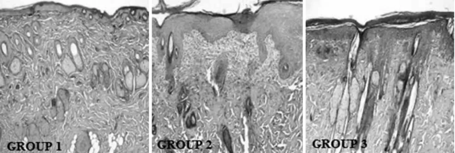
On histopathological examination, vascularization, collagenization, and formation of granulation tissue and inflammatory cell response were evaluated to follow the duration of healing of tissue samples (Figure 8).
Day
Group 1 Group 2 Group 3
1 2 3 4 5 6 7 8 9 10 11 12 13 14
Figure 6. Changes of the macroscopic edema scores of groups
according to days. -1 0 1 2 3 Ed em a s co re Ep ith eli aliz at io n s co re Day
Group 1 Group 2 Group 3
1 2 3 4 5 6 7 8 9 10 11 12 13 14
Figure 7. Changes of the macroscopic epithelialization scores
of groups according to days.
0 1 2 3
Figure 8. Microscopic images of wound area belonging to Group 1, Group 2 and Group 3 on Day 14 (Masson’s trichrome stain
1- Vascularization: In tissue samples examined,
vascularization degree was at the lowest level in Group 1 on Day 14. Treatment of Group 2 and 3 led to an increase in the vascularization degree. However, this increase was more significant in Group 2 (Table 1).
2- Collagenization: In tissue samples examined,
collagenization degree was at the lowest level in Group 1 on Day 14. Treatment of Group 2 and 3 led to an increase in the collagenization degree. However, this increase was more significant in Group 2 (Table 1).
3- Formation of granulation tissue: In tissue samples
examined, formation of granulation tissue was at the lowest level in Group 1 on Day 14. Treatment of Group 2 and 3 led to an increase in the formation of granulation tissue. However, this increase was more significant in Group 2 (Table 1).
4- Inflammatory cell response: In tissue samples
examined, inflammatory cell response was at the lowest level in Group 1 on Day 14. Treatment of Group 2 and 3 led to an increase in the inflammatory Figure 9. Proliferating cell nuclear antigen immunostaining of Group 1, Group 2 and Group 3. Proliferating cell nuclear
antigen positive cells (arrows) (immunoperoxidase technique hematoxylin counterstain ¥200).
Table 1. Vascularization, collagenization, and granulation tissue, and inflammatory cell response Vascularization Collagenization Granulation tissue Inflammatory cell response
Mean±SD Mean±SD Mean±SD Mean±SD
Group 1 2.1±0.1 2.4±0.1 2.1±0.1 1.3±0.1
Group 2 2.8±0.2** 2.9±0.2** 2.7±0.2** 1.8±0.1**
Group 3 2.5±0.2* 2.7±0.2* 2.5±0.2* 1.6±0.1*
SD: Standard deviation; Group 3; * p<0.01 compared to Group 1; Group 2; ** p<0.001 compared to Group 1; ** p<0.05 compared to Group 3, indicating a statistically significant difference; Mann-Whitney U test.
Figure 10. Cytokeratin immune staining belonging to Group 1, Group 2 and Group 3 (immunoperoxidase technique hematoxylin
cell response. However, this increase was more significant in Group 2 (Table 1).
B) Immunohistochemical findings
1- PCNA immunostaining findings: The PCNA
immunostaining was observed in hair and fat follicles more frequently in epithelial basal lamina cells. There was the least number of PCNA-positive cells in Group 1. Following treatment of Group 2 and Group 3, PCNA positivity significantly increased. The highest increase was observed in Group 2 (Figure 9, Table 2).
2- Cytokeratin immunostaining findings:
Cytokeratin immunostaining occurred only in cytoplasm of epithelial cells. Formation of epithelium was clearly exhibited and epithelialization degree measured via this staining. Epithelialization degree was minimal in Group 1 on Day 14. Treatment of Group 2 and 3 led to an increase in the epithelialization degree. However, this increase was more significant in Group 2 (Figure 10, Table 2).
DISCUSSION
In this study, we compared the morphological and histological effects of MFT and ES on burn injury in an experimental rat model. According to the gross examination findings, we found that the burn area, hyperemia, edema, and epithelialization produced more significant improvements in the MAT and ES, compared to the control group, particularly in the MAT-treated group. Similarly, based on the light microscopic and immunohistochemical findings, we observed greater improvement in MAT and ES groups compared to the control group with a higher rate with MAT than ES.
The main goals of burn rehabilitation are to protect the range of joint mobility, to prevent muscle atrophy, and to prevent deformity. In this respect, rehabilitation program should be started as soon as
possible and continued following discharge, as well. During early period of rehabilitation, the aim is to accelerate injury healing, to control infection and edema, and to provide mobility of joint and skin. To prove efficiency of physical treatment agents to reach this target, there are many studies of injuries with different etiopathogenesis and successful results
have been reported.[17-20] However, burn injury has
been emphasized in a very limited number of studies. In many experimental studies, there are contradictory results about the effects of pulsed and static magnetic fields on injury healing. Some of the studies have reported positive effects on injury healing, while some
others have advocated that they are not useful.[17,21]
Healing duration in burn injury varies depending on the other injury types. Burn leads to a reduction in the blood flow to damage area by damaging the tissue
blood vessels.[22] Besides, hematogenous filling seen at
the beginning of injury healing and provides limitation of damage does not occur in burn injury. Although the main mechanism of therapeutic effect of PEMFT on injury healing has not been clearly understood yet, it is considered that PEMFT demonstrates this effect by increasing the blood flow on applied area, if healing depends on the blood flow and enough oxygenation at
the microcirculatory level. [23] Also, it has been shown
that it has an ion exchange and enzymatic activity on
the membrane.[24] In our study, the PEMFT increased
injury healing by measurement of the injury area. It was found that the mean injury area was statistically smaller in the PEMFT group (159.18±33.38) compared to mean injury area of control group (214.02±28.3) on day 13.
In the study of Athanasiou et al.[25] evaluating
short-term PEMFT effects on full-thickness skin injury, injury healing was found to be more rapid in the PEMFT group during the first nine days. However, there was no significant difference between the control group and PEMFT group at the end of the experiment. There was an increase in angiogenesis, collagenization, and epithelialization in the PEMFT group under the light microscopic examination of injury healing. In another study, PEMFT caused early injury healing and provided short-term increase in tensile strength of injury; however, when healing was completed, there was no significant difference between the control group and PEMFT group in terms of the tensile
strength.[26] These aforementioned studies showed that
PEMFT accelerated injury healing at early stage, but in the long-term, it did not yield an additional healing or durability in the injury. Similarly, in our study, we Table 2. Comparison of PCNA indexes and epithelialization
degree
PCNA Index Epithelialization level
Mean±SD Mean±SD
Group 1 15.4±5.3 2.4±0.1
Group 2 42.3±7.9** 2.9±0.2**
Group 3 32.7±7.1* 2.7±0.1*
PCNA: Proliferating cell nuclear antigen; SD: Standard deviation; Group 3; * p<0.01 compared to Group 1, Group 2; ** p<0.001 compared to Group 1; ** p<0.05 compared to Group 3, indicating a statistically significant difference. Mann-Whitney U test.
observed that vascularization and collagenization were greater in the PEMFT group, compared to the other groups.
During inflammation phase of injury healing, leucocytes migrate to the injured area by increased blood circulation and vessel permeability of the injured area. Neutrophils come first to the injured area. When injury environment is adequately prepared by the cells, injury healing progresses to the third phase, called proliferative stage. During this stage, fibroblasts are mainly activated and endothelial cells are proliferated. Granulation tissue is formed in the injury as a fibrous filling by progression of fibroblasts from healthy tissue around injury to inside the injury. Also, in our study, there was a higher rate of granulation tissue in the PEMFT group, compared to the other groups. Taken together, we can suggest that injury healing was more rapid, more granulation tissue was formed by fibroblast migration, the PCNA index showing the proliferation phase of the cell cycle was much higher, and much more epithelialization was formed by cytokeratin staining in the PEMFT group, compared to the other groups. These findings indicate that PEMFT positively affects injury healing in the superficial burn injury model with partial thickness.
Under normal conditions, cells have a natural electric activity and they are notably sensitive to changes in electrical field. There are metabolic, immunological, and physiological changes, when electrical current
is applied to different cell cultures.[9] It has been also
experimentally proven that there is an increase in the tissue blood flow, bacteriostatic effect, protein synthesis, and migration of myofibroblast, fibroblast, and epithelial cells to the applied area (galvanotaxis)
by ES application.[10,11] In addition, edema can be
decreased by inhibiting ES microvascular protein
leakage.[12] When all these effects are combined,
positive results can be obtained in injury healing using ES.[9,10] In addition, ES provides an increase in the injury separation power in short-term by accelerating
injury healing.[4,13]
In the literature, it has been demonstrated that negative polarity has an antibacterial effect, increased blood flow, and provided debridement of necrotic material, although there are different opinions on positive (anode) and negative (cathode) polarity choice during application. In a study performed on an experimental burn injury model, re-epithelialization started two days earlier and there was more rapid new vessel formation in the anode side, compared to
the cathode side.[27] It is recommended that treatment
is firstly started with negative polarity and, then,
continued with positive and negative applications.[11]
In addition, pulsed ES was shown to be more effective, compared to continuous stimulation, and did not cause burning or irritation in injury, and also increased the
vascular flow.[28]
In an experimental study on 124 rats, the effect
of laser and ES in injury healing was investigated.[4]
Histopathological, biomechanical characteristics, and injury separation power of the injury were evaluated. Both ES and laser treatment were found to be effective on three phases of injury healing. During the inflammatory phase, there was a significant decrease in the count of macrophage and PNL in group treated with ES, compared to laser. Thus, the authors concluded that ES more shortened the inflammation stage and accelerated healing, compared to laser.
In the study of Chu et al.[29] on a burn model with
partial thickness, anodal direct current was applied to the injury area. Re-epithelialization was completed in 12 days in the group of direct current, while this period lasted for 16 days in the control group. Also, revascularization was more rapid, compared to the control group, although inflammation and fibrosis rates were lower. The authors concluded that direct current accelerated burn healing and limited tissue damage by decreasing inflammation and fibrosis. In another study on full-thickness burn injury, high-voltage pulse galvanic stimulation was applied to the injury area and a significant increase in the fibroblast amount and injury side contraction was observed in the treatment group, compared
to the control group.[30] Similarly, in our study,
vascularization, collagen synthesis, and formation of granulation tissue were higher, compared to the control group and lower, compared to the PEMFT group. There was also a significantly higher PCNA index and cytokeratin staining, compared to the control group. Based on these findings, we can suggest that, in the superficial injury model with partial thickness, ES positively affects injury healing, compared to the control group.
In conclusion, PEMFT and ES were found to be effective in an experimental burn healing model. The PEMFT was more effective treatment modality, compared to ES, in burn healing based on histopathological and immunohistochemical methods. Nonetheless, further large-scale and long-term studies are needed to confirm these findings.
Declaration of conflicting interests
The authors declared no conflicts of interest with respect to the authorship and/or publication of this article.
Funding
This work was supported by the Unit of Scientific Research Projects as project number 2010-77.
REFERENCES
1. Press B. Thermal, electrical and chemical injuries. Grabb and Smith’s Plastic Surgery. 5th ed. Philadelphia: Lippincott-Raven; 1997.
2. Weiss DS, Kirsner R, Eaglstein WH.Electrical stimulation and wound healing. Arch Dermatol 1990;126:222-5. 3. Patiño O, Grana D, Bolgiani A, Prezzavento G, Miño J,
Merlo A, et al. Pulsed electromagnetic fields in experimental cutaneous wound healing in rats. J Burn Care Rehabil 1996;17:528-31.
4. Demir H, Balay H, Kirnap M.A comparative study of the effects of electrical stimulation and laser treatment on experimental wound healing in rats. J Rehabil Res Dev 2004;41:147-54.
5. Sauer HD, Rudy D. The significance of low-frequency magnetotherapy for local treatment of burns. An experimental comparative approach (author's transl). Aktuelle Traumatol 1980;10:9-13. [Abstract]
6. Schlager A, Kronberger P, Petschke F, Ulmer H. Low-power laser light in the healing of burns: a comparison between two different wavelengths (635 nm and 690 nm) and a placebo group. Lasers Surg Med 2000;27:39-42.
7. Cambier DC, Vanderstraeten GG.Failure of therapeutic ultrasound in healing burn injuries. Burns 1997;23:248-9. 8. Yen-Patton GP, Patton WF, Beer DM, Jacobson BS.
Endothelial cell response to pulsed electromagnetic fields: stimulation of growth rate and angiogenesis in vitro. J Cell Physiol 1988;134:37-46.
9. Alvarez OM, Mertz PM, Smerbeck RV, Eaglstein WH. The healing of superficial skin wounds is stimulated by external electrical current. J Invest Dermatol 1983;81:144-8.
10. Kloth LC. Electrical stimulation for wound healing: a review of evidence from in vitro studies, animal experiments, and clinical trials. Int J Low Extrem Wounds 2005;4:23-44. 11. Kloth LC, McCulloch JM. Promotion of wound healing
with electrical stimulation. Adv Wound Care 1996;9:42-5. 12. Reed BV. Effect of high voltage pulsed electrical stimulation
on microvascular permeability to plasma proteins. A possible mechanism in minimizing edema. Phys Ther 1988;68:491-5.
13. Brown M, McDonnell MK, Menton DN. Polarity effects on wound healing using electric stimulation in rabbits. Arch Phys Med Rehabil 1989;70:624-7.
14. Schlager A, Oehler K, Huebner KU, Schmuth M, Spoetl L. Healing of burns after treatment with 670-nanometer low-power laser light. Plast Reconstr Surg 2000;105:1635-9. 15. Hsu SM, Raine L, Fanger H. Use of avidin-biotin-peroxidase
complex (ABC) in immunoperoxidase techniques:
a comparison between ABC and unlabeled antibody (PAP) procedures. J Histochem Cytochem 1981;29:577-80. 16. Chu Y, Yu D, Wang P, Xu J, Li D, Ding M. Nanotechnology
promotes the full-thickness diabetic wound healing effect of recombinant human epidermal growth factor in diabetic rats. Wound Repair Regen 2010;18:499-505.
17. Callaghan MJ, Chang EI, Seiser N, Aarabi S, Ghali S, Kinnucan ER, et al. Pulsed electromagnetic fields accelerate normal and diabetic wound healing by increasing endogenous FGF-2 release. Plast Reconstr Surg 2008;121:130-41.
18. Stiller MJ, Pak GH, Shupack JL, Thaler S, Kenny C, Jondreau L. A portable pulsed electromagnetic field (PEMF) device to enhance healing of recalcitrant venous ulcers: a double-blind, placebo-controlled clinical trial. Br J Dermatol 1992;127:147-54.
19. Sarma GR, Subrahmanyam S, Deenabandhu A, Babu CR, Madhivathanan S, Kesavaraj N. Exposure to pulsed magnetic fields in the treatment of plantar ulcers in leprosy patients--a pilot, randomized, double-blind, controlled clinical trial. Indian J Lepr 1997;69:241-50.
20. Salzberg CA, Cooper-Vastola SA, Perez F, Viehbeck MG, Byrne DW. The effects of non-thermal pulsed electromagnetic energy on wound healing of pressure ulcers in spinal cord-injured patients: a randomized, double-blind study. Ostomy Wound Manage 1995;41:42-4.
21. Milgram J, Shahar R, Levin-Harrus T, Kass P. The effect of short, high intensity magnetic field pulses on the healing of skin wounds in rats. Bioelectromagnetics 2004;25:271-7. 22. Kazakos K, Lyras DN, Verettas D, Tilkeridis K, Tryfonidis M.
The use of autologous PRP gel as an aid in the management of acute trauma wounds. Injury 2009;40:801-5.
23. Webb CY, Lo SS, Evans JH. Prevention of diabetic foot using low frequency magnetotherapy-Wound management; 2007. 24. Low J. Dosage of some pulsed shortwave clinical trials.
Physiotherapy 1995;81:611-6.
25. Athanasiou A, Karkambounas S, Batistatou A, Lykoudis E, Katsaraki A, Kartsiouni T, et al. The effect of pulsed electromagnetic fields on secondary skin wound healing: an experimental study. Bioelectromagnetics 2007;28:362-8. 26. Strauch B, Patel MK, Navarro JA, Berdichevsky M, Yu
HL, Pilla AA. Pulsed magnetic fields accelerate cutaneous wound healing in rats. Plast Reconstr Surg 2007;120:425-30. 27. Greenberg J, Hanly A, and Davis S. The effect of electrical
stimulation (RPES) on wound healing and angiogenesis in second degree burns. Proceedings of the 13th Annual Symposium on Advanced Wound Care; 2000, April 1-4; Dallas, TX.
28. Reich JD, Tarjan PP. Electrical stimulation of skin. Int J Dermatol 1990;29:395-400.
29. Chu CS, McManus AT, Mason AD Jr, Okerberg CV, Pruitt BA Jr. Multiple graft harvestings from deep partial-thickness scald wounds healed under the influence of weak direct current. J Trauma 1990;30:1044-9.
30. Cruz NI, Bayrón FE, Suárez AJ. Accelerated healing of full-thickness burns by the use of high-voltage pulsed galvanic stimulation in the pig. Ann Plast Surg 1989;23:49-55