Patient Report
Rectal duplications accompanying rectovestibular fistula: Report of
two cases
Arzu Pampal,1Asli Ozbayoglu,2Cem Kaya,2Yildiz Pehlivan,2Aylar Poyraz,3I. Onur Ozen,2Ferda E. Percin4and
Billur Demirogullari2 1
Department of Pediatric Surgery, Faculty of Medicine, Ufuk University and Departments of2
Pediatric Surgery,3
Pathology and4
Medical Genetics, Faculty of Medicine, Gazi University, Ankara, Turkey
Abstract Rectal duplication (RD) cysts are rare congenital anomalies that can be diagnosed with the presence of another opening in the perineum. They seldom accompany anorectal malformations (ARM). Two cases of RD accompanying ARM at opposite ends of the phenotypic spectrum, are described. A 3-month-old baby and a 2-year-old girl with ARM were scheduled for posterior sagittal anorectoplasty. The infant had an orifice at the anal dimple and the other had an orifice at the vestibulum posterior to the rectovestibular fistula. The infant presented with no other anomalies whereas the older one presented with an unusual coexistence of caudal duplication and caudal regression syndromes. Perioperatively both orifices were found to be related to retrorectal cysts, and were excised. Clinicians should always be alert when dealing with complex malformations. Because these malformations have variable anatomical and clinical presentations, they can represent a diagnostic and therapeutic challenge.
Key words anorectal malformation, caudal duplication anomaly, caudal regression syndrome, rectal duplication.
Duplication cysts are rare congenital malformations that can occur anywhere along the gastrointestinal tract. Rectum is an uncommon site for these cysts and rectal duplications comprise only 5% of all duplication cysts. They generally present as ret-rorectal masses, and a small proportion of the cysts communicate with the rectal lumen. They are generally cystic in nature, and present with different clinical features due to their size, localiza-tion, and possession of ectopic mucosa. These cysts tend to fistulate either through the rectum or anus or to the perineum. They could be interpreted mistakenly as fistulo-in-ano or perirec-tal abscess. Chronic constipation, intestinal obstruction, or a pro-lapsed mass through the rectum are the other main clinical features. Digital rectal examination followed by radiological evaluation is necessary in order to evaluate the anatomy and the extent of the cyst. Treatment consists of surgical removal of the cyst via posterior sagittal, endoanal/transanal, transcoccygeal or abdominal approaches according to the location. Surgical removal does not only relieve the symptoms but also reduces the risk of malignant transformation of the cyst.1,2
Cystic rectal duplications are rarely associated with anorectal malformations; they are usually associated with cloaca and ante-rior ectopic anus.2Nearly 100 rectal duplications are reported in
the literature and none is alike in terms of phenotypic spectrum.
The aim of this report is to describe two new cases of rectal duplication accompanying rectovestibular fistula. These cases also represent the two ends of the phenotypic spectrum: the first presented as rectal duplication and rectovestibular fistula with no other anomalies whereas the second is an example of an unusual coexistence of caudal duplication anomaly and caudal regression syndrome.
Case report
Case 1
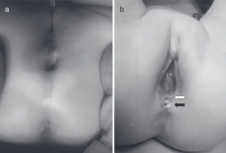
A newborn girl with imperforated anus and rectovestibular fistula was hospitalized and evaluated for accompanying anomalies. No cardiac, urinary, vertebral, and sacral anomalies were found. She was discharged with dilation of rectovestibular fistula and sched-uled for posterior sagittal anorectoplasty (PSARP) with a protec-tive colostomy at the age of 3 months. Preoperaprotec-tively, the surgical team encountered an opening at the anal dimple (Fig. 1a). Sur-gical exploration identified a 3 cm cyst located at the retrorectal region. It was found to be fistulated posterior to the vertical muscles. The cyst was meticulously dissected and excised. PSARP and a protective colostomy at the descending colon with Hartmann pouch were performed. Histopathological evaluation of the cyst indicated a rectal duplication cyst lined with squamous epithelium. At 6th months, the patient was scheduled for colos-tomy closure. An intraperitoneal closure was attempted. The patient is now 2 years old and having regular defecations under lactulose treatment.
Even though the surgical team planned a genetics consulta-tion, the parents refused to visit the medical genetics department. Correspondence: Arzu Pampal, MD, Ufuk Universitesi Tip Fakultesi,
Cocuk Cerrahisi ABD, Konya Yolu No:86-88 Balgat 06520, Ankara, Turkey. Email: [email protected]
Presented at the 18th International Pediatric Colorectal Club Meeting in Ankara, Turkey, 10–12 June 2011.
Received 23 September 2012; accepted 13 February 2013.
bs_bs_banner
Pediatrics International (2013) 55, e86–e89 doi: 10.1111/ped.12086
© 2013 The Authors
Case 2
A 2-year-old girl with imperforated anus was admitted for a pull-through. She had a divergent colostomy and a mucous fistula at the descending colon and two openings located at the vestibu-lum (Fig. 1b). She was evaluated for accompanying anomalies. Congenital bilateral dislocation of the hip, agenesis of the coccyx, lateral deviation of the sacrum, posterior fusion defect at S1 and spina bifida were found. Urinary evaluation indicated a right-sided non-functioning kidney with ureteric duplication. The ectopic ureter was found to enter the urethra just under the bladder neck and grade V vesico-ureteral reflux (VUR) to the ectopic ureter was also diagnosed. Her pull-through was post-poned several times due to recurrent urinary infections resulting from VUR. When it was performed, the posterior orifice at the vestibulum was found to be related to a cystic mass lying up to the coccyx. The cyst was excised and PSARP was performed. Histopathological evaluation indicated a rectal duplication cyst lined with squamous and columnar epithelium. Four months after PSARP, the patient underwent surgery for colostomy closure and right-sided nephro-ureterectomy. Six months after these proce-dures she was re-evaluated for recurrent urinary infections and found to have a hypocompliant and small bladder with grade II–III VUR on the left side. The patient was put on oxybutynin and antibiotic prophylaxis therapy. Endoscopic subureteric injec-tion (STING) was performed. She is now 4.5 years old, and has voluntary bowel movements (twice to three times per day) with minimal soiling (once in a day). She is under oxybutynin and antibiotic prophylaxis therapy for recurrent urinary tract infection.
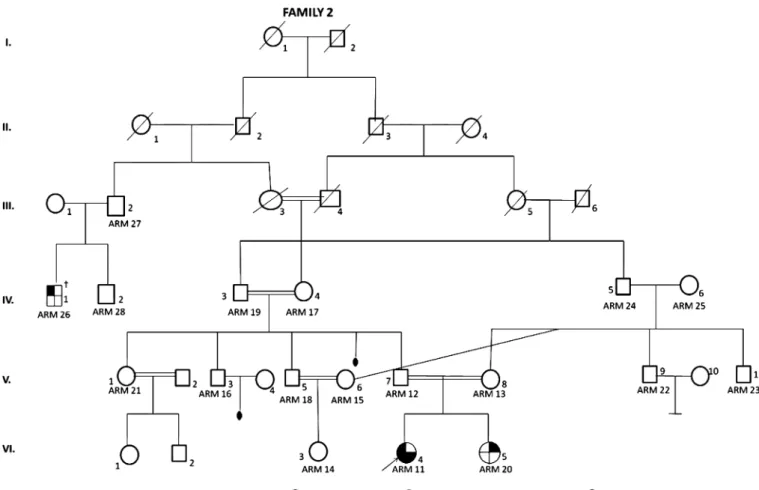
Given that the patient had atypical facial features (prominent forehead, epichantal folds, mild hypertelorism and high palate) and accompanying multiple congenital anomalies, the family attended a genetics consultation. The patient was the one of the daughters of healthy but consanguineous parents (Fig. 2). Her
prenatal history was unremarkable. The other daughter was also found to have unilateral renal agenesis. Chromosome analysis of the peripheral leukocytes of the patient using high-resolution binding showed a normal female karyotype (46, XX).
Discussion
Different theories have been suggested for the development of duplication cysts. One possible explanation is the abnormal split-ting of the notochord in the developing embryo that results in a variety of malformations including central nervous system, spinal and vertebral anomalies, and posterior enteric remnants. Persistence of embryonic diverticuli of the stomach, duodenum or ileum, and double vacuolization of the epithelium during plug-ging of the intestinal lumen are the other possible explanations.3
Given that the notochord normally separates during the fourth gestational week, the former theory is likely to be related to additional malformations. And because the vacuolization occurs between fifth and eighth gestational weeks, the latter two theories can explain isolated short segment duplications without ectopic epithelial lining. The present two cases are distinct from each other and also suggest the multifactorial etiologies of the forma-tion of duplicaforma-tion cysts.
Rectal duplications accompanying anorectal malformations are extremely rare. Apart from the tubular hindgut duplication cysts, cystic rectal duplications are believed not to be associated with other congenital anomalies. Only three cases of imperfo-rated anus and duplication cyst are presented in the literature.3–5
Two of these patients presented during infancy and the third patient presented at the age of 15. The 15-year-old girl, who underwent surgery for ARM in infancy, was evaluated for fecal incontinence and diagnosed as having Currarino’s triad. The presacral cyst was evaluated on pathology and reported to be a rectal duplication cyst. All three cases involved additional con-genital renal, vertebral and spinal anomalies. Also in a review of 174 patients with anorectal malformation, Stoll et al. described two cases of intestinal duplication6 but the authors mentioned
neither the locations of these cysts nor accompanying congenital anomalies. Casaccia et al. reviewed the literature and found that the incidence of accompanying gastrointestinal anomalies in anorectal malformations is approximately 15%.7In their review
they have noted the sonic hedgehog (SHH) proteins and their role in the embryogenesis of the human digestive system. The SHH genes are involved in the organization and morphology of the developing embryo and are expressed in the early gut endoderm, neural tube and throughout the notochord. SHH and its down-stream mediators are essential for development of the distal hindgut, and any mutations in this signaling pathway result in abnormal phenotypes at the anorectal region. The implications for the SHH gene in gastrointestinal anomalies accompanying ARM, however, have not been studied.
Caudal duplication anomaly (OMIM 607864) is a rare entity that involves duplication of diverse organs at the caudal region. It is associated with hypermethylation of the Axis inhibition protein 1 (AXIN1) promoter on chromosome 16p13.3.8Even though the
embryological origin is obscure, an insult to the migration of the
a b
Fig. 1 Preoperative images. (a) Patient 1: orifice of the rectal
dupli-cation cyst at the anal dimple; (b) patient 2: two orifices at the vestibulum (white arrow, rectovestibular fistula; black arrow, orifice of rectal duplication cyst).
Rectal duplication with ARM e87
© 2013 The Authors Pediatrics International © 2013 Japan Pediatric Society
caudal cell mass at the 23rd–25th gestational days or a sagittal symmetric pairing of axial structures of the caudal embryo are the proposed theories, especially for the genital and urinary duplications. Caudal regression syndrome (OMIM 607864) is another rare entity; this refers to the heterogeneous group of congenital caudal anomalies concerning caudal spine, spinal cord, hindgut, urogenital system and lower extremities. Some cases involved heterozygous mutation in the VANGL1 gene on chromosome 1p13. This constellation of anomalies is due to the failure of regression of the structures derived from the caudal mesoderm before the fourth gestational week and the etiology is thought to be related to maternal diabetes mellitus, genetic pre-disposition, and vascular hypoperfusion.9Either caudal
duplica-tion or caudal regression are usually found sporadically in society. The congenital anomalies involved in the present second case (imperforated anus, rectal duplication, ureteric duplication, VUR, sacral deviation, agenesis of coccyx, hip dislocation) fit the phenotype of caudal duplication anomaly and a probable caudal regression syndrome variant. That patient was the daughter of consanguineous (but healthy) parents and her sister also had unilateral renal agenesis. The medical genetics department also evaluated the pedigree of four generations (Fig. 2). It is probable that a major anomaly such as renal agenesis may have been
coincidental in the sibling, but it is more possible that the caudal anomaly in the patient herself was inherited autosomal recessively.
It is important to recognize rectal duplication and whether it accompanies anorectal malformation. These complex malforma-tions can represent a diagnostic and therapeutic challenge not only because of their rarity but also their variable anatomical and clinical presentation and other accompanying unexpected con-genital anomalies.
References
1 Knudtson J, Jackson R, Grewal H. Rectal duplication. J. Pediatr. Surg. 2003; 38: 1119–20.
2 Rajah S, Ramanujam TM, Anas SR et al. Duplication of the rectum: Report of four cases and review of the literature. Pediatr. Surg. Int. 1998; 13: 373–6.
3 Schalamon J, Schleef J, Höllwarth ME. Experience with gastro-intestinal duplications in childhood. Langenbecks Arch. Surg. 2000;
385: 402–5.
4 Shinkai M, Mochizuki K, Hirata Y et al. Anorectal malformation associated with a perineal protrusion of the rectal mucosa: Case presentation. Pediatr. Surg. Int. 2009; 25: 815–17.
5 Wang J, Shi C, Yu S et al. A rare association of rectal and genitouri-nary duplication and anorectal malformation. Chin. Med. J. (Engl.) 2003; 116: 1955–7.
Fig. 2 Patient 2: pedigree of four generations. ( ) Anal atresia; ( ) unilateral renal agenesis; ( ) congenital hip dislocation;
( ) vesico-ureteral reflux, duplication of ureter, agenesis of coccyx. †Anal atresia, not examined. e88 A Pampal et al.
© 2013 The Authors
6 Stoll C, Alembik Y, Dott B et al. Associated malformations in patients with anorectal anomalies. Eur. J. Med. Genet. 2007; 50: 281–90.
7 Casaccia G, Catalano OA, Bagolan P. Congenital gastrointestinal anomalies in anorectal malformations: What relationship and man-agement? Congenit. Anom. (Kyoto) 2009; 49: 93–6.
8 Becker K, Howard K, Klazinga D et al. Caudal duplication syn-drome with unilateral hypoplasia of the pelvis and lower limb and ventriculoseptal heart defect in a mother and features of VATER association in her child. Clin. Dysmorphol. 2009; 18: 139–41. 9 Singh SK, Singh RD, Sharma A. Caudal regression syndrome: Case
report and review of literature. Pediatr. Surg. Int. 2005; 21: 578–81. Rectal duplication with ARM e89
© 2013 The Authors Pediatrics International © 2013 Japan Pediatric Society