Elastofibroma dorsi is a rare benign fibroelastic lesion, mostly found in the periscapular region in elderly (1). It arises on the back, deep to the rhomboid and latissimus dorsi muscles, adjacent to the inferior angle of the scapula (2). The lesions are periscapular in 99%of the reported cases (2) and bilateral in 10-60% of cases (1-2).
Elastofibroma is well known by the pathologists but has received attention recently in the radiologic literature with the advent of computed tomography (CT) and magnetic resonance imaging (MRI). This is a report of two patients who were evaluated preoperatively by MR imaging and operated successfully with marginal resection after biopsy.
CASE REPORTS:
Case 1: A 45-year-old female, without a previous history of trauma presented with a painful mass located in the right inferior periscapular region, first noticed 7 months ago. Pain was radiating to the back and right shoulder. Physical examination revealed a tender, firm mass with a diameter of 10 cm, located at the inferomedial aspect of the right scapula. No associated lymphadenopathy was found. Routine laboratory tests were normal except a mild decrease in the haemoglobin level. An MR imaging examination was performed with a 1.5 T MR unit (GE Medical Systems, Milwaukee, WIS, U.S.A). T1-weighted spin echo (TR/TE = 600/18 msec) and T2-weighted FSE (TR/TE =4000/90
A. Tuba Karagülle*, İlhan Erden*, Ayşe Erden*, Demet Karadağ*
ELASTOFIBROMA DORSI: MAGNETIC RESONANCE
IMAGING FINDINGS IN TWO CASES
–––––––––––––––––––––––––
* University of Ankara, Ibn-i Sina Hospital, Department of Radiology, S hhiye, 06100 Ankara, Turkey
–––––––––––––––––––––––––––––––––––––––––––––––––––––––––––––––––––––––––––––––––––––––––––––––––––– Received: March 21, 2001 Accepted: July 12, 2001
SUMMARY
Elastofibroma dorsi is a pseudotumoral mass of unknown pathogenesis that is usually seen in elderly patients. We report two cases of histologically proven elastofibroma dorsi, each presenting with painful periscapular masses. Magnetic resonance imaging showed periscapular tumoral mass with low to intermediate signal intensity with interspersed areas of high signal intensity on both T1 and T2 weighted images. After I.V. administration of gadopentate dimeglumine, these lesions demonstrated mild enhancement. Awareness of the characteristic location and radiologic appearance of this benign lesion prevents unnecessary radical interventions.
Key Words: Elastofibroma,Magnetic Resonance maging, Chest, Neoplasms.
ÖZET
Elastrofibroma Dorsi: 2 Olgunun Manyetik Rezonans Görüntüleme Bulguları
Elastofibroma dorsi, patogenezi belli olmayan ve s kl kla ya larda g r len, periskapular yerle imli yalanc t m ral (ps dot m ral) kitle lezyonudur. Bu yaz da, histolojik olarak elastofibroma dorsi tan s konmu , iddetli periskapular a r ile gelen iki olguya ait manyetik rezonans g r nt leme bulgular n sunduk.
Anahtar Kelimeler: Elastofibroma-Manyetik Rezonans G r nt leme
msec) images were performed in axial and coronal imaging planes. On T1-weighted and T2-weighted images a soft tissue mass of 8x10x10 cm, deep to rhomboid and latissimus dorsi muscles, low to intermediate in signal intensity relative to the adjacent skeletal muscles with interspersed areas of high signal intensity was seen (Figure 1). Mild linear enhancement was observed in the soft tissue mass following the injection of gadopentate dimeglumine (Figure 2). A diagnosis of elastofibroma dorsi was suggested according to the findings on MR imaging. An incisional biopsy was performed. Histopathologic examination revealed the diagnosis of elastofibroma dorsi. The mass was removed by wide local excision.
Case 2: A 60-year-old male patient was admitted to our hospital with mild pain and swelling on the left periscapular region. There was no history of trauma. Physical examination revealed a firm mass measuring 8x3x3 cm at the inferior part of the left scapula. Routine laboratory findings were normal. An MR imaging examination was performed with a 1.5 T MR unit (GE Medical Systems, Milwaukee, WIS, U.S.A). T1-weighted (TR/TE = 708/229 msec) and
T2-weighted (TR/TE = 4000/80 msec) were obtained in axial and coronal imaging planes. A 8x3x3 cm soft tissue mass, inferior to left serratus anterior muscle, low to intermediate in signal intensity relative to the skeletal muscles with intervening hyperintense areas was seen (Figure 3). The mass
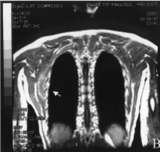
Figure 1: On T1-weighted coronal image a soft tissue
mass of 8x10x10 cm, deep to rhomboid and latissimus dorsi muscles, low to intermediate in signal intensity relative to the adjacent skeletal muscles with interspersed areas of high signal intensity was seen
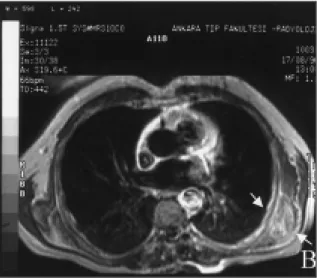
Figure 3: On T1-weighted axial image, a 8x3x3 cm
soft tissue mass, inferior to left serratus anterior muscle, low to intermediate in signal intensity relative to the skeletal muscles with intervening
hyperintense areas was seen
Figure 2: Mild linear enhancement was observed in
the soft tissue mass following the injection of gadopentate dimeglumine on T1-weighted coronal
demonstrated mild enhancement after I.V. administration of gadopentate dimeglumine (Figure 4). A presumptive diagnosis of elastofibroma dorsi was made according to MR imaging findings. The mass was removed by wide local excision. The histopathologic diagnosis was consistent with elastofibroma dorsi.
Discussion: Elastofibroma is a rare tumor-like process made up of a mixture of fibroelastic tissue and fat (2). It is a completely benign lesion, and has a subclinical nature as it has an obscure location and rarely causes severe pain. Hypertrophy and degeneration of elastin in a hypercellular background of mature collagen and fat lobules with no abnormal mitosis are the histologic hallmarks of the lesion (2). The lesion was reported as elastofibroma dorsi initially due to its typical location in the infrascapular and subscapular regions at the level of sixth through eighth ribs and deep in relation to serratus anterior, latissimus dorsi and rhomboid muscles. Although the pathogenesis of these lesions is not clear, it has been suggested that, elastofibroma is a reactive process between the scapula and chest wall, caused by mechanical friction during heavy manual labor (3). However, Stemmerman and Stout (4) found no correlation
between repeated trauma and pseudotumor formation. Instead, they proposed that degenerative changes in collagen was responsible for the development of the lesion. From the history of the current two cases, a mechanical cause for the progress of the lesion has not been found. Elastofibroma is more common on the right side, presumably due to the side of patient’s dominant handedness (1). In our report, it was on the right side in the first case and on the left side in the second.
The imaging features of elastofibroma dorsi have been characterized recently. Plain chest radiographs may show a soft tissue density mass in subscapular region (5). Computed tomography (CT) shows an inhomogenous soft tissue mass with attenuation almost the same as that of skeletal muscle and containing linear low-density streaks in most of the cases. But in some cases it could be relatively homogenous, with an attenuation less than that of muscle (6). Sonographic appearance of elastofibroma dorsi has been recently reported as layers of hypoechoic strands in an echogenic background (7).
Magnetic resonance imaging of elastofibromas may lead to a presumptive diagnosis of elastofibroma (3). A soft tissue with signal intensity similar to that of skeletal muscle, interlaced with streaks of fat tissue is the mainly observed signal characteristic of elastofibroma dorsi (1,6,8). Although atypical for elastofibromas, mild or marked enhancement have been reported in several cases (6,8). Histological examination did not reveal a difference between the tissue composition of the elastofibromas, showing mild or marked enhancement after the administration of gadopentate dimeglumine (8). However in cases with marked contrast enhancement, a strong vascularity was demonstrated on histologic workup (8). Although the basic nature of the contrast enhancement in elastofibromas is not well understood, one should know that marked contrast enhancement could be seen in elastofibromas (8).
Figure 4: The mass demonstrated mild enhancement
after gadopentate dimeglumine administration on T1-weighted axial image.
1. Naylor MF, Nascimento AG, Sherrick AD, McLeod RA. Elastofibroma Dorsi: Radiologic findings in 12 patients. Am J Roentgenol 1996; 167: 683-687.
2. Nagamine N, Nohara Y, Ito E. Elastofibroma in Okinawa: a clinicopathologic study of 170 cases. Cancer 1982; 50: 1794-1805.
3. Hoffman JK, Klein MH, McInerney VK. Bilateral elastofibroma: a case report and review of the literature. Clin Orthop Rel Res 1996; 325: 245-250.
4. Stemmerman GN, Stout AP. Elastofibroma dorsi Am Clin Pathol 1962; 37: 499-506.
5. Vande Berg B, Malghem J, Leflot JL, Lagneaux G, Maldague B. Case report: elastofibroma dorsi: pseudomalignant lesion. Clin Radiol 1996; 51: 67-69.
6. Kransdorf MJ, Meis JM, Montgomery E. Elastofibroma: MR and CT appearance with radiologic-pathologic correlation. Am J Roentgenol 1992; 159: 575-579.
7. Bianchi S, Martinoli C, Abdelwahab IF, Gandolfo N, Derchi LE, Damiani S. Elastofibroma dorsi: sonographic findings. Am J Roentgenol 1997; 169: 1113-1115.
8. Schick S, Zembsch A, Gahleitner A, et al. Atypical appearance of elastofibroma dorsi on MRI: case reports and review of the literature. J Comput Assist Tomogr 2000; 24: 288-292. 9. Sundaram M, McLeod RA. MR imaging of
tumor and tumorlike lesions of bone and soft tissue. Am J Roentgenol 1990; 155: 817-824. 10. Marin ML, Perzin KH, Markowitz AM.
Elastofibroma dorsi: benign chest wall tumor. J Thorac Cardiovasc Surg 1989; 98: 234-238. The differential diagnosis of a periscapular
lesion with a signal intensity similar to skeletal muscle is limited and includes lesions with decreased cellularity like extraabdominal desmoid, neurofibroma, cicatricial fibroma and malignant fibrous histiocytoma (9). Normal structures can also mimic elastofibroma dorsi, like the inferior fibers of the serratus anterior muscle, which has a globular appearance especially on CT sections. As elastofibroma contains strands of soft tissue of fat signal intensity differential diagnosis must also include lipomatous lesions such as lipoma or liposarcoma and lesions containing blood, such as hemangioma, hematoma and intralesional hemorrhage (8). The variable enhancement pattern of the lesion is not a particularly striking feature (8). Marked enhancement seen in some cases could mimic an inflammatory or malignant mass. But the characteristic location and
interspersed fat with typical T1-and T2-weighted signal intensities help the radiologist for the differential diagnosis. Another helpful feature for differential diagnosis is bilaterality of the lesion. Presence of a similar periscapular lesion on the contralateral side strongly eliminates malignancy from differential diagnosis (1).
In patients who are clinically symptomatic complete surgical excision is the preferred treatment. In asymptomatic individuals excision of the lesions exceeding 5 cm in diameter is recommended (10).
In conclusion, the diagnosis of elastofibroma dorsi can be made accurately on the basis of its characteristic location and signal intensity on MR imaging. Identification of a similar lesion on the contralateral side further supports the diagnosis and prevents nonessential extensive surgical resection.