THE LEVELS OF TRACE ELEMENTS AND ELECTROLYTES IN SERUM AND
CEREBROSPINAL FLUID OF PATIENTS WITH ACUTE STROKE
AKUT İNMELİ HASTALARIN SERUM VE SEREBROSPİNAL SIVILARINDA ESER
ELEMENT VE ELEKTROLİT DÜZEYLERİ
Emine KÖKSALDI1, Aysun HACIŞEVKİ2, Meral TORUN2
1 SSK Ankara Hospital, Department of Clinical Biochemistry, Dışkapı – Ankara/TURKEY 2Gazi University, Faculty of Pharmacy, Department of Biochemistry, 06330, Etiler-Ankara
TURKEY ABSTRACT
Trace metals have important influences on brain development and function. The trace element zinc is a necessary component of many metalloenzymes, some of which are of importance for the function of the central nervous system. Magnesium ions act as an endogenous vasodilators of the cerebral circulation. The aim of the present study was to determine the CSF(cerebrospinal fluid) and serum concentration of the metals zinc, copper and magnesium in acute stroke and to discuss the probable correlations of our findings with the clinical conditions. In our study, serum and CSF copper, zinc, magnesium, iron, sodium and potassium levels of 51 patients diagnosed as acute stroke patients and of 15 healthy subjects were measured. The effects of factors such as age, smoking status, drinking habits and sex on serum and CSF copper, zinc, magnesium, iron, sodium and potassium concentrations were evaluated statistically. These levels were statistically no significant when compared to that of control group except CSF copper. Our data indicate that there is no apparent association between serum and CSF copper, zinc, magnesium, iron, sodium and potassium levels and above factors. There is no statistically significant relationship between serum iron, sodium and potassium concentrations and CSF iron, sodium and potassium concentrations. Only significant correlation was found between serum and CSF magnesium concentrations. We investigated whether there was a change of trace element and electrolyte levels and traverse the blood-brain barrier in serum and CSF.
ÖZET
Eser elementler, beyin gelişimi ve fonksiyonunda önemli etkilere sahiptir. Çinko elementi santral sinir sistemi fonksiyonu için önemli birçok metalloenzimin gerekli bir komponentidir. Magnezyum iyonları serebral sirkülasyonun endojen vazodilatörü olarak etki göstermektedir. Bu çalışmanın amacı, akut inmede çinko, bakır, demir ve magnezyumun CSF ve serum konsantrasyonlarını tayin etmek ve klinik çalışmalarla bizim bulgularımızın olası korelasyonlarını tartışmaktır. Çalışmamızda akut inme teşhisi konmuş 51 hasta ve 15 sağlıklı kişinin serum ve CSF bakır, çinko, magnezyum, demir, sodyum ve potasyum düzeyleri ölçüldü. Yaş, sigara içme durumu, alkol kullanma alışkanlığı ve cinsiyet gibi faktörlerin serum ve CSF bakır, çinko, magnezyum, demir, sodyum ve potasyum konsantrasyonları üzerine olan etkileri istatistiksel olarak değerlendirildi. Bu düzeyler, kontrol grubu ile karşılaştırıldığında bakır hariç, istatistiksel olarak anlamlı bir farklılık gözlenmedi. Ölçülen parametrelerin serum ve serebrospinal sıvı düzeyleri ile yukarıdaki faktörler arasında görünürde bir ilişki yoktu. Yalnız magnezyuma ilişkin bir korelasyon bulundu. Çalışmamızda akut inmede eser element ve elektrolit düzeylerinin değişip değişmediğini ve kan-beyin bariyerini geçip geçmediğini serum ve serebrospinal sıvıda araştırdık.
Anahtar Kelimeler: Geçiş metalleri, bakır, çinko, magnezyum, demir, akut inme
INTRODUCTION
The development of a focal neurologic deficit is designated stroke if the cause of the deficit is thought to be the consequences of a local disturbance in the cerebral circulation. The main causes of these, frequently abrupt changes in brain circulation are a reflection of either obstruction of the cerebral blood flow or rupture of the wall of a vessel supplying the brain or spinal cord. (1). The significance of trace elements concentration in the serum and various body fluids has been demonstrated in a number of publications. However, trace elements determinations in the cerebrospinal fluid (CSF) as well as in the serum in neurological diseases has been reported only in a small number of communications. Recently, a greater emphasis has been given to the role of trace elements in the function of the nervous system both in normal and pathological conditions (2).
Trace metals have important influences on brain development and function (3). Zinc (Zn) is a necessary component of many metalloenzymes some of which are known to be of importance for the function of central nervous system (CNS). For example, RNA and DNA polymerase and carbonic anhydrase. Zinc deficiency in rats may lead to malformation of the CNS. Zinc deficiency has been suggested as an explanation for the CNS symptoms in liver cirrhosis, foetal alcohol syndrome, malabsorption and acrodermatitis enteropathica. Low serum or plasma concentrations of zinc have been found in multiple sclerosis and in chronic alcoholics with liver disease. High
concentrations have been found in multiple sclerosis and Pick’s disease. There are some reports of zinc concentrations in normal CSF and in CSF from patients with neurological diseases. There is a basic requirement for zinc in order for cells to function. Zinc is involved in nearly all aspects of cellular metabolism and plays a key role in numerous essential processes: protein synthesis, DNA and RNA metabolism, and carbohydrate and lipid metabolism. Zinc plays a crucial role in maintaining cell membrane structure and function and influences immune response, lysosomal enzyme release, and macrophage function. Based on these and other zinc-dependent effects, it is not surprising that considerable attention has been paid to zinc as an element of importance in inflammatory diseases (4-7).
Copper (Cu) acts as a prooxidant agent, although it is also essential for the antioxidant function of the protein ceruloplasmin and a necessary cofactor for the enzymatic activity of superoxide dismutase. Zinc has antioxidant activity, is also important for the activity of superoxide dismutase and has a stabilizing influence on membranes (8,9). Copper is essential for the antioxidant function of proteins such as caeruloplasmin and superoxide dismutase, but it acts as a pro-oxidant (for instance, towards brain lipids) when present in low molecular-mass loosely bound complexes. Iron (Fe) is the most important of the essential trace metals. An appreciable number of human diseases are related to iron deficiency or disorders of iron metabolism (9, 11).
Magnesium (Mg) ions act as endogenous vasodilators of the cerebral circulation and act pharmacologically as noncompetitive antagonists of the N-methyl-D-aspartate receptor by virtue of their role as endogenous voitage-sensitive blockers of the ion channel. The magnesium ion regulates cellular energy metabolism, vascular tone, and cell membrane ion transport (2).
The aim of the present study was to determine the CSF and serum concentration of the trace elements zinc, copper, iron, magnesium, sodium and potassium in acute stroke and to discuss the probable correlations of our findings with the clinical conditions.
MATERIALS AND METHODS
The study was performed in acute stroke patients admitted to SSK Ankara Hospital in Ankara. In our study, blood and cerebrospinal fluid (CSF) samples were collected from patients diagnosed as acute stroke (AS) (n=51) and from healthy subjects (n=15). Blood and CSF samples were stored at -20°C until the analysis. All the subjects including controls filled out a questionnaire about their age, sex, alcohol intake and smoking habit. Serum and CSF copper, zinc, iron,
magnesium levels were measured in control and acute stroke patient groups by using the colorimetric and ion selective electrote method (12). In this study, RA-XT otoanalyzer was used.
Statistical analyses were performed by using student’s-t test, Mann-Whitney U test and variance analysis. P value of <0.05 was considered as significant. A correlation analysis was carried out by simple regression test.
RESULTS AND DISCUSSION
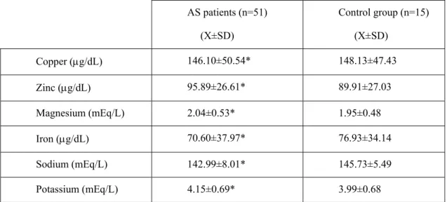
The means of serum copper, zinc, magnesium, iron, sodium and potassium levels in patients with acute stroke and in healthy subjects have been shown in Table 1.
Table 1. Serum trace element and electrolyte levels in acute stroke patients and control group.
AS patients (n=51) (X±SD) Control group (n=15) (X±SD) Copper (µg/dL) 146.10±50.54* 148.13±47.43 Zinc (µg/dL) 95.89±26.61* 89.91±27.03 Magnesium (mEq/L) 2.04±0.53* 1.95±0.48 Iron (µg/dL) 70.60±37.97* 76.93±34.14 Sodium (mEq/L) 142.99±8.01* 145.73±5.49 Potassium (mEq/L) 4.15±0.69* 3.99±0.68
* Not significantly different from controls (p>0.05) (student’s-t)
The means of CSF copper, zinc, magnesium, iron, sodium and potassium levels in patients with acute stroke and in healthy subjects have been shown in Table 2.
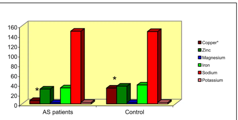
These levels were statistically no significant when compared with those of control group (p>0.05) except CSF copper (Fig 1). In our study, the effects of factors such as age, smoking status, drinking habits and sex on serum and CSF copper, zinc, magnesium, iron, sodium and potassium concentrations were evaluated statistically.
0 20 40 60 80 100 120 140 160 AS patients Control
Fig 1. CSF trace element and electrolyte levels in AS patients and controls.* Significantly different from controls (p<0.05)
Copper* Zinc Magnesium Iron Sodium Potassium
*
*
Table 2. Cerebrospinal Fluid trace element and electrolyte levels in acute stroke patients and control group.
AS patients (n=51) (X±SD) Control group (n=15) (X±SD) Copper (µg/dL) 7.02±2.07 (n=13) * 32.38±30.31 (n=3) Zinc (µg/dL) 30.41±30.98 (n=40) ** 36.27±26.61 (n=5) Magnesium (mEq/L) 2.49±0.55 ** 2.65±0.69 Iron (µg/dL) 32.96±29.52 ** 38.80±19.16 Sodium (mEq/L) 148.87±6.80 ** 148.08±5.01 Potassium (mEq/L) 3.089±0.55 ** 3.02±0.47
* Significantly different than controls (p<0.05)
** Not significantly different than controls (p>0.05) (Mann-Whitney U test for copper and zinc, student’s-t test for others)
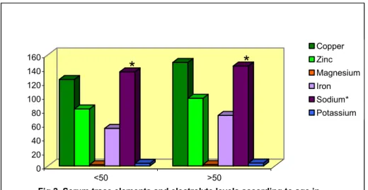
According to age, serum copper, zinc, magnesium, iron, potassium and CSF copper, magnesium, iron, sodium, potassium levels didn’t show any significant difference in patients with acute stroke (p>0.05). But there was a significant difference in terms of CSF zinc and serum sodium levels (p<0.05, Fig 2,3). On the other hand, in controls, there wasn’t statistically significant difference (p>0.05). The CSF zinc and copper levels couldn’t be evaluated because of the fact that they were observed in CSF of very small groups. Copper-dependent redox reactions are relevent to processes leading to many different forms of neurodegeneration. Indeed, damage of the same targets results in different diseases depending on the cell type that is affected, i.e., on the
concurring biochemical background. It should be kept in mind that many neurodegenerative diseases are typical of late life: aging is strictly interrelated with the susceptibility of neurons to oxidative stress and to apoptosis. A further observation is that the level of antioxidant enzymes is decreased during ageing, while the level of copper increases with age (13-14).
*
*
0 20 40 60 80 100 120 140 160 <50 >50Fig 2. Serum trace elements and electrolyte levels according to age in AS patients. *Significantly different from >50 ( p<0.05)
Copper Zinc Magnesium Iron Sodium* Potassium
*
*
0 20 40 60 80 100 120 140 160 <50 >50Fig 3. CSF trace element and electrolyte levels according to age in AS patients. *Significantly different from >50 ( p<0.05)
Copper Zinc* Magnesium Iron Sodium Potassium
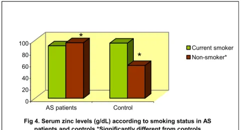
When the serum copper, magnesium, iron, sodium and potassium levels of controls and AS groups were evaluated in terms of smoking habits, there wasn’t any significant difference among the subgroups including current smoker and non-smoker (p>0.05). When the serum zinc levels of controls and AS patients including non-smokers were only assessed there was found a statistically
significant difference (p<0.05, Fig 4). According to smoking status, when comparing the CSF magnesium, iron, sodium and potassium levels of patient groups with acute stroke and control groups there wasn’t a significant difference (p>0.05). In addition, the CSF copper and zinc values couldn’t be evaluated because of the fact that they were found in a bit of control groups.
0 20 40 60 80 100 AS patients Control
Fig 4. Serum zinc levels (g/dL) according to smoking status in AS patients and controls.*Significantly different from controls
(p<0.05)
Current smoker Non-smoker*
*
*
According to sex, there wasn’t a significant difference in both AS patients and controls (p>0.05). The means of CSF copper and zinc values of healthy subjects couldn’t be evaluated because of the fact that they were only observed in a less number of woman and man. In cases, a significant difference wasn’t observed among the subgroups according to alcohol intake (p>0.05). The mean of these parameter values of control group couldn’t be evaluated because of not all the subjects taking alcohol.
There were no statistically significant relationship between serum iron, sodium and potassium concentrations and CSF iron, sodium and potassium concentrations (r=1.90, r=49.64, r=0.99, respectively). Significant correlation was only found between serum and CSF magnesium concentrations (r=74.39).
To produce physiological or pathophysiological effects on the brain, the metal must either traverse the blood-brain barrier in order to act directly on the neurones or glial cells, or alternatively it could have an indirect effect. The trace metal might also have an indirect effect by influencing the transport or regulation of another substance at the blood-brain barrier (3).
Iron, copper (15) and manganese act as prooxidant agents. Paradoxically, copper is also essential for the antioxidant function of the protein ceruloplasmin. Copper and manganese are constituents of the cytosolic Cu/Zn- and the mitochondrial Mn-superoxide dismutases, respectively, which could protect organism against oxidative processes. Zinc having antioxidant activity is a constituent of the cytosolic Cu/Zn-superoxide dismutase (16-18), and has a stabilizing influence on membranes. Many studies have shown increased iron concentrations in the substantia nigra of PD (Parkinson’s disease) patients. In addition, increased zinc (19), decreased copper (20-21) and normal manganese concentrations have been reported in the substantia nigra of PD patients (9,22).
The results of the previous study showed that the CSF and serum levels of iron, copper, and
manganese, and the serum levels of zinc were normal in AD (Alzheimer’s disease) patients when compared with age and sex-matched controls. However, the CSF zinc levels were
decreased in AD patients. There was no correlation between the serum and CSF iron, copper, zinc and manganese levels and the analyzed clinical features of AD (10). An unchanged concentration of Zn in the serum of Parkinson subjects has been previously reported (8,9) and supports the observation of no difference between PD subjects and controls in the investigation (8-11). No significant difference in zinc levels of PD subjects and controls was found also in urine and CSF. However, the latter fluid showed slightly lower values in PD patients, in agreement with Jimenez-Jimenez et al. (9).
Dexter et al. (23) reported an increase in zinc levels in substantia nigra, lateral putamen and caudate nucleus in patients with PD. The authors related this increase of zinc to an attempt of protection against oxidative stress. Other authors have not found alterations in zinc levels in parkisonians brain (20-21). Pall et al. (24) reported raised copper and normal iron concentration in cerebrospinal fluid of patients with parkinson disease and they deduced that it might be raised in brain. However, two groups reported decreased levels of copper in substantia nigra of parkinsonian patients (21). Serum levels of copper (25) and ceruloplasmin were normal in previous studies. A possible role of zinc deficiency in the pathogenesis of AD has been suggested (25), and some authors found decreased zinc concentrations in the brain of Alzheimer patients (26).
Several studies have shown increased zinc and decreased copper concentrations in the cerebrospinal fluid of Parkinson’s disease patients, although it is not clear whether these changes are the cause or consequence of neuronal death. There is conflicting results about the effects of trace elements on the neurological diseases. Our studies showed that the serum and CSF levels of copper, zinc, magnesium, iron, sodium and potassium did not differ significantly between patients with acute stroke and controls. We think that the studies including large number of controls and
cases will be useful for the epidemiological studies. Additional information from a large number of patients is necessary to corroborate these findings and clarify the clinical significance of the results.
REFERENCES
1. Barnett,H.J.M., Stein, B.M., Mohr, J.P., Yatsu, F.M.,“Stroke: Patophysiology, Diagnosis, and Management” Churchill Livingstone, Second Ed.,New York, Edinburgh, London, Melbourne, Tokyo, pp.125-145 (1992).
2. Muir, K.W., Lees, K.R., “A Randomized, Double-Blind, Placebo-Controlled Pilot Trial of Intravenous Magnesium Sulfate in Acute Stroke” Stroke, 26:1183-1188 (1995).
3. Bradbury M.W.B., “An Approach to Study of Transport of Trace Metals at the Blood-Brain Barrier” Progress.in Brain Research, 91:133-138 (1992).
4. Kapaki, E., Segditsa, J., Papageorgiou, C., “Zinc, Copper and Magnesium Concentration in Serum and CSF of Patients with Neurological Disorders” Acta Neurol. Scand., 79:373-378 (1989).
5. Palm, R., Strand, T, Hallmans, G., “Zinc, Total Protein, and Albumin in CSF of Patients with Cerebrovascular Diseases” Acta Neurol.Scand., 74:308-313 (1986).
6. Dexter, D.T., Sian, J., Jenner, P., Marsden, C.D., “Implications of Alterations in Trace Element Levels in Brain in Parkinson’s Disease and Other Neurological Disorders Affecting the Basal Ganglia” Advances in Neurology, 60:273-281 (1993).
7. Bogden, J.D., Troiano, R.A., Joselow, M.M., “Copper, Zinc, Magnesium, and Calcium in Plasma and Cerebrospinal Fluid of Patients with Neurological Diseases” Clinical Chemistry, 23(3):483-489 (1977).
8. Jimenez-Jimenez, F.J., Fernandez-Calle, P., Martinez-Vanaclocha, M., Herrero, E., Molina, J.A., Vazquez, A., Codoceo, R., “Serum Levels of Zinc and Copper in Patients with Parkinson’s Disease” Journal of the Neurological Sciences, 112:30-33 (1992).
9. Jimenez-Jimenez, F.J., Molina, J.A., Aguilar M.V., Meseguer, I., Mateos-Vega, C.J., Gonzales-Munos, M.J., Bustos, F., Martinez-Salio, A., Orti-Pareja, M., Zurdo, M., Martinez-Para, M.C., “Cerobrospinal fluid levels of transition metals in patients with Parkinson’s disease” Journal of Neural Transmission, 105: 497-505 (1998).
10. Forte, G., Bocca, B., Senofonte, O., Petrucci, F., Brusa, L., Stanzione, P., Zannino, S., Violante, N., Alimonti, A., Sancesario, G., “Trace and major elements in whole blood, serum, cerebrospinal fluid and urine of patients with Parkinson’s disease” Journal of Neural Transmission, 111:1031-1040 (2004).
11. Molina, J.A., Jimenez-Jimenez, F.J., Aguilar M.V., Meseguer, I., Mateos-Vega, C.J., Gonzales-Munos, M.J., Bustos, F., Porta, J., Orti-Pareja, M., Zurdo, M., Barrios, E., Martinez-Para, M.C., “ Cerebrospinal fluid levels of transition metals in patients with Alzheimer’s disease” Journal of Neural Transmission, 105:479-488 (1998).
12. Norbert W. Tietz, “Fundamental of Clinical Chemistry, Third Ed., W.B. Sounder Company , pp. 517-532, 614-619 (1987).
13. Rotilio, G., Carri, M.T., Rossi, L., Ciriolo M.R., “Copper-Dependent Oxidative Stress and Neurodegeneration” IUBMB Life, 50:309-314 (2000).
14. Morita, A., Kimura, M., Itokawa, I., “The effect of aging on the mineral status of female mice” Biol.Trace Elem.Res., 42:165-177 (1994).
15. Halliwell, B., Gutteridge, J.M.C., “Oxygen-radicals and the nervous system” Trens. Neurosci., 8:22-26 (1985).
16. Mercer, J.F.B., Llanos, R.M., “Molecular and cellular aspects of copper transport in developing mammals” J.Nutr., 133:1481S-1484S (2003).
17. Marttila, R.J., Lorentz, H., Rinne, U.K., “Oxygen toxicity protecting enzymes in Parkinson”s disease: increase of superoxide dismutase-like activity in the substantia nigra and basal nucleus” J.Neurol.Sci., 86:321-331 (1988).
18. Sagu, H., Cooksey, J., Dexter, D., Wells, F.R., Lees, A., Jenner, P., Marsden, C.D., “A selective increase in particulate superoxide dismutase activity in parkinsonian substantia nigra” J.Neurochem., 53:692-697 (1989).
19. Dexter, D.T., Wells, F.R., Lees, A.J., Agid, F., Agid, Y., Jenner, P., Marsden, C.D., “İncreased nigral iron content and alterations in other metal ions occuring in brain in Parkinson”s disease” J.Neurochem., 52:1830-1836 (1989).
20. Riederer, P., Sofic, E., Rausch, W.D., Schmidt, B., Reynolds, G.P., Jellinger, K., Youdim, M.B.H., “Transition metals, ferritine, glutathione, and ascorbic acid in parkinsonian brains” J.Neurochem., 52:515-520 (1989).
21. Uitti, R.J., Rajput, A.H., Rozdilsky, B., Bickis, M., Wollin, T., Yuen, W.K., “Regional metal concentrations in Parkinson”s disease, other chronic neurological diseases, and control brains” Can J.Neurol.Sci., 16:310-314 (1989).
22. Larsen, N.A., Pakkenberg, H., Damsgard, E., Deydorm, K., Wold, S., “Distribution of arsenic, manganese, and selenium in the human brain in chronic renal insufficiency, Parkinson”s disease, and amyotrophic lateral sclerosis” J.Neurol.Sci., 51:437-446 (1981). 23. Dexter, D.T., Jenner, P., Schapira, A.H.V., Marsden, D.,”Alterations in levels of ıron,
ferritin, and other trace metals in neurodegeneretive diseases affecting the basal ganglia” Ann.Neurol., 32:S94-S100 (1992).
24. Pall, H.S., Blake, D.R., Gutteridge, J.M., Williams, A.C., Lunec, J., Hall, M., Taylor, A., “Raised cerebrospinal-fluid copper concentration in Parkinson’s disease” Lancet, 238-241 (1987).
25. Burnet, F.M., “A possible role of zinc in the pathology of dementia” Lancet, 186-188 (1981). 26. Wenstrup, D., Ehmann, W.D., Markesbery, W.R., “Trace element imbalances in isolated
subcellular fractions of Alzheimer’s disease brains” Brain Res., 533:125-131 (1990).
Received: 03.09.2009 Accepted: 08.10.2009