C iP
S5X
Y 3 S
INTERACTION OF SYNDECAN-1 CYTOPLASMIC
DOM AIN W ITH CASK PDZ DOMAIN
A THESIS SUBMITTED TO
THE DEPARTMENT OF M OLECULAR BIOLOGY AND GENETICS
AND THE INSTITUTE OF ENGINEERING AND SCIENCE OF BILKENT UNIVERSITY
IN PARTIAL FULFILLMENT OF THE REQUIREMENTS
FOR THE DEGREE OF MASTERS OF SCIENCE
BY
ABDULLAH YALÇIN
I certify that I read this thesis and that in my opinion it is fully adequate, in scope and in quality, as thesis for the degree o f Master o f Science.
P ro f Dr. W a ^ e Criss
I certify that I read this thesis and that in my opinion it is fully adequate, in scope and in quality, as thesis for the degree o f Master o f Science.
Assoc. P ro f Gtinay Smith
I certify that I read this thesis and that in my opinion it is fully adequate, in scope and in quality, as thesis for the degree o f Master o f Science.
Ass:
Approved for Institute o f Engineering and Science.
QP
Ç5Z У 93
lo c o
0,53298
ABSTRACT
Syndecans are integral membrane proteoglycans which have important cellular functions. They are thought to function as co-receptors for various extracellular ligands like extracellular matrix molecules, cell-cell adhesion molecules, and growth fectors. It is shown that they modulate cell proliferation, cell adhesion, cell motility, and cell fate upon such interactions. Recent studies indicated that they also modulate signal transduction events by their core protein interactions. M ost o f these interactions are through their highly conserved cytoplasmic domains. They contain PDZ domain binding site at their carboxy terminal end, and identification o f PDZ protein interactions o f Syndecans may be important to explain their role in signaling. We aimed to identify a protein-protein interaction between Syndecan-1 cytoplasmic domain and PDZ domains o f CASK and AF-6 proteins with yeast two-hybrid technique. We showed that there is a detectable interaction between CASK and Syndecan-1 through these domains. Our results indicated that CASK PDZ domain binds to Syndecan-1 cytoplasmic domain when expressed in yeast, and this interaction may have important functions in signaling events.
Ö ZET
Sindekan proteinleri önemli hücresel faaliyetlerde görev alan hücre zan proteoglikanlarındandır. Sindekanların, hücre dışı matriks molekülleri, hücreler arası bağlantı molekülleri, ve bazı büyüme hormonları gibi değişik hücre dışı ligandları için yardımcı reseptör olarak görev aldıkları düşünülmektedir. Önceden bu tip fonksiyonları sayesinde, hücre çoğalmasında, hücreler arası bağlantılarda, hücre haraketlerinde, ve hücre kaderinin belirlenmesinde ayarlayıcı rolleri gösterilmiştir. Yakın zamandaki çalışmalarda ise bu moleküllerin hücre içindeki protein zinciri bölümlerinin görev aldığı etkileşmeler sayesinde sinyal iletimi fonksiyonlarını ayarladıklan gösterilmiştir. Bu etkileşmelerin büyük bir kısmı bu moleküllerin büyük oranda korunmuş olan sitoplasmik bölgeleri yardımıyla gerçekleşmektedir. Sindekanların karboksil uçlarında sinyal iletimi olaylarındaki rollerirli açıklamada çok önemli olabilecek PDZ bölgesi bağlanma alanları bulunmaktadır. Biz, mayada melezleme tekniği ile sindekanların sitoplasmik bölgelerinin görev aldığı yeni protein etkileşmelerini ortaya çıkarmayı amaçladık. Bunun için CASK proteini ve AF-proteininin PDZ bölgeleri ile sindekanların sitoplasmik bölgeleri arasındaki etkileşmeleri mayada melezleme tekniği ile denedik. Çalışmalanmızm sonunda CASK proteini ile sindekan-I proteini arasında bir etkileşme olduğunu gösterdik. Sonuçlarımız gösterdi ki CASK proteini PDZ bölgesi sayesinde sindekan-1 proteininin sitoplasmik bölgesine, maya hücresi içerisinde eksprese edildikleri zaman bağlanabilmektedir. Ve bu etkileşmenin sinyal iletimi olaylarında önemli fonksiyonları olması muhtemeldir.
ACKNOW LEDGEM ENT
1 would like to thank the following people for their kind support, and
invaluable friendship:
Günay Çizm eci-Sm ith, Uğur Yavuzer, Ergûn Pınarbaşı, M ehm et
··
··
O ztürk, Em ine Erçıkan Abalı, İşık Yuluğ, Tayfun Ozçelik, David
Sm ith, R engûl Ç etin Atalay, Can Akçalı, G ürol Tunçm an, Em re Sayan,
Berna Sayan, A hm et Uçar, Dilhan Ö ncel, A hm et M urat Denli, Sılay
K oçer, Arzu A talay, C em aliye Akyerli, Hani A l-O taibi, Tolga Çağatay,
Tuba Dinçer, Birsen Cevher, and the other m em bers o f the Bilkent
University M olecular Biology and G entics D epartm ent.
TABLE OF CONTENTS Page TITLE SIGNATURE PAGE ABSTRACT ÖZET ACKNOWLEDGEMENT TABLE OF CONTENTS LIST OF TABLES LIST OF FIGURES ABBREVIATIONS 111 VI XI XII CHAPTER 1 1- INTRODUCTION 1.1-SYNDECANS
1.1.1 - Syndecan core protein 1.1.2- Functions ofSyndecans
1.1.3- Cytoplasmic domains ofSyndecans 1.1.4- Core protein interactions ofSyndecans
1 1 1 2 4 5
1.2- W N T-FRIZZLED PATHW AY 1.2.1- Wnt signaling 1.2.2- Ligand binding 1.2.3- p-catenin vs. E-cadherin 1.2.4- Wnt signaling vs. Syndecan-1 1.3- CASK-TBR-1 PATHWAY
1.3.1- CASK and Syndecans 1.3.2- CASK in nucleus 1.4- PDZ DOMAIN PROTEINS
1.5- AF-6 AS A PDZ DOMAIN PROTEIN 1.6- HYPOTHESIS 8 10 12 13 14 14 15 18 20 22 8 CHAPTER 2
2- MATERIALS AND METHODS
2.1- Recombinant DNA manipulation techniques 2.1.1- Polymerase Chain Reaction (PCR) 2.1.2- Purification o f DNA
2.1.3- Restriction enzymes 2.1.4- DNA ligation
2.1.5- Culturing and handling bacteria 2.1.5.1- Bacterial strains 2.1.5.2- Storage of bacteria 2.1.5.3- Growth of E. coli strains
24 24 24 25 26 27 27 27 28 28 VII
2.1.5.4- Preparation o f competent bacteria 28 2 .1.5.5- Transformation o f plasmid DNA in bacterial cells 29 2.1.6- Isolation of plasmid DNA from bacteria 30 2.1.6.1- Small scale preparation of plasmid DNA 30 2.1.6.2- Medium scale purification of plasmid DNA 3 1 2.1.6.3- Large scale purification of plasmid DNA 32
2.1.6.3.1- Lysis by alkali 32
2.1.7- Quantification of double stranded DNA 33
2 .1 .8 - Plasmids 33
2.2- Yeast two-hybrid system 34
2 .2 .1 - Strains o f Yeast 36
2.2.2- Storage o f yeast 37
2.2.3- Growth of yeast 38
2.2.4- Transformation o f Plasmid DNA into Yeast using LiAc method 39
2.2.5- Colony-lift filter assay 40
2.2.6- Liquid Culture Assay Using ONPG as Substrate 41 2.2.7- Purification o f total protein from yeast 44 2.2.7.1- Preparation o f yeast cultures for protein extraction 44 22.1.1- Preparation o f protein extracts by Urea/SDS method 45
2.3- Constructs 46
2.4- Sequencing of DNA 47
2.5- Gel Electrophoresis 47
2.5.1- Agarose Gel Electrophoresis (AGE) 47
2.5.2- SDS-Polyacrylamide gel electrophoresis of proteins 48
2.6- Transfer of proteins from SDS-polyacrylamide gels to solid supports 50 2.7- Staining SDS-polyacrylamide gels with Coomassie Brilliant Blue 52 2.8- Staining proteins immobilized on solid surfaces with Ponceau S 52
2.9- Immunological detection of immobilized proteins (Western Blotting) 53 2.10- Detection of proteins immobilized on membranes 55
2 .1 1 - Equipment 55
2.12- Consumables 55
CHAPTER 3
3- RESULTS 56
3.1- Cloning of Syndecan-1 cytoplasmic domain in yeast vector pAS2-l 56 3.2- Cloning of PDZ domain o f CASK and AF-6 in yeast plasmid pACT2 59
3.3- Yeast two-hybrid assay 63
CHAPTER 4 4- DISCUSSION 4.1- Future Perspectives 70 74 REFERENCES 75 IN
Page Table 2.1: List o f the synthetic oligonucleotide primer sets used in this study 25
Table 2.2: Restriction enzymes used in this study 26
Table 2.3: Strains o f E.coli used in this study 27
Table 2.4: List o f Plasmids used in this study 34
Table 2.5: Yeast strains used in this study 37
Table 2.6: Dropout (DO) supplements 39
Table 2.7: Antibodies used in this study 54
Table 3.1: Lac Z phenotype o f the transformant yeast 64
Table 3.2: Control transformations for yeast-two hybrid assay 65
Table 3.3: Results o f Yeast two-hybrid assay 66
Page
Figure 1.1: Structure o f Syndean core protein 3
Figure 1.2: Core protein interactions o f Syndecans 7
F igure 1.3: Wnt signaling 11
F ig u re 1.4: Wnt pathway and CASK pathway 17
Figure 1.5: Binding consensus sequences o f some PDZ domain proteins 19
Figure 2.1: Yeast two-hybrid assay 36
Figure 2.2: Schematic representation of recombinant proteins 47
Figure 3.1: PCR amplification o f Syndecan-1 cytoplasmic domain 56 F ig u re 3.2: Restriction enzyme digestion of Sy 1 -pAS construct 57 F igure 3.3: Expression o f constructs cloned in pAS2-l vector 58 Figure 3.4: PCR amplification o f CASK PDZ domain 59 Figure 3.5: PCR amplification o f Af-6 PDZ domain 60
Figure 3.6: Restriction enzyme digestion of Csk-pACT construct 61
Figure 3.7: Restriction enz)nne digestion of AF6-pACT construct 61
F igure 3.8: Expression o f constructs cloned in pACT2 vector 63 Figure 3.9: Syndecan-1 cytoplasmic domain and CASK PDZ domain interaction 67
Figure 3.10: Comparison o f P-galactosidase expressions in different transformants 68 Figure 3.11: Liquid culture assay using ONPG as a substrate 69
LIST OF FIGURES
ABBREVIATIONS
amp. amplification
APS amonium persulfate
bisacrylamide N, N, methylene bis-acrylamide
bp base pairs
c-terminus carboxyl terminus
cDNA complementary deoxyribonucleic acid
kb kilobasepairs
kD kilo daltons
DMSO Dimethyl sulfoxide
dNTP deoxynucleotide triphosphate
DNA deoxyribonucleic acid
EDTA diaminoethane tetra-acetic acid
EtBr ethidium bromide
HSPG heparan sulfate proteoglycan
MCS multiple cloning site
ml milliliter
mg milligram
MQ MilliQ water
nm nanometer (1/109 of a meter) N-term in us amino terminus
MW molecular weight
OD optical density
РАСЕ polyacrylamide gel electrophoresis PBS phosphate buffered saline
PCR polymerase chain reaction
RNA ribonucleic acid
RNAse ribonucléase
rpm revolutions per minute
SDS sodium dodecyl sulfate
SDS-PAGE SDS- polyacrylamide gel electrophoresis
TAE tris-acetic acid-EDTA
TE tris-EDTA
TEMED N,N,N,N-tetramethyl-l ,2 diaminoethane Tris tris (hydroxymethyl)-methylamine
UV ultraviolet
X-Gal 5 -bromo-4-ch loro-3 -indo lyl-p -D-galactop;
1- INTRODUCTION
1 .1 -SYNDECANS
Syndecans are members of cell sürfece heparan sulphate proteoglycans (HSPG) consisting o f a single membrane spanning domain, a short cytoplasmic domain, and a highly variable extracellular domain. Heparan sulphate chains attachment sites are localized on the extracellular domain o f Syndecans. These attachment sites are highly variable among the four members o f the Syndecan femily whereas their locations and numbers are conserved through evolution for each single member o f the family.
1.1.1- Syndecan core protein
Syndecans are long unbranched proteoglycans consist of a core protein, to which long unbranched carbohydrate polymers namely glycosaminoglycans are covalently attached (Saunders et al. 1989, Mali et al. 1990). According to a common nomenclature Syndecans are designated numerically (from 1 to 4) with respect to their cloning order (Carey etal. 1992).
Syndecans are also cloned from some basic organisms such as Drosophila melanogaster, and Caenorhabditis elegans (Spring et al. 1994). In mice and humans, each o f the four syndecan genes is located on a different chromosome. Genes coding for Syndecans I, 3 and 4 have been cloned (Carey et al. 1997). They show a strikingly
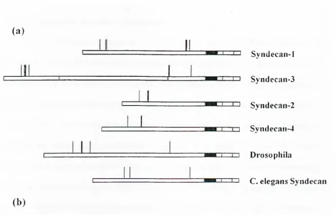
similar exon-intron organization, which supports the idea that the Syndecans arose by gene duplication from a single ancestral gene (Carey 1997). A schematic representation of all Syndecans is shown in Figure 1.1.
1.1.2- Functions of Syndecans
Large number of ligands have been identified that bind to Syndecans at their extracellular domains. These encompass an amazing variety o f molecules, including growth factors, extracellular-matrix proteins, cell-cell adhesion receptors, enzymes and other proteins (Schlessinger et al. 1995). In most cases the specificity of binding of these ligands to Syndecans appears to be relative and not absolute. For example, although bFGF binds to specific sugar sequences within heparan sulphate chains, every syndecan that has been examined appears to contain these sequences and binds bFGF (Lopez Casillas et al. 1993). On the other hand, bFGF has its own receptor bFGFR. These features appear to rule out the notion that Syndecans function as ligand-activated signaling receptors in the usual sense.
According to these notions Syndecans have been thought to function as co receptors that bring extracellular ligands into close proximities to their specific receptors. For a long time these membrane proteins are considered to be fine tuners of ligand-receptors interactions, and they are conceived to be modulators of cell adhesion, cell migration, and cell differentiation.
(a)
J i ^ Svndecan-1 ^ Syndecaii-3 ^ Svndecan-2 □ZD Syiidecan-4 I . .1 I Drosophila (b ) I C. elegans SyndecanSyn-3 KSILERKE VLVAVIVGG WGALFAAFLVTL LIY EMKKKDEGSYTLEEPKQASVT YQ KP DKQEEFYA
Syn-l QGLLDRKE VLGGVIAGG LVGLIFAVCLVAF MLY EMKKKDEGSYSLEEPKQANGG AYQ KP TKQEEFYA
Syn-4 DNLFKRTE VLAAVIAGG VIGFLFAIFLILL LVY EMRKKDEGSYDLGERKPSS AAYQ K APTK EFYA
Syn-2 SNIFERTE VLAALIVGG WGILFAVFLILL LVY RMKKKDEGSYDLG KKPI Y KK APTN EFYA
Dr. SSFFSQPG ILAAVI G G A W G L L C A IL^/"/MFIVY RMRKKDEGSYALDEPKRSPANNSY AKNA NNREFYA
C. e FHETLANG FYAA lAGGVLVAVITA I L L V L F W F RIRKKD-EGSYALDEPKQARPYASYGYTK ASTK EFYA
Figure l . l : S tructure of Syndean core protein
(a) Diagram illustrating the structures of the four vertebrate and two invertebrate Syndecans. The black boxes represent the transmembrane domains. Vertical lines above the boxes indicate positions of attachment o f heparan sulphate chains, (b) Amino acid sequences of syndecan transmembrane and cytoplasmic domains.
The cytoplasmic domains are short but highly conserved. The sequence o f the 13-amino-acid segment immediately following the transmembrane domain is essentially identical in all Syndecans, including the invertebrate forms (Cl region). All syndecan core proteins also contain an identical tetrapeptide sequence at their C-terminal ends (C2 region). The function o f these portions o f the cytoplasmic domain is unknown. Interrupting these highly conserved segments is a variable region that shows considerably less sequence similarity among syndecan types (V region). Interestingly, however, the variable-region sequences are highly conserved for specific Syndecans types in different species. The sequence o f syndecan-1 in this region is identical in human, mouse, rat and hamster; the corresponding region in syndecan-2 is identical in human, rat and Xenopus. Thus, once the four vertebrate syndecan genes diverged during evolution, there was strong selective pressure to maintain their particular cytoplasmic domain primary structures. This, plus the structural diversity of the ectodomains, suggests that different Syndecans have evolved to carry out similar, but non-identical, functions.
The conserved characteristic of cytoplasmic domain of syndecan proteins indicate that these domains have important cellular functions and may bind to different cytoplasmic proteins. These possible interactions may be important for the determination o f cell fate. Most probably Syndecan proteins regulate cell-matrix and cell-cell interactions outside the cell membrane and they also modulate cell signaling by signaling receptors through protein-protein interactions inside the cell via their highly conserved cytoplasmic domains.
N-syndecan (Syndecan-3) was previously isolated as a cell sur&ce receptor for heparin-binding growth-associated molecule (HB-GAM) and suggested to mediate the neurite growth-promoting signal from cell matrix-bound HB-GAM to the cytoskeleton o f neurites. However, it was unclear whether N-syndecan would possess independent signaling capacity in neurite growth or in related cell differentiation phenomena. Later it was reported that Syndecan-3 binds a protein complex containing Src family tyrosine kinases and their substrates and that Syndecan-3 acts as a neurite outgrowth receptor via the Src kinase-cortactin pathway (Kinnunen et al. 1998).
In 1995 it was sho\vn that S)mdecan-4 but not other Syndecans can be phosphorylated by Protein kinase C (PKC) which is an important protein in cell signaling events (Prasthofer et al. 1995, Baciu et al. 1995). It was shown that Syndecan-4 can be phosphorylated by PKC and is possibly a physiological substrate for PKC in cells. The core protein o f S3mdecan-4 can directly bind the catal)4ic domain of PKCalpha and potentiate its activation by phospholipid mediators. It can also directly activate PKCalpha in the absence o f other mediators. This activity resides in the sequence LGKKPIYKK in the center o f the short cytoplasmic domain, and other Syndecans lack this sequence and PKC regulatory properties. Syndecan-4 is a focal adhesion component, and this interaction may both localize PKC and amplify its activity at sites o f forming adhesions (Oh et al. 1997). This was the first report suggesting direct transmembrane signaling through cell surface proteoglycans.
Another novel cellular protein was identified that interacts with the cytoplasmic domain o f syndecan-4 but not with those o f the other three syndecan family members. The interaction involves both the membrane proximal and variable central regions ofthe
cytoplasmic domain. The protein is named as syndesmos. Syndesmos is ubiquitously expressed and can be myristylated. Consistent with its myristylation and syndecan-4 association, syndesmos colocalizes with syndecan-4 in the ventral plasma membranes of cells plated on fibronectin (Baciu et al. 2000). When overexpressed in NIH 3T3 cells, syndesmos enhances cell spreading, actin stress fiber and focal contact formation in a serum-independent manner.
A third protein which was identified by yeast two-hybrid screening that binds to the cytoplasmic domains o f the S5mdecans, is syntenin (Grootjans et al. 1997). It contains a tandem repeat o f PDZ domains that reacts with the FYA C-terminal amino acid sequence o f the Syndecans. Cells that overexpress eGFP-syntenin show numerous cell sürfece extensions, suggesting effects o f syntenin on cytoskeleton-membrane organization. It may be proposed that syntenin may function as an adaptor that couples Syndecans to cytoskeletal proteins or cytosolic downstream signal-effectors. This study was the first report suggesting PDZ proteins as interaction counterparts o f Syndecan proteins.
CASK, the rat homolog o f a gene (LIN-2) required for vulval differentiation in Caenorhabditis elegans, is expressed in mammalian brain, but its function in neurons is unknown (Hata et al. 1996). It is another member of PDZ protein family. Again by yeast two-hybrid screening, a specific interaction was identified between the PDZ domain of CASK and the COOH terminal tail of syndecan-2, a cell surface heparan sulphate proteoglycan (HSPG). The interaction was confirmed by coimmunoprecipitation from heterologous cells (Hsueh et al. 1998). In brain, syndecan-2 localizes specifically at synaptic junctions where it shows overlapping distribution with CASK, consistent with an interaction between these proteins in synapses. Cell surface HSPGs can bind to
extracellular matrix proteins, and are required for the action o f various heparin-binding polypeptide growth/dififerentiation factors. The synaptic localization o f CASK and syndecan suggests a potential role for these proteins in adhesion and signaling at neuronal synapses (Cohen etal. 1998).
All these interactions are indications of important cellular functions of Syndecan proteins other than their co-receptor functions. Some of the possible functions of such interactions may be association of focal adhesion complexes at cell to cell junctions, localizing membrane proteins or signaling molecules at specific membrane loci by communication with cytoskeletal proteins, or regulation o f special cellular complexes. Some possible signaling pathways are Wnt-frizzled pathway, and CASK-reelin pathway which are summarized in the proceeding parts of this thesis.
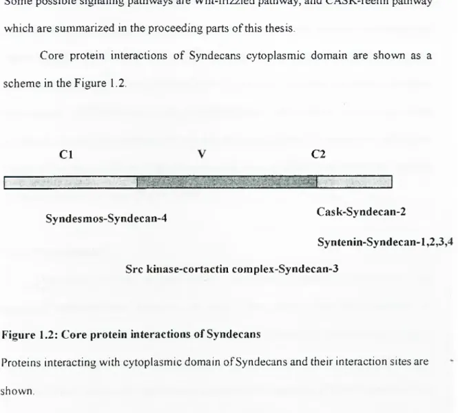
Core protein interactions o f Syndecans cytoplasmic domain are shown as a scheme in the Figure 1.2.
C l C2
Syndesmos-Syndecan-4 Cask-Syndecan-2
Syntenin-Syndecan-1,2,3,4 Src kinase-cortactin complex-Syndecan-3
Figure 1.2: Core protein interactions of Syndecans
Proteins interacting with cytoplasmic domain of Syndecans and their interaction sites are shown.
Wnt proteins are large family o f cystein rich glycoproteins that ftinction as ligands controlling development in various organisms ranging from nematode worms to mammals. At the cellular level Wnt regulates cell proliferation, cell morphology, cell motility, and cell fete. Wnt proteins serve as short range chemical messengers, paracrine agents. Wnt proteins are associated with cell sürfece and extracellular matrix, such that their effects tend to be spatially localized. The first mammalian Wnt gene, originally termed Int-1 was identified at a site o f murine mammary tumor virus (MMTV) integration in mammary tumors (Burrus et al. 1995). The Int-1 gene exhibited homology to wingless [wg], a Drosophila segment polarity gene, and subsequently the name Wnt was chosen for members o f this femily. At least sixteen mammalian Wnt genes have been identified so far. Wnt proteins elicit a variety o f cellular responses, including proliferation, differentiation, and morphogenesis, and control of developmental decisions such as axis formation in Xenopus embryos. Indeed, Wnts are now recognized as one of the major classes o f signaling proteins that regulate development and cell fate in multicellular organisms (Peifer 1996).
1.2- WNT-FRIZZLED PATHWAY
1.2.1- Wnt signaling
The action o f Wnts on target cells is mediated by binding to the frizzled (Fz) group of transmembrane receptors, o f which there are at least eight members in mammals. Structurally, Frizzled receptors have an extracellular Wnt-binding domain, seven-transmembrane-spanning sequences and an intracellular C-terminal tail (Bhanot et al. 1996). They form part of the seven-transmembrane-spanning (7TMS) superfamily o f
receptors. The regulation o f Wnt binding to Frizzled receptors is a key restriction point in Wnt signaling. The immediate downstream component o f the signal transduction pathway is disheveled (Dsh), an intracellular protein that may directly interact with Fz receptors. Members o f the Dishevelled family encode cytoplasmic proteins with no known enzymatic functions (Tsang et al. 1996). Sequence comparisons identified three regions o f homology: an N-terminal Dsh homology domain, a central region containing a basic and a PDZ domain, and a DEP domain upstream o f the non-conserved C- terminal sequence. The target o f Dsh is glycogen synthase kinase-3 (GSK-3), which is homologous to the Drosophila protein encoded by zeste-white3. GSK-3 is believed to be constitutively active and functions to inhibit the Wnt pathway in the absence o f Wnt signals. GSK-3 phosphorylâtes p-catenin, which enhances p-catenin turnover by a ubiquitin-mediated degradation pathway. P-Catenin is a member o f a multigene family o f proteins characterized by the presence o f 'Arm' amino acid repeats that mediate a range o f protein-protein interactions. Genetic analysis o f the Drosophila homologue Armadillo (Arm), showed that it functions downstream o f GSK-3/zw3. Considerable evidence suggests that the Wnt inhibition of GSK-3 activity prevents the turnover o f P- catenin, leading to its accumulation. In response to Wnt signals, GSK-3 activity is inhibited, leading to stabilization of the P-catenin protein. One o f the targets o f P-catenin is the transcription fector TCF, an HMG-box DNA-binding protein. Multiple TCF family members exist in mammals, including TCF-1, LEF-1, TCF-3, and TCF-4. P- Catenin forms a complex with TCF, converting it from a transcriptional repressor to an activator and thereby stimulating the expression o f target genes (Behrens et al. 1996). Although the genes in mammalian cells that are activated by TCFs in response to Wnt
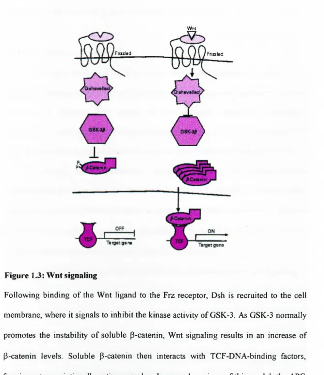
signaling are not well characterized, the c-myc proto-oncogene is one target that may mediate proliferative responses to Wnt signals (He et al. 1998). In several versions of this model, the APC (adenomatous polyposis coli) gene product has been proposed to function as a signaling intermediate between GSK-3 and ß-catenin. In this context, the finding that APC can bind both ß-catenin and GSK-3 suggested that APC may function as a signaling scaffold (Cadigan & Nusse 1997, Siegfried et al. 1994). A schematic representation o f Wnt signaling is shown in Figure 1.3.
1.2.2- Ligand binding
The W nt-binding domain' found at the N-terminus o f Frizzled femily members was recently identified in genes o f the Frzb family and in a splicing variant of collagen XVIH. The complexity of receptor-ligand specificity may be further increased by co receptors such as proteoglycans and other putative 'Wnt receptors'. Proteoglycans consist o f proteins linked to chains o f disaccharide repeats called glycosaminoglycans. Proteoglycans are highly charged and are found predominantly at the cell surface, where they function as low-affinity cell-surface receptors for a variety of ligands, including TGF-ß and FGF. Cell-culture studies showed that many W nt family members bind glycosaminoglycans (Reichsman et al. 1996) and that cell-surface glycosaminoglycans are required for Wg stabilization o f Arm in Drosophila Cl-8 cells. Cell-surface proteoglycans may increase the local concentration o f Wnts, leading to increased avidity and/or receptor clustering, as has been proposed in models for FGF-glycosaminoglycan interactions. Thus the loss o f heparin-like glycosaminoglycans may allow Wnt proteins to abnormally diffuse and reach a concentration at which they are no longer effective.
Wnt
Frizzled Frizzled
Figure 1.3: Wnt signaling
Following binding o f the Wnt ligand to the Frz receptor, Dsh is recruited to the cell membrane, where it signals to inhibit the kinase activity of GSK-3. As GSK-3 normally promotes the instability o f soluble p-catenin, Wnt signaling results in an increase of P-catenin levels. Soluble P-catenin then interacts with TCF-DNA-binding factors, forming a transcriptionally active complex. In several versions o f this model, the APC (adenomatous polyposis coli) gene product has been proposed to function as a signaling intermediate between GSK-3 and p-catenin (Dale T. C. Biochem J. 1998).
Alternatively, the ability o f W nt-Frizzled complexes to form or transduce their signals may be directly regulated by glycosaminoglycan interactions.
1.2.3- p-catenin vs. E-cadherin
P-Catenin encodes a 90 IcDa protein with several structural domains. The N- terminus contains the XGSK-3 phosphorylation sites. The central domain o f the protein contains 13 imperfect 'Arm' repeats that fold to make a superhelix containing a positively charged groove that is hypothesized to interact with acidic regions o f APC, TCF transcription factors and cadherin cell adhesion molecules (Huber et al. 1997).
Cadherins are Ca^^-dependent adhesion molecules that mediate cell-cell interactions at adhesive junctions. The intracellular domain o f cadherins interact with the cytoplasmic adaptor proteins p-catenin or plakoglobin. While complexed to cadherins, P-catenin is also able to interact with a-catenin, a cytoplasmic protein with similarity to vinculin, which links actin filaments to the adherens junctions.
Levels o f cadherin expression affected free p-catenin pools and signaling, in Xenopus embryos, ES cells and Drosophila embryos. However, as E-cadherin-deficient L-cells were able to respond to Wnt-1 by stabilizing P-catenin, it is unlikely that the reductions in E-cadherin expression were sufficient or were required for Wnt signaling. Cell adhesion may be a downstream target of Wnt signaling as the adhesive functions o f cadherins are regulated by interactions with P-catenin.
E-cadherin and lymphocyte-enhancer factor-1 (LEF-1) form mutually exclusive complexes with beta-catenin; the association of beta-eaten in with LEF-1 compete by the E-cadherin cytoplasmic domain. Similarly, LEF-1 and adenomatous polyposis coli
(APC) form separate, mutually exclusive complexes with beta-catenin. The potent ability o f E-cadherin to recruit P-catenin to the cell membrane and prevent its nuclear localization and transactivation is demonstrated using SW480 colon carcinoma cells (Orsulic et al. 1999).
1.2.4- Wnt signaling vs. Syndecan-1
Syndecans bind to various different ligands including oncogenic proteins. But there were no direct evidence that Syndecans are involved in tumorigenesis. In a recent publication it was reported that they may have role in mammary tumor formation caused by ectopic Wnt-1 proto-oncogene. In this study, S)mdecan-1-deficient mouse and transgenic mouse that express W ntl in mammary gland are crossed. Ectopic Wnt-1 expression is known to induce generalized mammary hyperplasia, followed by the development o f solitary tumors (Ramakrishna et al. 1993). It is shown that in Sdcl-/- mice, Wnt-1-induced hyperplasia in virgin mammary gland was reduced by 70%, indicating that the Wnt-1 signaling pathway was inhibited. These results provide both genetic and biochemical evidence that syndecan-1 can modulate Wnt signaling, and is critical for Wnt-1-induced tumorigenesis o f the mouse mammary gland (Alexander et al. 2000).
CASK is identified as a neurexin, a neuronal cell surface protein binding counterpart in synaptic junctions o f neurons (Hata et al. 1996). It shows a great homology to Drosophila PSD95 protein. Later a C. elegans homolog o f CASK LIN-2 is identified which co-localizes with EOF receptors in cell membranes (Kaech et al. 1998). It belongs to Membrane Associated Guanylate Kinase (MAGUK) protein family. Members o f this protein family all share some characteristics like being composed o f multiple protein domain such that C terminal calcium/calmodulin binding domain,SH3 domains and guanylate kinase domains. Guanylate kinase domain indicates that these proteins may bind to ATP through these regions and display kinase activities. Yet, no significant kinase activity could be shown to guanylate kinase domain of CASK.
MAGUK proteins are possibly involved in localization of multi protein complexes to cell sürfece proteins. These protein complexes may be involved in signal transduction events and are probably regulated by these scaffold proteins.
CASK is a membrane-associated protein that contains domains found in Ca^^ activated protein kinases and in proteins specific for intercellular junctions, suggesting that it may be a signaling molecule operating at the plasma membrane, possibly in conjunction with neurexins.
1.3.1- CASK and Syndecans
Another important domain o f CASK is its PDZ like domain showing a similarity with drosophila PSD95 protein. This domain is also thought to be involved in protein- protein interactions o f CASK. Later, CASK was reported to interact directly with
1.3- CASK-TBR-1 PATHWAY
Syndecan-2 protein via its cytoplasmic domain through this PDZ domain. It is hypothesized that PDZ domain of CASK may interact with all other Syndecan proteins via their carboxy terminal EFYA sequence. In brain, Syndecan-2 localizes specifically at synaptic junctions where it shows overlapping distribution with CASK, consistent with an interaction between these proteins in synapses.
It is logical to think that other members o f Syndecan family of proteins are also interacting with CASK through the same domain. Yet no such study is reported indicating that CASK is interacting with a syndecan protein other than Syndecan-2. Cell surface HSPGs can bind to extracellular matrix proteins, and are required for the action o f various heparin-binding polypeptide growth/dififerentiation factors. The s)maptic localization of CASK and sjmdecan suggests a potential role for these proteins in adhesion and signaling at neuronal synapses.
1.3.2- CASK in nucleus
A recent study reported that CASK interacts with a T-box protein (Tbr-1) which is a transcription factor. Tbr-1 binds DNA at a specific sequence (T-box) and activating the transcription of a protein named reelin (Hsueh et al. 2000). The interaction is through c a s k’s mysterious guanylate kinase domain. This was the first function attributed to guanylate kinase domain of MAGUK proteins. However the interaction was specific to CASK alone and other MAGUK proteins did not show any interaction with Tbr-1 protein.
The paradoxical finding of interaction o f a membrane associated MAGUK protein CASK, interacting with a transcription factor which normally resides in nucleus is solved when it is demonstrated that CASK localizes to nucleus in Tbr-1 over
expressing cells. It was also shown that CASK activates the transcriptional activating function of Tbr-I protein through this interaction.
Tbr-I is a T-box transcription factor involved in forebrain development. CASK enters the nucleus and binds to a specific DNA sequence (the T-element) in a complex with Tbr-I. CASK acts as a coactivator of Tbr-I to induce transcription o f T-element containing genes, including reelin, a gene that is essential for cerebrocortical development. This findings show that a MAGUK which is usually associated with cell junctions has a transcription regulation function.
Another important finding is regulation of this pathway by syndecan expression. Syndecan-3 over expression diminished the translocation of CASK to nucleus. This type o f regulation can be observed in Wnt pathway where P-catenin translocation to nucleus is regulated by E-cadherin expression. Probably in CASK pathway Syndecans have similar functions to E-cadherin functions in Wnt pathway. S)mdecans are known to have important functions in developmental processes. This phenomenon may be a good example how these membrane glycoproteins are involved in developmental events.
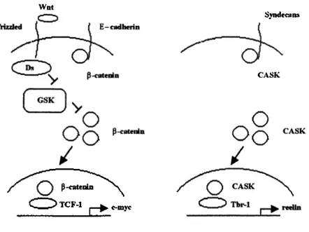
CASK pathway resembles Wnt pathway in many ways indicating that both may have similar components and are regulated by similar mechanisms (Bredt 2000). A comparative diagram o f both pathway is presented in Figure 1.4.
Wat P-catenin TCF-1 SyndecaiM CASK c-myc
Figure 1.4: Wnt pathway and CASK pathway
Wnt pathway is shown as a diagram in the left picture. The CASK pathway is shown in the right picture. The involvement o f P-catenin in the pathway resembles the involvement o f CASK in the other pathway. The other components o f the CASK pathway are not characterized yet, but may have similar characteristics to Wnt pathway.
Many cytosolic signaling proteins and cytoskeletal proteins are composed o f modular units o f small protein-protein interaction domains like SH2 domains, SH3 domains or PDZ domains. As the name implies, PDZ domain have high homology with the drosophila PSD95 proteins, disc-large gene encoded Dig protein, and ZO-1 protein. The first mammalian homolog identified was p55 protein which show a silent G/T polymorphism. PDZ domain containing proteins bind directly to the carboxy terminal residues o f transmambrane proteins (RufFet al. 1996). For example PSD95 and Dig bind to similar carboxy terminal sequences on Shaker type K ' channels (Kim et al. 1995). So PDZ domain proteins are thought to be necessary for clustering of transmembrane proteins to specific locations in cells.
In a peptide library screening assay it was identified that there is a consensus peptide sequence for PDZ domain binding (Songyang et al. 1997). In this consensus it was identified that PDZ domains of most of the proteins tend to bind to a hydrophobic amino acid at the carboxy terminal end. It was concluded that an internal consensus sequence can not bind to PDZ domains even they match perfectly with the obtained consensus sequence. However the most important finding in this study is the PDZ domains can be divided into two major groups according to their interaction partner specificity. According to this finding it was shown that type 11 PDZ domains which are the PDZ domains of p55, CASK, Tiam-1, and also AF-6 prefer a hydrophobic residue especially a phenylalanine residue at the -2 location from the carboxy terminal end. This was against the general belief that PDZ domains préféra hydroxyl group aminoacid like serine, threonine, and tyrosine at this specific location. The optimal residues predicted
1.4- PDZ DOMAIN PROTEINS
by peptide library screening can be rationalized on the basis of crystal structure studies performed to identify PDZ domain binding (Doyle et al. 1996).
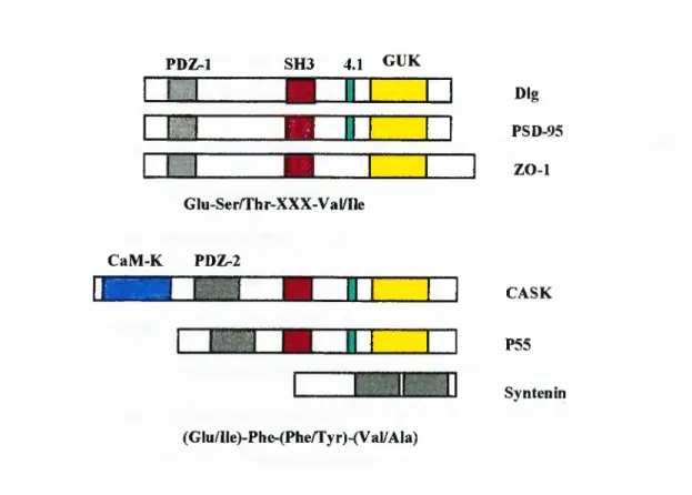
Structures and binding consensus sequences of some PDZ domain proteins are shown in Figure 1.5. PDZ-1 SH3 4.1 GUK Z B m 1 1 IBB Dig PSD-95 ZO-1 GIu-Ser/Thr-XXX-Val/Ile CaM-K PDZ-2 & 'TS P « |'''
E
3 CASK P55 Syntenin (Glu/Ile)-Phe-(Phe/Tyr)-(Val/Ala)Figure 1.5: Binding consensus sequences o f some PDZ domain proteins
Domain structures o f some PDZ domain proteins are shown in the figures. CaM-K: Calcium/Calmodulin kinase. 4.1: 4.1 protein binding domain. GUK: Guanylate kinase homology domain. Type I PDZ domains bind to a C Terminal consensus sequence o f Glu-Ser/Thr-XXX-Val/Ile, whereas Type II PDZ domain proteins bind to a C Terminal consensus sequence (Glu/Ile)-Phe-(Phe/Tyr)-(Val/Ala) that match perfectly to C Terminal amino acid sequences of Syndecan proteins.
AF-6 protein was identified and cloned as a proto-oncogene in acute myeloid leukemia (AML). It was identified to be the fusion partner protein o f ALL-1 protein caused by a chromosomal translocation [t(6,l)(q23,q27)](Prasad et al. 1993, Saito et al. 1998). It was observed that this protein contains shared motifs with proteins involved in signal transduction, namely PDZ motifs. It was also mapped to minimal deletion region in epithelial ovarian cancer but it was shown that AF-6 sequences maps distal to these region (Saha et al. 1995). Later drosophila, C. elegans, and mouse homolog of AF-6 were identified and named as canoe, Ce-AF-6, and afadin respectively (Kuriyama et al. 1996, Watari et al. 1998, Mandai et al. 1997). Canoe is an important protein for drosophila development and is involved in Notch signaling (Miyamoto et al. 1995).
AF-6 contains a putative Ras binding domain, which shows great homology to Raf-Ras activating domain. It was shown to bind to Ras protein and another PDZ domain protein, ZO-1 in vivo. Unlike p-catenin, AF-6 is localized to tight junctions of epithelial cells and ZO-1 is known to be an important component o f this type of cell-cell junctions (Yamamoto et al. 1997). Ras binding property o f AF-6 is regulated with its ZO-1 interaction and activated Ras disturbs AF-6 localization to tight junctions. Its localization at junctional cell-cell contact sites is also confirmed by showing AF-6 interaction with JAM (Junctional Adhesion Molecule) (Ebnet et al. 2000).
AF-6 was shown to bind to GTP-activated Ras but not to mutant Ras which lacks Its GTP binding domain. All these data indicate that AF-6 is a potential regulator of Ras
1.5- AF-6 AS A PDZ DOMAIN PROTEIN
pathway which is an important pathway for determination of cell fate (Yamamoto et al. 1999).
AF-6 was also shown to bind to cell surface receptor tyrosine kinases (RTK) epinephrine receptors, EphB3 via their PDZ like domain. Their interaction with Eph receptors is regulated by the phosphorylation o f the receptors. It was shown that they are phosphorylated in vivo upon binding to epinephrine receptors. Receptor tyrosine kinases are important integral membrane proteins involved in signal transduction processes. This result also show that PDZ like domains modulate signal transduction like other protein- protein interaction domains SH3 and PTB (Hock et al. 1998).
A very important finding about AF-6 came only recently reporting that AF-6 co-localizes and interacts with deubiquitinating enzyme FAM, the human homolog o f drosophila faf protein (Taya et al. 1998, Kanai-Azuma et al. 2000). Drosophila homolog o f FAM, faf (fat facets) is known to be an important protein in developmental processes. This enzyme regulates the signal transduction events by regulating libiquitination of some proteins. FAM and faf probably are involved in stabilization o f proteins by inhibiting the ubiquitination and therefore degradation. The importance of ubiquitination pathway in signaling events is reported for Wnt pathway. It is shown that P-catenin is stabilized by deubiquitination enzyme FAM and it can be protected from GSK induced degradation (Taya et al. 1999). This may be a potential pathway by which these type of signaling events are modulated. This shows that AF-6 is also regulated by a similar pathway.
1 .6 -H Y PO TH ESIS
PDZ domain containing proteins play an important role in signaling events as cytoplasmic modulators o f signaling pathways. They all localize to specific peripheral membrane locations like post-synaptic complexes, cell to cell adhesion locations, and tight junctions via protein-protein interactions of their PDZ domains. It is known that they bind to carboxy terminal o f different integral membrane proteins via these domains. They are thought to modulate the formation of such complexes and localize integral membrane proteins to such locations. Many important PDZ domain containing proteins are known to involve in signaling events, Dsh, PSD95 and AF-6 are some o f them.
Syndecans are integral membrane proteoglycans that are involved in signaling events. They interact with various cellular proteins via their core proteins. Some of these proteins are PKC and CASK. We hypothesize that, Syndecan proteins which are known to bind to two PDZ domain containimi proteins (CASK and syntenin) may bind to other similar proteins such as Dsh and AF-6, since their consensus binding sequence match perfectly with the carboxy terminal o f Syndecan family of proteins (EFYA).
It was recently reported that Syndecan-1 is involved in Wnt induced tumorigenicity in mammary cancer (Alexander et al. 2000). This report was the first finding suggesting that a Syndecan protein is involved in tumorigenesis. It is obvious that core protein interactions of Syndecan-1 may be important in this and other possible functions o f Syndecan-1. CASK is a recently identified transcriptional activator (Hsueh et al. 2000). Although terminal cytoplasmic region of Syndecan-2, which is responsible for the CASK-PDZ domain interaction is identical to the same region of Syndecan-1 there is no direct evidence yet, that Syndecan-1 binds to CASK protein. AF-6 is shown
there is no direct evidence yet, that Syndecan-1 binds to CASK protein. AF-6 is shown to localize to nucleus as a fusion partner of MLL gene product but the importance o f this localization is not identified yet (Joh et al. 1997). AF-6 protein has type IIPD Z domain like CASK and syntenin which is the Syndecan-2 protein interacting domain. According to our hypothesis that all Syndecan proteins bind to type II PDZ domain containing proteins, we predicted that Syndecan-1 interacts with PDZ domains o f CASK and AF-6 through its cytoplasmic domain. To test this hypothesis we used Yeast two-hybrid assay to identify Syndecan-1 cytoplasmic domain with PDZ domains o f CASK and AF-6.
2- MATERIALS AND METHODS
2.1- Recombinant DNA manipulation techniques
2.1.1- Polymerase Chain Reaction (PCR)
Syndecan-1 cytoplasmic domain, CASK-PDZ domain, and AF-6-PDZ domain were amplified by polymerase chain reaction using Human Fetal Brain cDNA as template.
A standard 100 ul reaction set up in a 0.2 ml PCR tube
primers 0.1-1 uM of each primer (reverse and forward) dNTP 0.2 mM diluted from the 10 mM stock
buffer IX diluted from the lOX stock template 10 ng of genomic or plasmid DNA Made up to 100 ul with MilliQ water
enzyme 2.5 unit of Taq DNA polymerase
Tubes were placed on ice. To perform a hot-start PCR the thermal cycler was preheated to approximately 90°C before placing the tubes into it. Reaction conditions were as follows: Dénaturation: 30 seconds at 94 "C, Annealing: 30 seconds at 60 "C, Extension: 30 seconds at 72 ”C, 35 cycles.
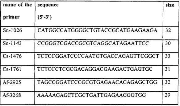
List o f the synthetic oligonucleotide primer sets used in this study are summarized in Table 2 ,1.
Table 2.1: List of the synthetic oligonucleotide primer sets used in this study
name o f the primer sequence (5’-3’) size Sn-1026 CATGGCCATGGGGCTGTACCGCATGAAGAAGA 32 Sn-1143 CCGGGTCGACCGCGTCAGGCATAGAATTCC 30 Cs-1476 TCTCCGGATCCCCAATGTGACCAGAGTTCGGCT 33 Cs-1761 TCTCCCTCGCGACAGGACGAAGACTGAGTGC 31 Af-2925 TAGCCGGATCCCGCGTGAGAACACAGAGCTGG 32 Af-3268 AAAAAGAGCTCGCTGATTGAGAAGGGTGG 29 2.1.2- Purification o f DNA
Purification o f PCR products and linearized plasmids were done either as described below or by the MBI DNA extraction kit (#K0513) according to the manufacturer’s instructions.
PCR products were purified by precipitating the DNA with ammonium acetate. 1/10 volume o f lOX STE buffer, equal volume o f 4M ammonium acetate and 2.5 volume o f 100% (v/v) ethanol were added to the PCR products sequentially. The reaction tube was centrifuged at 13000 rpm for 20 minutes at room temperature to pellet DNA. The supernatant was removed carefully and discarded. The pellet was washed
with 200 111 o f 70% (v/v) ice-cold ethanol. The pellet was dried and DNA was resuspended by using TE buffer.
2.1.3- Restriction enzymes
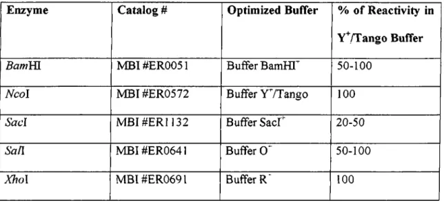
DNA restriction reactions were carried out in a total volume of20-100 ul with 5- 10 units of restriction enzyme, lul of boiled RnaseA (Img/ml) was used where appropriate. The volume o f the reaction buffers were arranged to be 1X and the enzyme volume was never greater than 1/10th of the reaction volume so as to prevent star activity due to the high glycerol concentration. Restriction enzymes used in this study are shown in Table 2.2.
Table 2.2: Restriction enzymes used in this study
Enzyme Catalog # Optimized Buffer % o f Reactivity in
V^/Tango Buffer
5a/wHI MBI#ER0051 Buffer BamHI'^ 50-100
Ncol MBI#ER0572 Buffer YVTango 100
S a d M BI#ER1132 Buffer Sacr 20-50
Sail MBI#ER0641 Buffer O' 50-100
Xho\ MBl#ER0691 Buffer R' 100
2.1.4- DNA ligation
DNA fragments, that were cut from agarose gel and recovered by MBl DNA extraction kit (#K 0513) were used for ligation. For cloning (where both vector and insert DNA has protruding ends) approximately 1 ;4 ratio o f vector and insert DNA was mixed with the T4 DNA ligase buffer and 1 to 3 units of T4 DNA ligase (MBI #EL 0011). The reaction was carried out either for 2 hours at room temperature or for 16 hours at 16°C. For protruding end ligation insert;vector ratio could be chosen as 1/1,2/1, or 3/1. The reaction was stopped directly by heating at 65°C for 10 minutes or directly used to transform competent bacterial cells.
2.1.5- C ulturing and handling bacteria 2.1.5.1- B acterial strains
Strains oiE.coli used in this study are summarized in Table 2.3. T able 2.3: Strains of E.coli used in this study
Strain genotype usage reference
D H 5a supEAA A/ncU169
{i^macZAM 15) hs(iR\ 1 recA 1 endA\ gyrA96 thi-\ relAl
Host for plasmid DNA
H anahan(1983)
H B lO l supE44 hsdS20{ryi mn') recA 13 ara- \4 proA2 lacYl galK2 rpsLlO xyl-5
mtl-\
Host for plasmid DNA Boyer and Rouland- Dussoix (1969); Bolivar an Backman (1979) JM 109 ? [iralB6 lacf<\ lacZAM\5
pro A H ] recA 1 end A1 gyrA 96 {NaEj thi hsdR17(r/^m·*·/;;) supE44 relA 1 ihi A{lac-proA/i)
Host for plasmid DNA
Yanisch-Perron et al. (1985)
2 .1.5.2- Storage of bacteria
Strains o f E.coli were stored in glycerol (long term storage). Glycerol cultures of E.coli were prepared by adding 0.81 ml o f fresh culture to 0.91 ml o f sterile 80% glycerol in a sterile screw capped tube. The tubes were vortexed and were then frozen and stored at -70°C.
2.1.5.3- Growth of E. coli strains
All strains were grown in LB medium or on LA medium supplemented with the appropriate antibiotics.
Solid and liquid mediums:
LB medium : 0.5% Yeast extract, 1% Bacto-tiyptone, 1% NaCl.
LA medium ; 0.5% Yeast extract, 1% Bacto-tryptone, 1% NaCl and 1% agar. Supplemented with appropriate antibiotics.
Antibiotics:
Ampicillin with a final concentration of 50 ug/ml was used in this study. 2.1.5.4- Preparation o f competent bacteria
Cells were made competent using a modification o f the CaCl2 method described by Maniatis et al., 1982.
5 ml LB was inoculated using a single colony from a freshly grown plate o f the li. coli strain to be used, and was incubated at 37^C for approximately 2 hours, until the ODeoo of the culture is 0.3-0.4. The culture was then cooled on ice for 5 minutes, 1 ml aliquots were added to microcentrifuge tubes and then the cells were pelleted by centrifugation (1 minute at 13,000 rpm ). The cells were resuspended in 0.5 ml of 50 mM
CaCh
by gentle vortexing, before being placed on ice for 30 minutes. The cellswere pelleted by centrifugation (1 minute at 13,000 rpm ), and the supernatant was discarded. The pellet was resuspended in 0.1 ml o f C aC b with gentle vortexing. The cells were then stored on ice until required for transformation. Alternatively competent cells were prepared and stored at -70°C until required. 500 ml of LB was seeded with a 10 ml o f overnight culture and grown to an OD5 0 0 - 0-6. Cells were harvested by
centrifugation at 5000 rpm for 10 minutes at 4®C, before being incubated on ice for 20 minutes. The cells were harvested as before, resuspended in 25 ml of 50 mM C aC b /20% glycerol and aliquoted into microcentrifuge tubes before being frozen. Samples were stored at -70°C and were viable for at least 2 months. Cells were thawed on ice prior to the addition o f DNA.
2.1.5.5- Transformation of plasmid DNA in bacterial cells
The DNA to be transformed (usually a 1 ul of ligation mixture or approximately 100 ng o f plasmid DNA) was added to the 100 ul of competent cells, mixed gently and incubated on ice for 30 minutes. The cells were then heat-shocked at 42^C for 90 seconds and chilled by placing on ice for 2 minutes. 1 ml o f pre-warmed LB was then added and the suspension was incubated at 37°C for 1 hour. Each sample was pelleted by centrifugation at 13,000 rpm for 2 minutes, resuspended in 100-200 ul o f LB and plated onto selective medium and incubated overnight at 37^C to allow the growth of the transformants.
This protocol is based on the alkaline lysis method o f Birnboim and Doly (1979).
The transformed bacterial strain containing the plasmid o f interest was grown at 37®C overnight in 5 ml of LB+antibiotic. 1.5 ml o f the bacterial culture was pelleted by centrifugation for 1 minute (bench-top microfuge, 13,000 rpm) in a 1.5 ml microfuge tube. After the removal of the supernatant, the cells were resuspended in 0.1 ml of ice- cold solution I and stored for 5 minutes at room temperature. 0.2 ml o f solution II was mixed by inversion, the tube was then stored on ice for 5 minutes. Bacterial chromosomal DNA and proteins were precipitated by the addition o f 0.15 ml of ice-cold solution HI. The mixture was left on ice for 5 minutes, then centrifuged at 13000 rpm in a bench-top centrifuge for 5 minutes to pellet the host DNA and proteins. The supernatant was mixed with an equal volume o f phenol-chloroform (1:1) and centrifuged in a bench-top microfuge at 13000 rpm for 3 minutes to separate the two phases. The top phase was removed and plasmid DNA was precipitated by mixing it with 2.5 volumes o f 95% ethanol, and pelleted by centrifugation for 10 minutes (bench-top microfuge, 13,000 rpm) after keeping the mixture at 4°C for 15 minutes. The supernatant was discarded and the pellet was left for 15-20 minutes at room temperature to dry and then resuspended in 20-30 ul of TE buffer containing 10 ug/ml RNaseA. Samples were stored at 4°C.
2.1.6- Isolation o f plasmid DNA from bacteria
Solution I
50 mM glucose
25 mM Tris Cl (pH 8.0) 10 mM EDTA (pH 8.0)
Solution I can be prepared in batches o f approximately 100 ml, autoclaved for 15 minutes at 10 Ib/square in. on liquid cycle, and stored at 4°C.
Solution II
0.2 NaOH (freshly diluted from 10 N stock) 1% SD S
Solution III
5 M potassium acetate 60 ml glacial acetic acid 11.5m l
H2 O 28.5 ml
The resulting solution is 3 M with respect to potassium and 5 M with respect to acetate. TE buffer
pH 7.4 lOmMTris Cl (pH 7.4) Im M ED TA (pH 8.0)
2.1.6.2- Medium scale purification of plasmid DNA
All midi-preparations were carried out by using the kit supplied by Macherey- Nagel (cat # 740 573) according to the manufacturer’s instructions.
The bacterial strain containing the plasmid of interest was first grown in a 30 ml culture with necessary antibiotic until late log phase (OD5 0 0 o f 0 .6 ) then inoculated into 500 ml LB medium for 10-12 hours. The cells were harvested by centrifugation at 4000 rpm for 15 minutes at 4°C in a Sorvall GS3 rotor (or equivalent). The supernatant was discarded and allowed to drain away in a upside down position.
2.1.6.3.I- Lysis by alkali
This protocol is based on the alkaline lysis method ofB im boim and Doly (1979) and the solutions were described above in the mini preparation section. The bacterial pellet was resuspended and washed in 18 ml solution I.
40 ml o f freshly prepared solution II was added, the lid o f centrifuge tube was closed and the bottle was inverted several times. The bottle was kept at room temperature for 5-10 minutes.
20 ml o f ice cold solution IH was added, the lid o f the bottle was closed and shaked several times. The bottle was stored on ice for 10 minutes.
The cell lysate was centrifuged at 4000 rpm for 15 minutes at 40C. The rotor was stopped without braking.
The supernatant was filtered through 4 layers of cheesecloth into a 250 ml centrifuge bottle. 0.6 volume o f isopropanol was added and mixed well, in order to precipitate the nucleic acids. The bottle was kept at room temperature for 10 minutes.
Nucleic acids were recovered by centrifugation at 5000 rpm for 15 minutes at room temperature in a Sorvall GS3 rotor (or equivalent). Salt may precipitate if the centrifugation is performed at 4°C. The supernatant was poured off gently and the bottle
2.1.6.3- Large scale purification o f plasmid DNA
bottle was inverted so as to allow all fluid to drain away. The pellet and the walls o f the bottle was washed with 70% ethanol at room temperature. After draining off ethanol, by the help of a Pasteur pipette attached to vacuum, beads o f liquid that were attached to the walls of the bottle were removed. The inaccessible ethanol was left to evaporate at room temperature. The pellet was dissolved in 3 ml of TE (pH 8.0).
2.1.7- Quantification o f double stranded DNA
The amount o f DNA in samples were determined by reading the absorbency of the samples at 260 nm. An OD260 o f 1 corresponds to a concentration o f 50 ug/ml for double stranded DNA, 40 ug/ml for single stranded DNA and 20 ug/ml for oligonucleotides (Maniatis e ta l, 1982).
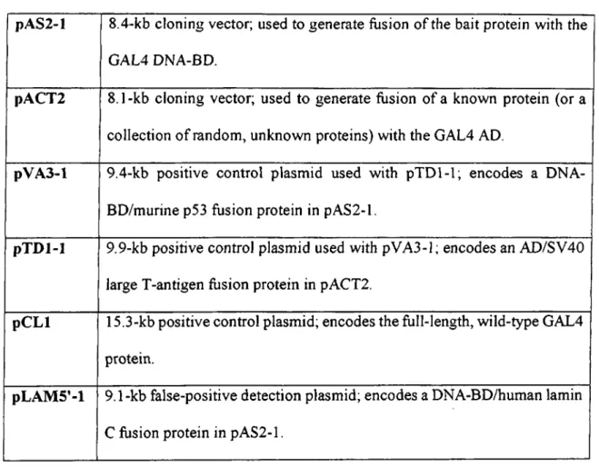
2.1.8- Plasmids
Plasmids used in this study are listed in Table 2.4. All plasmids are yeast vectors designed for expression o f exogenous genes in yeast. They also contain P-lactamase gene serving as ampicillin resistance gene in order to amplify and store them in bacteria.
Table 2.4: List of Plasmids used in this study
pA S2-l 8.4-kb cloning vector; used to generate fusion o f the bait protein with the GAL4 DNA-BD.
pACT2 8.1-kb cloning vector; used to generate fusion of a known protein (or a collection o f random, unknown proteins) with the GAL4 AD.
pV A 3-l 9.4-kb positive control plasmid used with pTD l-1; encodes a DNA- BD/murine p53 fusion protein in pAS2-l.
pT D l-1 9.9-kb positive control plasmid used with pVA3-l; encodes an AD/SV40 large T-antigen fusion protein in pACT2.
p C L l 15.3-kb positive control plasmid; encodes the full-length, wild-type GAL4 protein.
pLA M 5’- l 9.1-kb false-positive detection plasmid; encodes a DNA-BD/human lamin C fusion protein in pAS2-l.
2.2- Yeast two-hybrid system
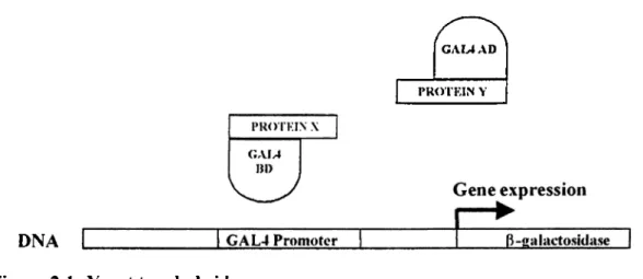
Yeast two-hybrid assay was used in this study to show Syndecan-1 interaction with PDZ domains o f CASK and AF-6. Yeast two-hybrid assay was chosen since it is a recently developed, powerful, and easily used protein assay which is frequently used in similar studies (Fields & Song 1989, Frederickson 1998). MATCHMAKER Two- Hybrid System 2 (#K 1604-1) was used which is a GAL4 based two-hybrid system that provide a transcriptional assay for detecting specific protein-protein interactions in yeast. Yeast two-hybrid system is either used to screen a cDNA library to identify unknown
interactions among the total population of cellular proteins, or it is used to detect specific protein-protein interactions between two known proteins.
Yeast two hybrid system is based on the fact that many eukaryotic transacting transcription activators are composed o f separable, functionally distinct and independent domains. These domains are usually DNA binding domain (BD) that recruits the transcription factor to specific DNA sequences and transactivating domain (AD) that interacts with RNA polymerase II complex and initiates transcription. GAL4 is a yeast transcription factor composed of these two (BD and AD) domains. When these two domains are separated from each other by recombinant DNA techniques and expressed in the same cell they can not bind to each other and form a functional transcription factor. However these two domains can be placed in close proximity by the means o f protein-protein interaction and functional transcriptional factor can be formed. Reporter gene expression can be obtained by such means, hence the reporter gene expression can be utilized as an indicator of protein-protein interaction. In MATCHMAKER system two functional domains of GAL4 transcription factor is supplied in two different yeast expression vectors such that two domains o f GAL4 can be expressed as two fusion proteins in yeast. One of the vectors (pAS2-l) express DNA binding domain of GAL4 protein which binds to DNA at specific sequences. The other vector expresses transactivating domain of GAL4 protein which activates the transcription o f the genes by interacting with transcription machinery.
Two different proteins can be cloned in these two vectors fused to BD and AD o f GAL4. So that upon the interaction between these proteins reporter gene activation can be detected. A schematic representation of yeast two-hybrid assay is shown in the following figure. (Figure 2 .1 )
GAM AD
PROTKIN Y
DNA
Figure 2.1 : Yeast two-hybrid assay
Schematic representation of yeast two-hybrid assay. Protein-protein interaction between Protein X and Protein Y leads to construction o f functional GAL4 transcription factor. Which can transactivate a reporter gene (ß-galactosidase) under the control o f GAL4 promoter sequence.
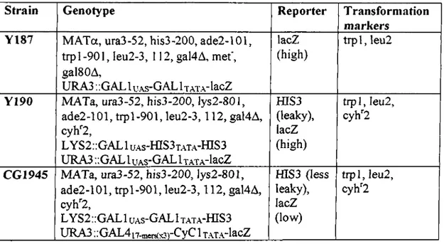
2.2.1- Strains o f Yeast
The Saccharomyces cerevisiae strains used in this study and their specifications are listed in Table 2.5.
The GAL I upstream activating sequence (UAS) and the three tandem copies of the GAL4 17-mer consensus sequence [GAL4 i7-mors(x3)] are both responsive to the GAL4 transcriptional activator. The trp 1, leu2 , his3, gal4 and gal80 mutations are all deletions. Table 2.5: Yeast strains used in this study
Strain Genotype Reporter T ransforniation
markers Y187 M ATa, ura3-52, his3-200, ade2-101,
trp 1-901, leu2-3, 112, gal4A, m ef, galSOA,
URA3 ..GAL 1 u.xs-GAL 1 xA T.ylacZ
lacZ (high)
tr p l, leu 2
Y190 MATa, ura3-52, his3-200, lys2-801, ade2-101, tr p l-901, le u 2 -3 ,112, gal4A, cyh"2 , LYS2::GAL1 UAS- fflS 3T .\T A -fflS 3 URA3 :;GALluAS~GALİTATA~lacZ_____ fflS3 (leaky), lacZ (high) trpl, leu2 , cyh'^2
CG1945 MATa, ura3-52, his3-200, }ys2-801,
ade2-101, tr p l-901, le u 2 -3 ,112, gal4A, cyh''2 ,
LYS2;;GAL1 UAS- GALİ TATA- fflS3
U R A 3 ..OAL4|7-mers(x3)~^y^ ^ T.AT.A’lacZ fflS3 (less leaky), lacZ (low) tip l, leu2 , cyh^2 2.2.2- Storage o f yeast
Yeast strains were stored in YPD medium with 25% glycerol at -70"C. Transformed yeast strains were stored in the appropriate SD medium to keep selective pressure on the plasmid. To recover frozen strains, a small portion o f glycerol stock was streaked onto YPD or appropriate SD agar plate.
2.2.3- G row th of yeast
Yeast strains were grown in liquid YPD medium or appropriate SD medium for preparing liquid cultures. They were grown on solid media by using YPD-Agar plates or SD-Agar plates according to their transformation status.
YPD m edium : 20 g/L Difco peptone
10 g/L Yeast extract
20 g/L Agar (for solid media only)
950 ml o f H2O is added and pH is adjusted to 5.8 and autoclaved.
After cooling glucose was added to a final concentration o f 2% as carbon source. SD m edium :
6.7 g Yeast Nitrogen Base without amino acids 20 g Agar for solid media only
850 ml H2O
100 ml o f the appropriate sterile lOX dropout solution
pH is adjusted to 5.8 and autoclaved. After cooling glucose was added to a final concentration o f 2 % as carbon source.
lOX D ro p o u t (DO) supplem ents:
Filter sterilized solution o f the following amino acids in H2O (Table 2.6).
T able 2 .6 : D ropout (DO) supplem ents
L-lsoleucine 300 mg/L 1-7383
L-V'aline 1500 mg/L V-0500
L-Adenine hemisulfate salt 200 mg/L A-9126
L-Arginine HCl 200 mg/L A-5131
L-Methionine 200 mg/L M-9625
L-Phenylalanine 500 mg/L P-5030
L-Threonine 2000 mg/L T-8625
2.2.4- Transformation o f Plasmid DNA into Yeast using LiAc method
1 ml o f YPD or SD was inoculated with several colonies of yeast, 2-3 mm in diameter and was vortexed for 5 minutes to disperse any clumps. For host strains previously transformed with another autonomously replicating plasmid, the appropriate SD medium was used. This was then transfeired into a flask containing 50 ml o f YPD or the appropriate SD medium. It was incubated at 30°C for 16-18 hr with shaking at 250 rpm to stationary phase (ODeoo >1.5). 30 ml o f the overnight culture was then transferred to a flask containing 300 ml o f YPD.
This culture was incubated at 30°C for 3 hr with shaking (230 rpm). At this point, the ODgoo was 0.4-0.6 . Cells were placed in 50-ml tubes and centrifuged at 1 ,0 0 0 X g for 5 min at room temperature (2 0 -2 1°C). Supernatant was discarded and 25-50 ml of sterile ТЕ or distilled H2O was added to the tube. Resuspended thoroughly by vortexing. Cells were pooled in one tube and centrifuged at 1 ,0 0 0 x g for 5 min at room temperature. Supernatant was discarded.
Cells were resuspended in 1.5 ml o f freshly prepared, sterile 1X T E /1X LiAc. 0.1 mg o f plasmid DNA and 0.1 mg o f Salmon testes carrier DNA (Sigma #D-9156) was added to a fresh 1,5-ml tube and mixed. 0.1 ml of yeast competent cells were added to each tube and mixed well by vortexing. Then 0.6 ml o f sterile PEG/LiAc solution was added to each tube and vortexed at high speed for 10 sec to mix. Incubated at 30°C for 30 min with shaking at 200 rpm. 70 ul o f DMSO was added. Mixed well by gentle inversion. Heat shocked for 15 min in a 42°C water bath. Cells were then chilled on ice for 1 - 2 min.
Cells were centrifuged for 5 sec at 14,000 rpm at room temperature. The supernatant was removed and were resuspended in 0.5 ml of sterile IX TE buffer. Plated
100 ul on each SD agar plate that would select for the desired transformants. PEG/LiAc Solution:
PEG 3350 (Sigma #P-3640) 40%
TE Buffer IX
LiAc 0.1 M
p H ; 7.5
2.2.5- Colony-lift filter assay
Fresh colonies were used (i.e., grown at 30°C for 2 -4 days), 1-3 mm in diameter. Z buffer/X-gal solution was prepared freshly as described below.
For each plate of transformants that were assayed, a sterile Whatman #5 or VWR grade 410 filter was presoaked by placing it in 2.5-5 ml of Z buffer/X-gal solution in a clean 100- or 1 50-mm plate.