IMMUNOTHERAPEUTIC ACTION OF EXTRACELLULAR VESICLES AND
EFFECTS OF TLR SIGNALING TO IMMUNE DYSFUNCTION OF SCI PATIENTS
A DISSERTATION SUBMITTED TO
THE GRADUATE SCHOOL OF ENGINEERING AND SCIENCE OF BILKENT UNIVERSITY
IN PARTIAL FULFILLMENT OF THE REQUIREMENTS FOR THE DEGREE OF
DOCTOR OF PHILOSOPHY IN
MOLECULAR BIOLOGY AND GENETICS
By Gözde Güçlüler
ii
Immunotherapeutic Action of Extracellular Vesicles and Effects of TLR Signaling to Immune Dysfunction of SCI patients
By Gözde Güçlüler March, 2017
We certify that we have read this dissertation and that in our opinion it is fully adequate, in scope and in quality, as a thesis for the degree of Doctor of Philosophy.
________________________ İhsan Gürsel (Advisor) ________________________ Tamer Yağcı ________________________ Kamil Can Akçalı
________________________ Özgür Şahin ________________________ Murat Alper Cevher Approved for the Graduate School of Engineering and Science
__________________ Ezhan Karaşan
iii
ABSTRACT
Immunotherapeutic Action of Extracellular Vesicles and Effects of TLR Signaling to Immune Dysfunction of SCI Patients
Gözde Güçlüler
Ph.D. in Molecular Biology and Genetics Supervisor: İhsan Gürsel
March, 2017
The primary aim of this thesis is to extend the breadth of in vivo application of externally loaded exosomes as prophylactic or therapeutic carriers against disease treatment. Exosomes are secreted from all cells and could be purified from all bodily fluids; however, engineering of exosomes to carry specific ligands post-purification is a daunting task. Herein, we show that lyophilization of exosomes together with the biological cargo alone or in combination of CpG ODN motifs, model protein antigen ovalbumin or lipidic ligand alpha-galactosylceramide (αGC) followed by controlled reconstitution successfully internalizes these cargos within exosomes. Furthermore, the bioactivity of the loaded ligand(s) surpasses the unloaded free ligand activities. When tested in vivo, exosome incorporated ligand(s) proved to be significantly effective against model tumors such as E.G7 thymoma or established melanoma models. The mechanism behind this elevated immune activity is the ability of exosomes to be delivered to target cells and boost immune antigen dependent immune activation. Our in vitro findings revealed that encapsulation of CpG ODN into exosomes enhances immunostimulatory activity of CpG ODN than free form as evidenced by superior levels of cytokines like IL6, IL12 and Type-I and II interferons. This magnified immune activity might be partly due the increased APC activation observed as elevated CD86/MHCII surface marker expression. Immunization of C57/Bl6 mice with exosomal CpG ODN plus OVA induced strong Th1-biased anti-OVA response. Following thymoma induction in naive and OVA-immunized animals, >85% of exosomal vaccine treated mice cleared tumors whereas almost all naive animals were positive for tumor. This data suggests that CpG ODN encapsulation into exosomes improve immunostimulatory activity, provide better anti-OVA immunity thereby contribute effective tumor clearance in mice. A second aim of this thesis was to establish that it is feasible to load exosomes with more than two ligands. Next, invariant natural killer T (iNKT) cell ligand αGC was included within exosomes as the third element next to OVA and CpG ODN. Initial in vivo
iv
studies revealed that exosomes containing αGC were significantly more potent in inducing antigen dependent immune responses in comparison to free form of CpG ODN, OVA and αGC. In therapeutic tumor vaccine model, two exosome injections (@d: 9 and d: 15) were done to B16-OVA tumor bearing animals and tumor regression was followed. Mice that had triple exosomal ligands significantly reduced tumors compared to mice treated with non-exosomal ligands. This study confirmed that exosomes with triple ligands could be effectively control established tumor development.
In this thesis, the elucidation of the involvement of extracellular vesicles (EVs) on the pathogenesis of autoimmune/autoinflammatory diseases was studied. The underlying mechanism in BD pathogenesis is still unclear. We found that one of the human cathelicidin group members, antimicrobial peptide LL37 along with EVs were elevated in active BD patients` plasmas. Strikingly, majority of plasma LL37 was associated with circulating EVs. We found that there was a strong correlation between i) LL37 level, ii) EV #/ml plasma and iii) cytokine production.
In the last part of this thesis, one of the possible mechanisms of immune dysfunction contributing to severe neurological deterioration of chronic spinal cord injured (SCI) patients was unearthed. We aimed to investigate whether there is a correlation between susceptibility to infections of chronic SCI patients within the context of impaired innate recognition of pathogen associated molecular patterns (PAMPs). Our data implicated that although there was no dysfunction of B cell, or CD4+ Treg activity, but sensing TLR7 and TLR9 ligands by monocytes and pDCs were ablated in patients with SCI, leading to lower IFNγ and IP10 production along with co-stimulatory molecule expression, that could explain the immunological dysfunction in patient with SCI contributing to persistent complications.
Keywords: Extracellular vesicles, Microvesicles, Exosomes, Vaccine carrier, Anti-tumor therapy, Behçet’s Disease, LL37, Spinal cord injury
v
ÖZET
Hücredışı Keseciklerin İmmünoterapötik Etkinlikleri ve TLR Yolaklarının Omurilik Hasarlı Hastalarda Görülen İmmün Disfonksiyona Etkileri
Gözde Güçlüler
Moleküler Biyoloji ve Genetik Bölümü, Doktora Tez Danışmanı: İhsan Gürsel
Mart, 2017
Bu tezin temel amacı, dıştan yüklenmiş eksozomların hastalıklara karşı koruyucu ve terapötik taşıyıcılar olarak kullanımının in vivo uygulanabilirliğinin genişletilmesidir. Eksozomlar bütün hücreler tarafından salınmakta ve bütün vücut sıvılarından saflaştırılabilmektedir. Fakat saflaştırma sonrası eksozomların istenilen ligandlar ile yüklenmesi pek denenmiş bir yöntem değildir. Bu çalışmada, eksozomların kendi biyolojik kargolarının yanında CpG ODN dizinleri, model protein antijeni ovalbumin ve yağsı ligandlardan alpha-galactosylceramide (αGC) molekülü ayrı ayrı ya da birlikte kullanarak önce liyofilizasyonu yapılıp sonrasında da kontrollü rekonstrüksiyonu sonucunda bu kargoların eksozomlara başarılı bir şekilde yüklendiği gösterilmiştir. Eksozoma yüklenmiş ligandların immünolojik etkinliğinin yüklenmemiş serbest ligandların etkinliğinden daha yüksek düzeyde olduğu belirlenmiştir. In vivo çalışmalarda eksozoma yüklenmiş ligandların E.G7 timoma ve B16 melanoma tümör modellerinde oldukça etkili olduğu saptanmıştır. Gözlenen immün etkinlik artışının arkasındaki mekanizma, eksozomların hedef hücrelere kargoyu bozulmadan taşıyabilmesi ve antijene bağlı immün tepkinin gelişmesini sağlamasına bağlıdır. In vitro çalışmalar, CpG ODN’lerin eksozomlara yüklenmesiyle, serbest CpG ODN’lere göre immün uyarıcı etkinliğini artırdığını IL6, IL12, tip I ve II interferonlar gibi sitokinlerin yüksek seviyelerde salınmasını sağladığını gösterilmiştir. Bu artan immün etkinlik kısmen CD86/MHCII yüzey markörlerinin artışıyla gösterilen antijen sunum hücrelerin aktivitesindeki artıştan kaynaklanıyor olabilir. C57/B16 farelerin eksozomal CpG ODN ve OVA ile aşılanması güçlü bir anti-OVA Th1 tepkisine yol açmıştır. Aşılanmamış ve aşılanmış hayvanlarda timoma tetiklendiğinde, eksozomal aşı ile aşılanmış farelerin 85%’inde tümör gerilemişken, neredeyse bütün aşılanmamış hayvanlarda tümör gelişimi görülmüştür. Bu bulgular, CpG ODN’lerin eksozomlara yüklenmesiyle oluşan
vi
immün uyarıcı mekanizmanın, güçlü anti-OVA bağışıklığı sağladığını ve farelerde anti-tümör tepkisini geliştirdiğini göstermektedir.
Bu tezin ikinci bir amacı, eksozomların ikiden fazla ligand ile yüklenmesinin uygulanabilir olduğunu göstermektir. İnvaryant doğal öldürücü T (iNKT) hücrelerinin ligandı αGC, eksozomlara OVA ve CpG ODN’lerin yanında üçüncü ligand olarak katılmıştır. Ön in vivo çalışmalar, αGC içeren eksozomların antijene bağlı immün tepkiye yol açmada ligandların serbest formlarina kıyasla çok daha etkili olduğunu ortaya çıkarmıştır. Terapötik tümör aşılaması modelinde, B16-OVA tümörü taşıyan hayvanlara 9. ve 15. günlerde iki eksozom enjeksiyonu yapılmış ve tümörlerde gerileme gözlenmiştir. Üç ligandla yüklenmiş eksozomlarla tedavi edilen farelerde eksozoma yüklenmemiş ligandlarla tedavi edilen farelere göre tümörlerde daha ciddi bir azalma görülmüştür. Bu çalışma, üçlü ligandlarla yüklenmiş eksozomların tümör gelişimini etkili bir şekilde geriletebildiğini ortaya koymuştur. Bu tezde ayrıca, hücre dışı keseciklerin (EV) otoimmün ve otoinflamatuar hastalıkların patogenezindeki rolü araştırılmıştır. Behçet Hastalığı’nın (BH) patogenezinin altında yatan mekanizma hala belirsizdir. Çalışmalarımız, BH hastalarının plazmalarında EV’lerin yanısıra bir cathelicidin grubu üyesi antimikrobiyal peptid olan LL37’nin yüksek miktarda bulunduğunu göstermiştir. Çarpıcı bir şekilde, plazmadaki LL37’nin büyük çoğunluğunun dolaşımdaki EV'lerle asosiye olduğu tespit edilmiştir. Bu bağlantı hem EVlerin monositlere ve plazmasitoid dendritik hücrelere sevkiyatının artmasına, hem de IL1β, IL6, IP10, IFNα gibi sitokinlerin üretiminin işaret ettiği üzere immün aktivasyonunun tetiklenmesi ve sürdürülmesine aracılık etmekte, böylece şiddetli ve uzun süreli BH patolojisine katkıda bulunmaktadır. Çalışmalarımızda LL37 seviyesi, plazmadaki EV miktarı ve sitokin üretimi arasında güçlü bir korelasyon olduğu bulunmuştur.
Bu tezin son kısmında, kronik omurilik hasarlı hastalarda şiddetli nörolojik bozulmaya katkı sağlayan immün bozukluğun muhtemel bir mekanizması açığa çıkarılmıştır. Kronik omurilik hasarlı hastalarda enfeksiyona yatkınlık ile patojen ilişkili moleküler desenlerin (PAMP) tanınmasındaki bozukluklar arasında bir korelasyon olup olmadığı araştırılmıştır. Omurilik hasarlı hastalarda, B hücreleri ve CD4+ Treg hücrelerinin etkinliğinde bir bozukluk olmamasına rağmen, monositler ve plazmasitoid dendritik hücreler tarafından TLR7 ve TLR9 ligandlarının tanınmasının
vii
işlevsiz olduğu, bunun da düşük IFNγ ve IP10 üretimi ile yardımcı uyaran molekül ifadesinde azalmaya yol açtığı belirlenmiştir. Bu mekanizma, omurilik hasarlı hastalardaki immünolojik bozukluğa bağlı kalıcı komplikasyonlara olan katkısını açıklayabilir.
Anahtar Kelimeler: Hücredışı kesecikler, Mikrokesecikler, Eksozom, Aşı taşıyıcı sistemler, Anti-tümor terapi, Behçet hastalığı, LL37, Omurilik hasarı
viii
ix
Acknowledgements
First and foremost, I would like to express my gratefulness to my advisor Prof. Dr. İhsan Gürsel for his support, trust, patience and guidance. It was a privilege to be his student and learn the basis of being how to be a good scientist along with all the values he thought to me.
I would like to also thank to Prof. Dr. Mayda Gürsel deeply from my heart for supporting me in life and science based perspectives.
I would like to thank to my thesis committee members; Prof. Dr. Tamer Yağcı and Prof. Dr. Kamil Can Akçalı for sharing their experience, academic knowledge and guidance throughout the progress of my thesis project. Moreover, I am thankful to Assist. Prof. Dr. Özgür Şahin and Assist. Prof. Dr. Murat Alper Cevher for accepting to become my thesis jury members.
I want to share my gratefulness with all past and new members of Thorlab; Fuat Cem Yağcı, Arda Gürsel, Mehmet Şahin, İhsan Dereli, Yusuf İsmail Ertuna, Merve Deniz, Elif Senem Kayali, , Hakan Köksal, Begüm Yıldız, Özlem Bulut, Gizem Kılınç, Havva Özgen Kılgöz, Muzaffer Yıldırım, Gizem Tincer König, Kübra Almacıoğlu and Banu Bayyurt Kocabaş. Particularly, I would like to thank to Tamer Kahraman for his scientific support and friendship during my PhD studies in Thorlab. It was a privilege to work with these helpful and nice people. Also, I am thankful to MG Group members Bilgi Güngör, Esin Alpdündar and Cihan Ayanoğlu for their support and friendship.
I would like to also extend my gratitude to Assoc. Prof. Susanne Gabrielsson (from Karolinska Institutet, Stockholm, Sweden) for accepting me as a guest researcher to work in her group. Also, I want to thank to all members of Gabrielsson’s research group especially to Pia Larssen for her support during and after my visit in Gabrielsson’s laboratory in Karolinska Institutet.
It was a privilege to be a part of MBG family and work with amazing people. I believe that their friendship will remain in the rest of my life. I am thankful to Dilan Çelebi Birand, Merve Mutlu, Gülşah Dal Kılınç, Deniz Cansen Yildirim, Banu
x
Bayyurt Kocabaş, Ayşegül Örs, Özlem Tufanlı, Şeyma Demirsoy, Büşra Yağabasan, Füsün Doldur Ballı, Gurbet Karahan and Nilüfer Sayar Atasoy.
Particularly, I would like to thank to Defne Bayık and Verda Bitirim for their friendship with full of support and guidance. They never let me feel alone during this journey.
I want to share my thanks to MBG veterinarian Dr. Gamze Aykut for her help and guidance in animal experiments and technicians Abdullah Ünnü and Turan Dastandır for their support in technical issues.
I would like to share my gratefulness to Begüm Han Horuluoğlu for her precious friendship during all good and bad days. She will remain my best friend throughout of my life.
Finally, I want to thank to my parents Nurten and Sefa, my sister Gaye for their support, trust and guidance. Without their endless love, I couldn’t accomplish to complete my PhD work. I hope I can always make them proud.
xi
Contents
Chapter 1 Introduction ... 1
1.1. Extracellular Vesicles ... 1
1.1.1. Types of extracellular vesicles ... 1
1.1.2. Biogenesis of EVs ... 3
1.1.3. Composition and cargo of EVs ... 4
1.2. Role of EVs in Different Physiological Conditions ... 7
1.2.1 Modulation of immune responses by EVs ... 8
1.2.2. Role of EVs in disease pathogenesis ... 12
1.2.2.1. EVs in cancer ... 12
1.2.2.2. EVs in inflammatory diseases ... 14
1.3. Application of EVs ... 15
1.3.1 Diagnostic and prognostic application of EVs ... 15
1.3.2. Therapeutic application of EVs as delivery tools ... 16
1.4. Immunostimulatory ligands ... 18
1.4.1. CpG ODN and their applications ... 18
1.4.2. α-Galactosylceramide ... 21
1.5. Behçet’s Disease and Pathogenesis ... 23
1.6. Spinal Cord Injury and Disease Pathogenesis ... 24
1.7. Aims of this thesis ... 26
Chapter 2 Materials and Methods ... 28
xii
2.1.1. Cell culture media and general solution components ... 28
2.1.2. Ligands ... 28
2.1.3. ELISA reagents ... 29
2.1.4. Western blot antibodies and solutions ... 30
2.1.5. Flow cytometry antibodies ... 31
2.2. Methods ... 33
2.2.1 Patients and Controls ... 33
2.2.1.1 SCI patients and controls ... 33
2.2.1.2. BD patients and controls ... 35
2.2.2. Cell Culture and Animals ... 37
2.2.2.1. Isolation of PBMCs and plasma from peripheral blood ... 37
2.2.2.2. Animals ... 37
2.2.2.3. Preparation of single cell suspension from mice spleens ... 38
2.2.2.4. Culture of RAW264.7 murine macrophage cells for supernatant collection ... 38
2.2.2.5. Preparation of PECs ... 39
2.2.2.6. Cell counting ... 39
2.2.3. Isolation of EVs and Exosomes ... 40
2.2.3.1. Isolation of exosomes from cell culture ... 40
2.2.3.2. Isolation of EVs from peripheral blood plasma ... 41
2.2.3.3. Purification of EVs by sucrose cushion ... 42
xiii
2.2.3.5. Determining protein concentration of exosomes ... 43
2.2.4. Characterization of Exosomes ... 43
2.2.4.1. Bead based flow cytometry technique to determine the specific EV and exosomal markers ... 43
2.2.4.2. Western blot for determining the presence of exosomal markers ... 44
2.2.4.3. Size distribution analysis of exosomes by AFM ... 45
2.2.4.4. Size distribution analysis of EVs by DLS ... 45
2.2.5. TLR9 and iNKT Ligand Loading within Exosomes ... 45
2.2.6. Cell Stimulation Studies ... 46
2.2.6.1. Stimulation of mouse splenocytes with free and exosomal CpG ODN 46 2.2.6.2. Stimulation of OT1 mice splenocytes with exosomal TLR9 and iNKT ligands ... 46
2.2.6.3. Stimulation of SCI patients and healthy control PBMCs with different immunostimulatory ligands ... 47
2.2.7. Flow Cytometry Studies ... 47
2.2.7.1. Intracellular cytokine staining ... 47
2.2.7.2. Staining of Cell Surface markers ... 49
2.2.7.3. BrdU staining ... 50
2.2.8. ELISA Studies ... 51
2.2.8.1. Cytokine ELISA ... 51
2.2.8.2. Anti-OVA IgG ELISA ... 51
2.2.8.3. Anti-OVA ELISA ... 52
2.2.9. ELISPOT ... 52
xiv
2.2.10.1. Immunization and E.G7 challenge study ... 53
2.2.10.2. In vivo cell proliferation study ... 53
2.2.10.3. Tumor therapy study ... 53
2.2.11. Statistical Analysis ... 54
Chapter 3 Results ... 55
PART-I: Utilization of Exosomes as a Prophylactic Anti-Cancer Vaccine ... 55
3.1. Use of CpG ODN Loaded Exosomes as a Potential Anti-cancer Therapeutic Agent ... 55
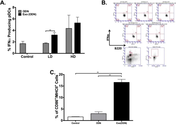
3.2.1. Encapsulation into exosomes increases immunostimulatory activity of CpG ODN ... 55
3.1.2. Exosomal CpG ODN activates APCs ... 56
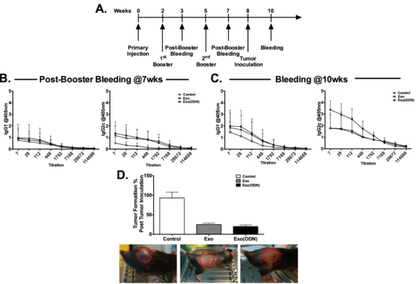
3.1.3. CpG ODN encapsulated into exosomes triggers Th1-biased anti-OVA immunity and prevents tumor formation. ... 58
PART-II: Utilization of Exosomes as a Therapeutic Anti-Cancer Vaccine ... 60
3.2. Inclusion of αGC and CpG ODN within Exosomes Contribute to Immunogenicity of Cancer Vaccine ... 60
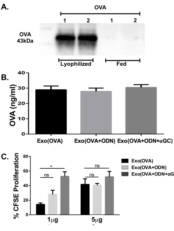
3.2.1. Exosomes isolated from RAW264.7 cells retain their exosomal properties after lyophilization. ... 60
3.2.2. Lyophilization is a more effective loading technique than feeding and moreover it enables to load exosomes with more than two molecules. ... 62
3.2.3. Encapsulation of D-type CpG ODN and αGC into exosomes induces OVA specific T-cell proliferation. ... 64
3.2.4. Antigen specific T cell responses and antibody production are more pronounced by ODN and αGC loaded into exosomes than their free form. ... 66
3.2.5. Therapeutic exosome vaccine harboring OVA+CpG ODN+αGC significantly regresses established tumors. ... 67
xv
PART-III: Role of EVs in Behçet’s Disease Pathogenesis ... 69
3.3. Role of LL37 Associated Plasma Extracellular Vesicles in Behçet’s Disease69 3.3.1. Vesicles isolated from plasma possess all universal EV characteristics. 69 3.3.2. BD patients have higher numbers of plasma EVs ... 70
3.3.3. Internalization of EVs by PBMCs induces IL6 production ... 72
3.3.4. Elevated levels of plasma LL37 in BD patients induces IL6 cytokine production and most of it is associated with EVs ... 73
3.3.5. Association of LL37 with EVs mediates cellular internalization and immune activation ... 76
PART-IV: Effects of TLR Signaling to Immune Dysfunction of Spinal Cord Injured Patients ... 78
3.4. Impaired TLR7 and TLR9 Induced Immune Activation in Chronic Spinal Cord Injured Patients Contributes to Immune Dysfunction ... 78
3.4.1. Healthy and SCI individuals have similar responses to different immune cell stimulants. ... 78
3.4.2. Healthy and SCI donor PBMCs respond similarly to PMA-Ionomycin stimulation. ... 79
3.4.3. SCI patients have altered co-stimulatory and MHCII expression in response to TLR7 and TLR9 ligand stimulation. ... 81
3.4.4. SCI patients have altered IFNγ production in response to TLR7 ligand stimulation. ... 83
3.4.5. SCI donors have impaired monocytes response to TLR9 ligand stimulation. ... 84
Chapter 4 Discussion ... 86
4.1. Utilization of exosomes in cancer therapy ... 86
xvi
4.3. Contribution of impaired TLR7 and TLR9 pathways to immune dysfunction in SCI 92
Appendix ... 113 Curriculum Vitae and Publications ... 123
xvii
List of Figures
Figure 1.1. Different types of EVs secreted from cells. ... 2
Figure 1.2. Biogenesis mechanisms of extracellular vesicles. ... 4
Figure 1.3. Composition of extracellular vesicles. ... 5
Figure 1.4. EVs modulate immune responses. ... 10
Figure 1.5. Representative modulatory effects of exosomes on immune cells. ... 11
Figure 1.6. Activated signaling pathways upon recognition of CpG ODNs. ... 19
Figure 1.7. Potential therapeutic uses of CpG ODNs.. ... 21
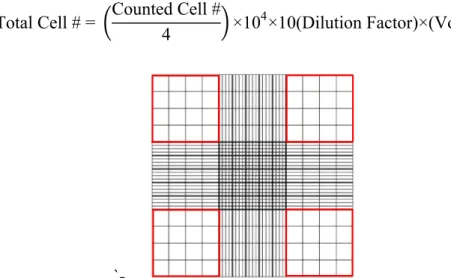
Figure 2.1. Representative picture of grids in hemocytometer. Cells residing within the red squares were included in the counting. ... 40
Figure 3.1. Immunostimulatory activity of CpG-ODN is elevated upon encapsulation into exosomes. ... 56
Figure 3.2. Exosomal CpG ODN enhances CpG ODN mediated immune activation. ... 57
Figure 3.3. Exosomal CpG ODN immunization promotes Th1 biased immunity and tumor clearance. ... 59
Figure 3.4. Characterization of exosomes isolated from RAW264.7 cell line. ... 61
Figure 3.5. Exosomes can be loaded with different antigens and adjuvant at the same time. ... 63
Figure 3.6. Exosomal CpG ODN and αGC induces OVA specific T cell proliferation. ... 65
Figure 3.7. Antigen specific T-cell responses and antibody production are more pronounced by CpG ODN and αGC loaded into exosomes than their free form. ... 67
Figure 3.8. Exosomal antigens and adjuvant contributes prevention of tumor development and survival. ... 68
Figure 3.9. EVs isolated from plasma carry exosomal markers and their size is in expected range ... 70
Figure 3.10. Total EV concentration is highly elevated in BD and majority of EVs is originated from platelets ... 71
Figure 3.11. BD patients’ EVs are internalized by cells and induce IL6 production ... 73
xviii
Figure 3.12. LL37 plasma level is highly elevated in BD patients’ plasma and it is
associated with EVs ... 76
Figure 3.13. LL37 association mediates EV internalization by PBMCs and IL6
production can be abrogated with the addition of anti-LL37 ... 77
Figure 3.14. Healthy and SCI donors show similar responses to different immune
cell stimulants. ... 79
Figure 3.15. Increased number of IFNγ producing T-cells in response to
PMA-Ionomycin stimulation by both healthy and SCI donor PBMCs. ... 80
Figure 3.16. SCI patients have altered co-stimulatory and MHCII expression in
response to TLR7 and TLR9 ligand stimulation. ... 82
Figure 3.17. IFNγ production by healthy and SCI donor PBMCs is different in
response to TLR7 ligand stimulation. ... 83
Figure 3.18. Altered SCI patient monocyte response to TLR9 ligand stimulation. .. 85
Figure A1. Exosomes retain cardinal exosomal markers after lyophilization…….113 Figure A2. Non-lyophilized and lyophilized exosomes are taken up by PECs at
similar levels.………...………114
Figure A3. Percent of immune cells 7 days after immunization with free and
exosomal form of OVA+ODN+αGC………...115
Figure A4. Percent number of B cell subtypes 7 days after immunization with free
and exosomal form of OVA+ODN+αGC………....116
Figure A5. Characterization of EV fractions obtained by FPLC Separation …….117 Figure A6. Different immune cells internalize EV/LL37 complex at different
levels……….118
Figure A7. Representative FACS analysis plots of IFNγ producing T cells ……..119 Figure A8. Representative FACS analysis plots of CD83+ and HLA-DR+ cells....120
Figure A9. Representative FACS analysis plots of IFNγ producing T-cells.……..121 Figure A10. Representative FACS analysis plots of IP10 producing
xix
List of Tables
Table 1.1. Differential characteristics of EVs [42]. ... 7
Table 2.1. ELISA reagents and their working concentrations. ... 29
Table 2.2. Western blot reagents and their working concentrations ... 30
Table 2.3. Specificities of the FACS antibodies ... 32
Table 2.4. Baseline characteristics of the patients and control group. ... 34
Table 2.5. Hematological characteristics of the patients and the control group. ... 35
xx
Abbreviations
αGC aBD alpha-Galactosylceramide active BDAFM Atomic Force Microscopy
AICD Activation induced cell death
AIM-2 Absent in melanoma 2
allo Allogeneic
ALP Alkaline phosphatase
AnV Annexin-V
APC Antigen presenting cell
ATP Adenosine three phosphate
BD Behçet's Disease
BSA Bovine serum albumin
CCL C-C motif chemokine ligand
CD Cluster of differentiation
CeA Carcinoembryonic antigen
CFSE Carboxyfluorescein succinimidyl ester
DC Dendritic cell
DIC Differential interferance contrast
DLS Dynamic Light Scattering
DNA Deoxynucleic acid
EDTA Ethylenediamine tetraacetic acid
EGFR2 Epidermal growth factor receptor 2
EGFRvIII Epidermal growth factor receptor variant III
EV Extracellular vesicles
FACS Flourescence assosicated cell sorting
FasL Fas ligand
gp100 Glycoprotein 100
GvHD Graft versus host disease
xxi
ICAM1 Intercellular adhesion molecule 1
IFN Interferon
Ig immunoglobulin
IL Interleukin
IP-10 Interferon gamma-induced protein 10
iBD inactive BD
L1CAM L1 cell adhesion molecule
LEAF Low Endotoxin, Azide-Free
LPS Lipopolysaccharide
MART1 Melanoma antigen recognized by T cells 1
MCP-1 Monocyte chemoattractant protein-1
MDA-5 Melanoma differentiation-associated
protein 5
MD-DC Monocyte derived DC
MDSC Myeloid-derived suppressor cell
MHC Major histocompatibility complex
miRNA MicroRNA
MMP Metalloproteinase
mRNA messenger RNA
MSC Mesenchymal stem cell
MSC-EV Mesenchymal stem cell derived EV
MV Microvesicle
MVB Multivesicular bodies
MyD88 Myeloid differentiation primary response
88
NK Natural killer
NKG2D Natural killer group 2D
NS Not significant
ODN Oligodeoxynucleotide
OVA Ovalbumin
xxii
PBS Phosphate buffered saline
pDC Plasmocytoid dentritic cell
PDGF Platelet-derived growth factor
PMV Platelet derived MV
PNPP p-nitrophenyl phosphate
PS Phosphatidylserine
RA Rheumatic arthritis
RANTES Regulated on activation, normal T cell expressed and secreted
RNA Ribonucleic acid
RT Room Temperature
siRNA small interfering RNA
SLE Systemic lupus erythematosus
SSc Systemic sclerosis
TF Tissue factor
TGFβ transforming growth factor β
Th T-helper
TLR Toll-like receptor
TMB 3,3′,5,5′-Tetramethylbenzidine
TNFα Tumor necrosis factor α
Treg Regulatory T cell
TSG101 Tumor susceptibility gene 101
1
Chapter 1
Introduction
1.1. Extracellular Vesicles
Secretion of membrane-enclosed vesicles to the extracellular environment has been reported many decades ago. Membrane vesicles are secreted into the extracellular environment by almost all cell types from different organisms from eukaryotic to prokaryotic and this process has gained an important role since they have been found as key players in several cellular activities from cell-to-cell signaling to regulation of immune responses. It is possible to isolate extracellular vesicles (EVs) from any bodily fluids such as blood, saliva, urine, breast milk, amniotic fluid, ascites, cerebrospinal fluid, and semen. These EVs differ from each other in their biogenesis, size and biological functions and they are named as apoptotic vesicles, membrane particles, ectosomes, microvesicles and exosomes according to their distinctive features [1].
1.1.1. Types of extracellular vesicles
Starting from 1970s, presence of EVs outside of the cells including solid tissues, biological fluids, tumor cells and platelets were documented by several groups [2-7]. This group of vesicles was first shown to be released by cells directly outward
2
budding of the plasma membrane (PM). In 1980s, a new EV secretion pathway was proposed describing the formation of vesicles approximately 50 nm in diameter initially within multivesicular bodies (MVBs) and release following fusion of MVBs with the PM [8, 9]. Then, in 1987, the term exosomes were first mentioned by Johnstone et al. to describe the formation and secretion of the EVs originated from endosomal pathway [10]. Today, it is known that EVs are heterogeneous populations composed of different types of vesicles classified according their sub-cellular origin and size (Fig 1.1).
Figure 1.1. Different types of EVs secreted from cells.
There are different types of EVs depending on their formation mechanism and size. EVs can be formed by direct outward budding of PM like microparticles or by fusion of MVBs with the PM leading to release of vesicles called exosomes (adopted from [20]).
EVs are in the size range between 150-1000 nm and formed by direct budding from PM. They are nomenclatured with the terms ectosomes, shedding vesicles, microparticle and microvesicles according to their cell origin and sizes [11]. On the other hand, the term exosome is used for the vesicles with smaller size range
3
between 30 and 100 nm formed within MVBs [5]. EVs are composed of lipid bilayer enclosing proteins and RNA as cargo proteins. They have a great impact on cell-to-cell communication as they are internalized by recipient cell-to-cell via receptor-ligand interaction or by endocytosis and/or phagocytosis or even fuse with the target cell’s membrane. These effects of EVs are dependent on the state of the cells that they are originated from and their cargo as it is proposed that they carry functional genetic materials such as mRNA, miRNA as cargo molecules [1, 11, 12].
1.1.2. Biogenesis of EVs
The formation of MVBs is a highly dynamic process from start to end along with involvement of internalization and recycling of membrane compartments [11, 13]. In this process, early endosomes generated by inward budding of the PM mature into the late endosomes; intracellular ligands and cellular compartments accumulate intraluminal vesicles (ILV) formed by inward budding of endosomal membrane changing the definition of endosomes to MVBs [14].
In general, MVBs are destined to fusion with the lysosomes to degrade the endosomal contents such as lipids, proteins and cytosol. On the other side, the fate of some MVBs, bearing tetraspanin molecule CD63, LAMP1 and LAMP2 (lysosomal-associated membrane proteins 1 and 2), can be released from the cells by fusion of MVBs with the PM. These different pathways for MVB formation propose that there are different subpopulations of MVBs within a cell. MVBs can differ in their morphology depending on the appearance and the size of the ILVS that they enclose [1, 15, 16].
There are two mechanisms proposed how ILVs are formed in MBVs (Fig 1.2). Endosomal sorting complex required for transport (ESCRT)-dependent mechanism is the best described explanation of the highly dynamic process of ILV formation. ESCRT is composed of almost thirty proteins that assemble into 4 different complexes ESCRT-0, I, II and III associated with several proteins such as ALIX, VTAI and Tsg101 [17]. All these complexes have different role in endocytic pathway including recognition and sequestration of transmembrane proteins in the endosomal membrane, membrane deformation into sorted cargo and vesicle excretion [17].
4
There are also evidences showing that ILVs and MVBs can be formed in an ESCRT-independent mechanism. This is proposed by different studies showing inactivation of four ESCRT complexes does not abrogate the formation of ILVs and MVBs [18]. These findings indicate that ILVs and MVBs can be generated by both ESCRT-dependent and inESCRT-dependent mechanisms. However, this different mechanistic formation can lead to different consequences in the release yield and composition of EVs [19].
Figure 1.2. Biogenesis mechanisms of extracellular vesicles.
EVs are generated by ESCRT-dependent or independent pathways (adopted from [21]).
1.1.3. Composition and cargo of EVs
Although PM-derived EVs and exosomes have similar biogenesis pathways, they show distinctive physicochemical and molecular features (Fig 1.3). When their properties are compared, exosomes bear the hallmarks of ILVs and their composition
5
is more likely closer to endosomal composition, whereas EVs are tend to show more PM characteristics [1]. Since exosomes are derived from the ILVs of which size range between 30 and 100 nm, exosomes released following MVB fusion with the PM would have a slightly larger size reaching to maximum 150 nm. In contrast, size of EVs released to the extracellular milieu following direct outward budding of PM can diverse between 150 nm and 1000 nm [20]. Sedimentation of exosomes and EVs is also different due to their different size ranges. While exosomes can be isolated by ultracentrifugation at 100000xg, larger vesicles should be isolated at lower speed from 10000xg to 50000xg [22, 23, 24]. In addition to ultracentrifugation, using sucrose gradient would be another alternative for isolation of exosomes that have density between 1.13 and 1.19 g/ml in sucrose [22]. Since studies showed that populations obtained by ultracentrifugation are composed of heterogeneous vesicles, each fraction should be characterized separately.
6
EVs are generated by ESCRT-dependent or independent pathways (adopted from [1]).
Exosomes contain biological contents as proteins, lipids and genetic materials. However, it has been shown that these contents are specific set of proteins, RNAs and lipids [21, 25]. Almost all exosomes are enriched in cytoplasmic proteins such as tubulin, actin, annexins and Rab proteins that can be find in the surface or in the lumen of exosomes [26]. Proteins associated with MVB formation such as Alix and Tsg101 are enclosed by exosomes [27, 28]. In 1998, it was found out that tetraspanins (CD63, CD81, CD82, CD53, CD37) were highly enriched on exosomes secreted from human B cells [29]. Other studies also showed that exosomes from other sources contain tetraspanin molecules [30]. Exosomes from different cell types also have been shown to carry Hsp70 and Hsp90 heat-shock proteins [10, 31]. Studies revealed that exosomes secreted from B cells carry MHC class I and MHC class II molecules [16, 32]. Exosomes have proteins on their membrane associated lipid rafts and also their membrane is enriched in cholesterol, sphingomyelin and hexosylceramides. Saturated and monounsaturated fatty acids also exist in exosomes [26]. In addition to their protein and lipid contents, they enclose genetic materials as mRNA and miRNA. This was first discovered in 2004 from blood of Hepatitis C virus (HCV) infected individuals that the virus envelope proteins were associated with exosomes [33]. It was also shown that mRNAs and miRNAs are transferred between cells via exosome-mediated mechanism (24). Following studies supported the existence of these genetic materials in exosomes from different cell types and body fluids [34-36]. Moreover, it has been revealed that exosomes also contain various types of small noncoding RNA molecules such as structural RNAs, tRNA fragments and small interfering RNAs [37, 38]. It is important to identify the exosome cargo by biochemical and biological techniques, since this kind of discovery elicits the understanding of the mechanisms associated with the exosomes in physiological and pathological conditions.
When PM derived EVs such as microvesicles are investigated, it has been documented that they share similar characteristics with exosomes. However, the most substantial difference of MVs from exosomes, apart from their larger size, is that they have similar surface composition to the cells that they are originated from.
7
Both exosomes and PM derived EVs have exposed phosphotidylserine (PS) on their surface. PS is a negatively charged molecule that tends to be located in the inner leaflet of the lipid bilayer in live cells in the presence of ATP by flippase [39, 40]. This feature makes it possible to identify the EVs by Annexin-V (AnV) staining [41]. Characteristics of EVs are summarized in Table 1.1. These physical and biochemical characteristics of exosomes and PM-derived EVs provide EV researcher to differentiate between different types of EVs.
Table 1.1. Differential characteristics of EVs [42].
Exosome Ectosome Microparticle (MP) Microvesicles (MV) Others Apoptotic bodies (AP) Large vesicles (LV) Size 30-100 nm 100-1000 nm AB: 1-4 µM LV: 2-50 µM Origin MVB fusion with PM Budding from PM Budding from PM
Markers Tetraspanins ((CD9, CD63, CD81) Alix, TSG101, HSP70) Annexin V, Integrin, Selectin, CD40 ligand, metalloproteinase AB: Annexin V, DNA, Caspase 3, Histones LV: Annexin V, ARF6, GAPDH Isolation method Ultracentrifugation (100000xg) Ultracentrifugation (10000–50000xg) Ultracentrifugation (6000–10000xg)
1.2. Role of EVs in Different Physiological Conditions
When EVs were first identified during monitoring of the loss of transferrin receptor, they were thought as just cell debris [8, 9]. However, when Raposo et.al. found out that Epstain-Barr virus (EBV) transformed B cell lines derived exosomes are enriched by MHC class II molecules and can present antigens to T cells [12], several important functions were started to be defined for EVs. It has been reported that EVs play crucial roles in vital cellular events from cell-to-cell communication to modulation of immune responses via transferring their cargo molecules. However, there is a great effort exposing the involvement of MVs to the pathogenesis of several diseases.
8
1.2.1 Modulation of immune responses by EVs
Since studies showed that EVs enclose both antigenic peptides and peptide-MHC complexes, they were thought that EVs could induce the immune activation [43]. It was revealed that exosomes isolated from tumor cells lines or patients’ tumors have tumor antigens and in vitro settings showed that they trigger T cell activation when there is DCs in the environment [44, 45]. The most common antigens carried by tumor derived exosomes are transmembrane proteins (human EGFR2, carcinoembryonic antigen CeA) or endosomal components such as MART1, GP100 and tyrosine-related protein 1 of the cells that exosomes are originated from.
In addition to activation of immune responses by antigens, EVs can directly activate T cells by peptide-MHC class complexes displayed on their surface, too. Numerous studies documented that exosomes derived from DCs trigger cytotoxic T cell activation even if they are alone or incubated with MHC Class I expressing DCs [46-49]. This suggests that exosomes carry physiologically functional MHC class I molecules. However, studies with immature and mature DCs pointed out that mature DC-derived exosomes are more potent in inducing T cells then those derived from immature DCs, since they carry co-stimulatory molecules [50]. However, the only way to activate CD8+ T cells with exosomes derived from tumor cells is to incubate cells with exosomes in the presence of DCs having the right MHC haplotype [44,45]. In addition to presence of MHC Class I molecules, exosomes isolated from APCs could also carry a large amounts of MHC Class II molecules [16, 51]. It was shown that these exosomes can transmit peptide-MHC Class II complexes to the DCs lacking MHC Class II molecule and then these DCs can trigger antigen specific CD4+ T cells [52, 53]. Like CD8+ T cell activation, exosomes derived from immature DCs are more potent activators of CD4+ T cells [52, 54]. Not only exosomes, PM derived microvesicles derived from mature DCs can also induce activation of allogeneic T cells via transfer of MHC molecules to DCs [55].
Immune activating properties of vesicles secreted from other cell types have been reported, too. It was shown that platelet-derived exosomes trigger proliferation, survival and chemotaxis of hematopoietic cells [56]. Moreover, these exosomes were shown to be functional in induction of pro-inflammatory cytokine secretion from
9
monocytes and activated B cells through CD40L present in exosomes [57, 58]. Furthermore, it was determined that EVs derived from macrophages infected with pathogens such as Mycobacterium tuberculosis, Salmonella enterica subsp. enterica,
serovar Typhimurium or T. gondii carry microbial antigens and they induce
activation of both CD4+ and CD8+ T cells in in-vitro and in-vivo settings [58, 59]. It was also documented that B- and T-cells can be activated by pro-inflammatory exosomes derived from cell cultures infected by Mycoplasma orale and Mycoplasma
arginini [60].
Studies also showed that EVs derived from cells can also exhibit immune suppressive profile [43]. Especially tumor derived exosomes were identified with immune suppressive characteristics. It was shown that EVs secreted from tumor cells of patients provide immune escaping by CD95L presence on their surface via triggering T cell apoptosis [61]. Also, exosomes isolated from tumors inhibit cytotoxic profile of T and natural killer (NK) cells [62, 63]. It was shown that this inhibitory capacity of tumor derived exosomes on NK and CD8+ T cells could be
correlated with the presence of NK2GD ligands and transforming growth factor-β (TGF-β) on the membrane of exosomes [63, 64]. Moreover, exosomes can trigger activation of regulatory T cell (Treg) signaling pathways and thus prevent interleukin 2 (IL2) mediated T cell proliferation [64]. Moreover, exosomes derived from tumor cells can induce generation of myeloid-derived suppressor cell (MDSC) because of activated Treg signaling by TGF-β dependent pathway attenuating the differentiation of myeloid precursors to DCs [65-67]. Collectively, tumor derived vesicles can play crucial role in the modulation of immune responses with dual activation mechanisms.
10
Figure 1.4. EVs modulate immune responses.
These include transmission of tumor-antigens to dendritic cells (DC), which may cross-present these to T cells, or may tolerize T cells in an antigen specific manner. EV-antigens may sequester antibodies, impacting antibody-dependent killing of tumor cells. Differentiation of myeloid (CD14+) cells to functional antigen presenting cells is inhibited by EV supporting the generation of myeloid derived suppressor cells (MDSC). T lymphocytes may undergo apoptosis, or downregulate functionally important receptors, and a subset of CD4+CD25-high cells may be
induced into regulatory cells, that suppress proliferation to mitogenic stimuli. NK cell functions may also be impaired through loss of activating surface receptors. (adopted from [68]).
Other than activating properties of immature DC derived exosomes, they also exhibit tolerogenic effect on cells. In a mouse septic shock model, it was shown that CD95L or IL10 harboring DC derived exosomes could reduce the inflammation mediated by macrophages [69, 70]. Moreover, tumor derived exosomes were shown suppressing immune responses in the absence of co-stimulatory molecules and promoting tumor development in mice vaccination model with exosomes [62]. Studies showed that exosomes isolated from Treg cell mediate immune suppression due to the presence
11
of CD73 [30]. These exosomes decrease the antigen presenting function of DCs thus led to suppression of cytotoxic activity of CD8+ T cells. In addition, presence of CD25 and CTLA-4 molecules on exosomes secreted from Tregs suggests that the suppression of CD4+ and CD8+ T cell activities by Treg derived exosomes could be mediated by CTLA-4 dependent pathway [71].
Collectively, both in vitro and in vivo findings propose that EVs can play dual role in the modulation of immune responses (Fig 1.5). Depending on the state of the donor cells, exosomes encounter different compositions leading to activation of different immunological pathways in the recipient cells.
Figure 1.5. Representative modulatory effects of exosomes on immune cells.
Exosomes induce immunostimulatory and immunosuppressive pathways depending on the cells they are originated from. (Adopted from [72]).
12
1.2.2. Role of EVs in disease pathogenesis
1.2.2.1. EVs in cancer
EVs secreted from tumor cells play critical roles in communication between tumor and stromal cells even between distant microenvironments. They regulate invasion, angiogenesis, coagulation and immune suppression of tumors [73].
Active communication between neighbor cells and their microenvironment is crucial for during primary tumor formation. Tumor cells were identified with critical roles in cell-to-cell communication between tumor cells and their surrounding counterparts. During primary tumor formation, oncogenic molecules are transferred between tumor cells by tumor-secreted EVs. It was shown that microvesicles derived from EGFRvIII expressing glioma cells were shown expressing EGFRvIII and they transmit this molecule to other malignant cells [74]. Following internalization of EVs by the recipient cells, mitogen-activated protein kinase (MAPK) and protein kinase B (PKB/Akt) are activated by EGFRvIII which triggering cancer growth [75]. EVs derived from tumors do not affect only other tumor cells, they also trigger different signaling pathways in adjacent stromal cells like fibroblasts and endothelial cells. It was shown that glioblastoma-derived microvesicles advocate proliferation of endothelial cells and growth of primary tumors as they transmit their cargo molecules mRNA, miRNA and angiogenic [35]. Pancreatic cancer-derived exosomes were also shown that they induce expression of angiogenic proteins in endothelial cells expressing tetraspanin 8 [76]. Contribution of tumor derived exosomes to tumor growth, vascularization and invasion was determined by fibroblast to myofibroblast transition mediated by TGF-β expressing tumor derived exosomes [77, 78]. Exosomes derived from breast cancer were shown promoting TGF-β expression, vascular endothelial growth factor (VEGF), stromal cell-derived factor 1 (SDF-1), and C-C motif chemokine ligand 5 (CCL5) in adipose tissue derived mesenchymal stem cells [79].
In addition to tumor derived vesicles, stromal cell derived exosomes can also induce the tumor growth. It was shown that tumor motility and invasion can be advocated by breast-cancer-associated fibroblasts by activation of the Wnt-planar cell polarity
13
(Wnt-PCP) signaling cascade [80]. These data suggest that the role of EVs in tumorigenesis is bidirectional between tumor cells and their microenvironment. Studies showed that extracellular matrix (ECM) remodeling influence the tumor invasion. This influence was shown to be promoted by the contribution of tumor derived EVs. Exosomes secreted from tumor cells having fibronectin were shown to induce cell motility [81]. It was documented that microvesicles from tumor cells transfer EMMPRIN to fibroblasts and induces the MMP production leading to tumor metastasis [82]. EVs derived from endothelial cells stimulated with VEGF and fibroblast factor 2 (FGF-2) triggered tumor invasion via initiating proteolysis [83]. Thrombotic complications are one of the major causes leading death in cancer patients [84]. Recognition of cancer cells by immune system is hampered in case of coagulation and platelet accumulation at tumor location and tumor growth and metastasis is promoted [85]. It was shown that microvesicles associated with coagulation are derived from platelets, inflammatory cells, and cancer cells [86]. In cancer patients, it was found out that increased risk of coagulation is strongly associated with elevated levels of circulating microvesicles expressing tissue factor (TF) thrombosis [86-88]. Microvesicles derived from pancreatic cancer cells were shown containing physiologically active F- and P-selectin glycoprotein ligand 1 (SELPLG). When these vesicles are delivered to injury site, they induce the inhibition of bleeding [87]. Collectively, these data propose that tumor derived EVs play important roles in coagulation promoting progression and metastasis of tumor [87, 88].
Accumulating evidences also showed that tumor derived EVs play modulatory role in immunosuppression that leads to progression of cancer. It was shown that tumor derived microvesicles carrying Fas ligand or TRAIL induces apoptosis of immune cells [89, 90] Tumor derived exosomes were also determined that they can suppress the cytotoxic activity of NK cell [91].
14
1.2.2.2. EVs in inflammatory diseases
Accumulating evidences suggest that EVs do not only play important role in tumor formation, they also influence the progression of inflammation involving in communication between the infected cells and interaction between host cells and bacteria [92].
It has been the most investigated question whether EVs can trigger or provide the maintenance of pathological autoimmune response as a target of autoreactive recognition. It was shown that EVs enclose several autoantigens such as HSPs, histones, and α-enolase [93]. Moreover, EVs were identified with a potential role in rheumatoid arthritis (AR), since it was found out that they carry citrullinated proteins (CD5 antigen-like precursor and fibrinogen components) [94, 95]. Studies with RA patients showed that immune recognition is mediated by antigens associated with EVs. RA pathogenesis is promoted by formation of pro-inflammatory immune complexes between large microvesicles derived from synovial fluid of patient with RA with citrullinated protein-specific, vimentin-specific and fibrinogen-specific autoantibodies [94].
RA associated EVs are particularly microvesicles originated from platelets (PMV). It was shown that level of PMVs is elevated in patients with RA compared to healthy individuals. Moreover, elevated number of PMVs in circulation inflammatory joints is strongly associated with disease severity [95-99]. In RA studies, disease progression is mediated by pro-inflammatory cytokines carried by PMVs [100]. Also, microvesicles of RA patients contributes to cardiovascular comorbidities [99, 102]. Of note, microvesicles isolated from synovial fluids of patients with RA were shown to induce inflammatory cytokine and chemokine production such as IL8, IL6, CCL5 (also known as RANTES), CCL2 (also known as MCP1), vascular endothelial growth factor A (VEGF) and intercellular adhesion molecule 1 (ICAM-1) by cultured synoviocytes [103]. PMVs also trigger synoviocyte activation.
Besides RA, PMV level was found elevated in patients with systemic lupus erythematous (SLE) [104, 105]. However, none of the studies with SLE patients show any correlation between the elevated PMV number and disease severity [98].
15
On the other hand, studies showed that increased number of circulating EVs contributes to thrombosis and induces cardiovascular risk in SLE patients. Studies showed that the composition of EVs in SLE were different from the EVs from RA, systemic sclerosis (SSc) and healthy individuals. They were shown to have immunoglobulins, complement and other opsonizing molecules [105]. EVs from patients with SLE were also found correlated sustained inflammation and disease pathogenesis similar to RA pathogenesis [106].
Also, elevated numbers of EVs were documented in patients with systemic sclerosis (SSc) mediating contribution of activated cell populations to pathogenesis [107] Endothelial cell apoptosis which has been suggested to be a primary pathogenic event in SSc might be substantially modulated by high concentrations of circulating EVs [108].
1.3. Application of EVs
1.3.1 Diagnostic and prognostic application of EVs
EVs are secreted from almost cell types and it is possible to isolate them from all bodily fluid such as plasma, serum, saliva, urine. Biological cargo and composition of EVs can differentiate according to the physiological condition of the cell that they are originated from. As it was mentioned above, EVs contributes to pathogenesis of several diseases like cancer and inflammatory diseases, there are several studies investigating whether EVs can be used as diagnostic marker for diseases.
There are accumulating evidences revealed that levels of microvesicles are elevated in malignant effusions, serum, and urine from cancer patients [8]. Moreover, the prognosis and survival of cancer patients were shown to be strongly associated with different levels of platelet derived microvesicles [109, 110]. Microvesicles enclosing tissue factor (TF) or mucin were determined as diagnostic marker for adenocarcinomas [111]. These evidences suggest that MVs can be considered as potential diagnostic molecules for several cancer types due to their specific composition.
In addition to protein composition, MVs from patients with cancer were shown carrying specific mRNA and miRNA molecules. It was shown that oncogenic
16
mRNA molecules HER-2/neu and MAGE-1 HER-2/neu were highly expressed in MVs from patients with gastric cancer [112]. Of note, it was also revealed that MVs from glioblastome patients had cancer specific mRNA and miRNA [35].
Morphology of MVs can be also used as a diagnostic marker as it was evidenced by the comparison of MVs from patients with prostate cancer and healthy individuals by electron microscopy. This study showed that urine of the prostate cancer patients contains cup-shaped exosomes whereas healthy urine has prostasomes [113].
In another study, it was shown that size of the MVs in plasma of patients with preeclampsia changes in addition to the elevated number [114].
Collectively, MVs can be used as diagnostic markers by analysis of them in three classes as following; i) MV quantity, (ii) MV protein composition, and (iii) MV miRNA or mRNA composition [115].
1.3.2. Therapeutic application of EVs as delivery tools
EVs play important roles in cell-to-cell communication and modulation of immune response due to their differential composition and capability of transferring cargo molecules. These evidences suggest that particularly exosomes are potential therapeutic agents due to their stable structures.
EVs can be used to modulate immune responses in pathological conditions such as autoimmune and inflammatory diseases depending on the state of origin cells. Particularly, EVs derived from mesenchymal stem cells (MSC) (MSC-EVs) are the most extendedly investigated population among EVs for therapeutic and regenerative purposes [116]. In an experimental acute injury study, it was shown that tubular injury was prevented via transmission of anti-apoptotic signals by MSC-EVs [117, 118]. Moreover, it was shown that levels of EVs are increased in MSC rich fragments and they provided the protection against tissue destruction [117]. In another study, it was shown that M2 type macrophage polarization could be mediated by MSC-EVs [119]. Furthermore, inhibitory effect of MSC-EV on NK cells was shown in patients with graft versus host disease (GvHD) [120].
17
Due to their immunosuppressive activity, Tregs are widely studied to prevent graft rejection. However, studies revealed that inflammatory environment inhibits immunosuppressive activity of Tregs following administration [121]. Since exosomes derived from Tregs are more resistant to inflammatory environment and they retain their immunosuppressive profile, there are several studies trying to utilize Treg exosomes in prevention of graft rejection. In one of them, it was shown that survival and function of kidney transplants was elevated following adoptive transfer of exosomes derived from Treg [122].
EVs derived from mature DCs activate T cells when expressing appropriate MHC class molecules and co-stimulatory molecules on them. It was shown that vesicles isolated from DCs pulsed with tumor antigen significantly contributed to tumor specific T cell response mediated tumor clearance in mice [123]. In couple of clinical trials, EVs having tumor-associated peptides were isolated from monocytes-derived DCs of patients were used for anti-therapeutic treatment of melanoma and non-small cell lung carcinoma [124-126]. In another study, it was found out that when monocyte derived DCs were treated with IFNγ, EVs from these cells were shown having more improved stimulatory activity due to elevated amount of CD40, ICAM1, and B7 molecules. According to this finding, EVs derived from tumor antigen pulsed IFNγ-matured MD-DCs were used for the treatment of non-small lung carcinoma in phase II clinical trial [127]. Collectively, these findings strongly support the attempts to develop therapeutic strategies using EVs isolated from appropriate cell type at an appropriate state.
In addition to use of EVs in therapeutic applications as they are isolated, there are also studies showing it is possible to modify exosomes to deliver different types of therapeutic agents to the cells [128]. Accumulating evidences point out that exosomes can deliver their cargo in an intact form to the recipient cells and modulate several cellular activities [34, 129, 49, 25, 130]
Two main strategies have been developed so far for utilization of exosomes as drug delivery systems. One of them is in vitro loading which required encapsulation of molecule of interest into exosomes via membrane breaching [131, 132] or physical entrapment [133, 134]. In vitro loading strategy was first introduced by mixing exosomes isolated from EL-4 cells with curcumin at room temperature. It was shown
18
that encapsulation into exosomes increased the anti-inflammatory effect of curcumin [133]. Moreover, this complex was investigated in another study and it was revealed that curcumin encapsulated into exosomes mediated LPS-induced brain inflammation passing through the blood-brain barrier [134]. It was also documented that siRNA and miRNA molecules could be also loaded into exosomes with similar approach and their encapsulation into exosomes involves in the tumor clearance [135, 136]. On the other hand, in vivo loading approach suggests that cells fed or stably transfected with the molecule of interest secrete exosomes expressing these molecules [137, 138]. Both techniques have limitations regarding to the loading of any molecule on demand with high yielding efficiency.
1.4. Immunostimulatory ligands
1.4.1. CpG ODN and their applications
Bacterial DNA is known inducing activation of immune cells. It was shown that this activation capacity is due to the sequences of unmethylated CpGs flanked by two purines and two pyrimidines on 5’ and 3’ side, respectively [139]. The frequency of CpG sequences in mammalian DNA is 20-fold less than bacterial DNA, thus the immune suppression capacity by methylation is less observed. This difference between mammalian and bacterial DNA ended up with development of an evolutionary conserved recognition system that discriminates self and non-self-bacterial DNA by Toll-like receptor 9 (TLR9) [140].
Synthetic ODNs mimicking bacterial DNA were first identified with immune activation function due to their sequences containing CpG motifs in 1992 (Yamamoto 1992). They induce secretion of pro-inflammatory and T-helper (Th) 1 biased cytokines [141]. All CpG ODN types contain at least 8 base pair and at least one unmethylated CpG motif flanked by two 5’ purines (Pu) and two 3’ pyrimidines (Py) to be functional in immune stimulation in mice [139]. However, human PBMCs are less responsive to CpG ODNs that stimulates mice immune cells [142].
There are 2 mainly types of CpG ODNs as D-type and K-type which are also known as A and B type. There is also a recent type of CpG ODN called C-class. K-type
19
ODNs can have more than one CpG motif that makes them more potent immunostimulatory, whereas D-type ODNs can form nanoparticle like structures since they have poly G tail [142]. Due to these structural differences, their fate and action mechanisms also differ. D-type ODNs are recognized by TLR9 on early endosomes and entrapped in early endosomes upon internalization in human pDCs. Then, they activate MyD88 dependent signaling cascade and trigger innate immune responses via inducing type-I IFN secretion [143]. In contrast to D-type ODNs, K-type ODNs are relocated in late endosomes and they activate adaptive immunity through DC generation [144]. In Fig 1.5, sub-cellular localization and immunostimulatory action of D- and K-type CpG ODNs are represented.
20
D-type ODNs are localized into early endosomes, whereas K-type ODNs are transferred to the late endosomes. Due to their structural differences, they activate different signaling pathways. (Adopted from [145]).
TLR9 ligands CpG ODNs have a great potential to be used as vaccine adjuvants, since they are capable of alarming immune system through activation of both innate and adaptive immunity. Upon recognition, they activate immune cells directly and also triggering signaling cascades that starts production of inflammatory cytokines. It has been shown that CpG ODNs directly activates human B cells and pDCs [146]. Their indirect effect is observed in maturation and proliferation of macrophages, T cell and NK cells [145]. They also activate APCs via upregulating co-stimulatory molecule expressions such as MHC class II, CD80, CD86 and CD40 [139, 148, 149]. Moreover, they induce humoral immunity by activating B cells that produces IgM along with IL10 and IL6 [150]. Of note, they are characterized by inducing secretion of IL12 and IFNγ as member of Th1 biased immunity from pDCs. It was also shown that pDCs start to produce several cytokines including IL6, IL-1β, IL18 and TNFα upon activation by CpG ODNs [151].
Due to their profound immune stimulatory capacity, CpG ODNs are studied as strong candidates for clinical use against cancer, protective agent against infectious diseases, as novel vaccine adjuvants and anti-allergic therapeutic agents to restore Th2 dominant immune response (Fig 1.6). Clinical studies showed that utilization of CpG ODNs in vaccines as adjuvant elicited antibody mediated immune responses against several pathogens such as influenza virus, anthrax, leishmania, measles virus, orthopox virus, tetanus toxoid and lymphocytic choriomeningitis virus [151-154], However, there are obstacles in their in vivo applications due to serum protein adsorption and nuclease attack.
21
Figure 1.7. Potential therapeutic uses of CpG ODNs. (Adopted from [141]). 1.4.2. α-Galactosylceramide
Invariant natural killer T (iNKT) cells are a specialized T cell subset that recognizes self and bacterial lipid antigens presented by monomorphic MHC class I-like CD1d expressed by antigen presenting cells (APC) [155, 156]. Following activation with lipid antigen, iNKT cells play a dual role as both protective and pathogenic, by mediating both innate and adaptive immunity [157]. Alpha-galactosylceramide (αGC) is a glycolipid ligand for both murine and human iNKT cells and it induces rapid activation of iNKT cells in vivo leading to Th1, Th2 and Th17 specific cytokines production (IFN- γ, IL-4 and IL-17, respectively) that can mediate the immune responses against pathogens and tumors [158-161]. Due to profound role of iNKT cells in activation of immune system, there are several studies trying to explore their role in health and disease conditions using αGC in several animal models and human trials [156, 162]. According to pre-clinical trials, following activation, iNKT cells starts to produce IFNγ at earlier time points and then they
22
trigger secretion of IFNγ from NK cells and IL12 from DCs [163]. After these encouraging findings, αGC has been tested in clinical trials for treatment of cancer patients. Surprisingly, none of these trials ended up with successful outcomes [164]. The underlying reasons behind these failures were thought to be correlated with low number of iNKT cells in patients or anergy upon multiple administration αGC [165, 166]. However, other studies showed that dendritic cells and B cells could efficiently activate iNKT cells bypassing iNKT cell unresponsiveness [167-169]. In addition to human studies, there are several studies showing potent anti-tumor activity of αGC when presented by DCs to iNKT cells in cell based mouse models [170]. DCs expressing mammary tumor–associated Ag Her-2 and loaded with αGC were shown that they could trigger anti-tumor responses. Moreover, the activity of these DCs was amplified upon combination with gemcitabine overcoming the immunosuppression [171]. In different mouse tumor models, DC based strategies were used to increase the anti-tumor activity of αGC in both preventive and therapeutic settings [172]. It was shown that DCs expressing OVA along with CCL21 via genetically modification could efficiently demolish OVA-expressing tumors [173].
In addition to based techniques, there are some efforts to elicit the iNKT cell-dependent anti-tumor immunity via delivery of αGC by using biodegradable vectors. This approach suggests that encapsulation of αGC into nanovectors smaller than 1 mm can be superior to soluble αGC increasing uptake by APCs and avoiding side effects. It has been reported that silica microspheres coated with αGC containing lipid bilayers were targeted to DCs and macrophages [174, 175]. It has been also documented that PLGA (poly(lactic-co-glycolic acid)) can be also used to activate iNKT cells through APC uptake [176, 177].
Overall, the potential of iNKT cells to be as an important actor and the adjuvant properties of αGC in the eradication of malignancies cannot be underestimated. However, clinical trials have been showed that there is a necessity to develop new innovative approaches to pronounce the anti-tumor potential of iNKT cells and use of αGC.
![Figure 1.7. Potential therapeutic uses of CpG ODNs. (Adopted from [141]).](https://thumb-eu.123doks.com/thumbv2/9libnet/5793218.117852/43.892.249.706.124.518/figure-potential-therapeutic-uses-cpg-odns-adopted.webp)