Nanotechnology
TOPICAL REVIEW
Self-assembled peptide nanostructures for
functional materials
To cite this article: Melis Sardan Ekiz et al 2016 Nanotechnology 27 402002
View the article online for updates and enhancements.
Related content
Smart Internal Stimulus-Responsive Nanocarriers for Drug and Gene Delivery: pH-sensitive micro/nanocarriers
M Karimi, P S Zangabad, A Ghasemi, M R Hamblin
-Challenges and Breakthroughs in Recent Research on Self-Assembly
Katsuhiko Ariga, Jonathan P Hill, Michael V Lee et al.
-Peptide amphiphile self-assembly
Aysenur Iscen and George C. Schatz
-Recent citations
Tailor-Made Functional Peptide Self-Assembling Nanostructures
Moran Amit et al
-Reductionist Approach in Peptide-Based Nanotechnology
Ehud Gazit
-Vertical Alignment of Size-Controlled Self-Assembled Diphenylalanine Peptide Nanotubes Using Polyethersulfone Hollow Fiber Membranes On Silicon
Giti Emtiazi et al
-Topical Review
Self-assembled peptide nanostructures for
functional materials
Melis Sardan Ekiz, Goksu Cinar, Mohammad Aref Khalily and
Mustafa O Guler
Institute of Materials Science and Nanotechnology, National Nanotechnology Research Center(UNAM), Bilkent University, Ankara, 06800 Turkey
E-mail:[email protected]
Received 29 April 2016, revised 18 June 2016 Accepted for publication 5 July 2016
Published 31 August 2016 Abstract
Nature is an important inspirational source for scientists, and presents complex and elegant examples of adaptive and intelligent systems created by self-assembly. Significant effort has been devoted to understanding these sophisticated systems. The self-assembly process enables us to create supramolecular nanostructures with high order and complexity, and peptide-based self-assembling building blocks can serve as suitable platforms to construct nanostructures showing diverse features and applications. In this review, peptide-based supramolecular assemblies will be discussed in terms of their synthesis, design, characterization and application. Peptide nanostructures arecategorized based on their chemical and physical properties and will be examined by rationalizing the influence of peptide design on the resulting morphology and the methods employed to characterize thesehigh order complex systems. Moreover, the
applicationof self-assembled peptide nanomaterials as functional materials in information technologies and environmental sciences will be reviewed by providing examples from recently published high-impact studies.
Keywords: peptide, nanomaterials, self-assembly, nanofiber (Some figures may appear in colour only in the online journal) 1. Introduction
Molecular self-assembly is an emerging and powerful tool in the synthesis of functional nanoscale structures as a bottom-up fabrication method. It is defined as the spontaneous organization of molecules into stable, well-defined structures under equilibrium conditions through noncovalent interac-tions. Self-assembly is ubiquitous in many biological systems and results in the formation of a variety of complex biological structures found in nature. These complex and elegant examples inspire scientists to create similar artificial systems by using the building blocks of life, such as amino acids, lipids, nucleic acidsand saccharides. In most cases, thermo-dynamically stable structures are formed through enthalpic and entrophic interactions that involve both the assembling subunits and the surrounding solvent molecules [1].
Naturally, many key developments have paved the way forprogress inthis sophisticated field, such as the discovery of close-packed forms of amphiphilic molecules, the arrangement of monolayers by long chain hydrocarbon amines and the distribution of ordered monolayers of alka-nethiolate molecules on gold substrates[2].
In this review, we describe peptide synthesis strategies, peptide self-assembly and the relation of the peptide design to the corresponding self-assembled nanostructures. In addition to the synthesis and functionalization of peptide nanos-tructures, we also provide an overview of the characterization techniques that have been developed for understanding the interactions between individual peptide subunits and their effecton the physical and chemical properties exhibited by the self-assembled system. The peptide nanostructures are considered to be attractive candidates for a broad range of
biomedical applications, and have already been reviewed extensively [3–6]. Here we present recent applications of
peptide-based nanomaterials by providing specific examples such as peptide-templated inorganic nanomaterials, semi-conducting peptide nanowiresand catalytic peptides.
2. Peptides: synthetic approaches
Peptides consisting of natural or synthetic amino acids are interesting building blocks for the construction of supramo-lecular assemblies. These simpler structures aid us in under-standing more complex systems present in nature. Scientists employ a variety of approaches in the synthesis of peptide building blocks and strive for the production of the peptide of interest while minimizing or eliminating other possible by-products. Synthetic approaches utilized for the fabrication of peptide molecules can be classified into three main classes: solid phase strategies, ring-opening polymerization techni-ques and genetic engineering[7,8].
Solid phase peptide synthesis(SPPS) is the most widely used method for thesynthesis of small to mediumsize pep-tides. In this technique, the peptide sequence is grown, step-by-step, on an insoluble polymeric resin through the sequential addition of individual amino acids. Deviations from the intended amino acid sequence are prevented through use of modified amino acids with unreactive N-termini, which can be activated only after treatment with a deprotecting agent [9]. Amino acids used for the couplings bear orthogonal
protecting groups. The orthogonal chemistry approach enables two or more protecting groups to be used con-currently without affecting each other in situations where one of the functional groups requires manipulation. General fea-tures associated with these protecting groups can be con-sidered as (i) their ease of introduction into the functional group;(ii) their stability in different reaction conditions; and (iii) their safe removal at the end of the synthetic process [10].
Prior to the coupling, there is a need to activate the carboxylic group of the last amino acid in the sequence by using phos-phonium, aminium, uronium or carbodiimide-based reagents that catalyse the formation of amide bonds. Although carbo-diimide derivatives lead to an increase in the degree of racemization during the activation of amino acids, this effect can be minimized by using additives, especially N-hydroxy derivatives, which suppress the formation of N-acylurea by shifting the reaction towards the formation of the active ester form of the amino acid[11]. At the end of the synthesis, the
peptide is cleaved from the resin with an acid treatment. Microwave-assisted SPPS is another option for synthe-sizinglonger peptides with increased reaction rates and purities. Typically, coupling reagents and additives used in microwave-assisted SPPS are identical to those applied in conventional SPPS, and peptide couplings are carried out at temperatures in the range50 °C–80 °C unless amino acids carry a potential risk of epimerization [12]. Setbacks
encountered in solution phase synthesis, such as time con-suming isolation and purification of intermediate peptides, are overcome with SPPS. With the SPPS strategy, it is possible to
control not only the amino acid sequence but also the C- and N-terminal functionality of the created peptide [13].
There-fore, this technique is eligible for the synthesis of linear, branched, dendritic or cyclic peptides [14, 15]. In addition,
different chemical moieties, such as fatty acids, lipids, sac-charides, nucleotides, polymers, drugs, aromatic unitsand dyescan be incorporated into the peptide backbone through linkage chemistry while the peptide is still on the resin. -Further progress in peptide synthesis is native chemical ligation, which bypasses the limitations of SPPS with respect to the size and solubility of the peptides, and entails the chemoselective conjugation of two unprotected peptides (an α-thioester-containing peptide and an N-terminal cysteine-ended peptide) throughthioester linkage. These peptides are first individually synthesized using SPPS, and subsequently linked to each other in thesolution phase to create large-sized peptides and even complex proteins [16].
On the other hand, the preparation of high molecular weight monodisperse polypeptides with precisely defined primary structures can be achieved by protein engineering, also called genetic engineering. This strategy allows the synthesis of not only structural proteins but also de novo designed proteins as a result of the cellular expression of artificial genes [17]. In contrast to chemical synthesis
meth-ods, this technology can be considered as a biological tool for thepreparation of peptide-based materials, and involves insertion of a synthetically designed gene into a circular DNA plasmid to produce the desired polypeptide or protein in bacterial or eukaryotic expression systems [18]. One of the
advantages of this methodology is that a broad range of nonproteogenic amino acids or functional groups can also be integrated for the preparation of artificial proteins [19].
Polypeptides with high molecular weights, narrow polydispersity and retained chiral integrity can be synthesized by the ring-opening polymerization of amino acid N-carboxyanhydrides(NCA) in a controlled manner [20]. For
the polymerization, there are two widely accepted mechan-isms:normal amine and the activated monomer mechanisms. In both mechanisms, a range of nucleophiles and bases such as amines and metal alkoxides are used for the initiation of polymerization [21]. Although NCA polymerization suffers
from a lack of control over the exact primary peptide sequence, in contrast to SPPS or genetic engineering, it allows the synthesis of polypeptides in high yields and large quan-tities [22]. In some cases, side reactions that occur during
NCA polymerization may result in failure in the production of homo and block polypeptides, which can be prevented by introducing various metal- and organo-catalysts [20]. In
addition, some applications require using high purity mono-mers to obtain optically pure polypeptides with predictable molecular weights and low polydispersity.
Several other chemical methodologies have been devel-oped to synthesize peptide- and protein-based hybrids. Anchorage of two components has been achieved through thiol–maleimide or alkyne–azide couplings as well as by imine and hydrazone linkages, atom transfer radical poly-merization and chemoselective peptide ligation methods [8,23, 24]. Although each strategy has its own limitations,
with recent advances in synthetic strategies, sophisticated peptidic architectures can be created by using these versatile chemical tools.
3.Self-assembly of peptides
Amino acids serve as a diverse biochemical toolbox for the construction of peptide-based materials, exhibiting a broad range of physicochemical properties with regards to their charge, hydrophobicity, size and polarity. Consequently, peptide materials containing different amino acid sequences can be designed to serve a variety of biological functions. This diversity is further enriched by the introduction of nonproteinogenic groups into the peptide backbone. Since the synthesis methods, structures and properties of peptides are wellknown, peptide-based materials can also be utilized as model systems to gain insight intobiological self-assembly mechanisms in nature [25]. Peptide self-assembly is mainly
governed by noncovalent interactions. These interactions not only drive the self-assembly process but also stabilizethe secondary structure of peptides and proteins[26].
3.1. Forces drivingpeptide self-assembly
Noncovalent interactions such as hydrogen bonding, hydro-phobic, electrostaticand van der Waals interactions, π–π stacking and coordination bonds are the main contributors of molecular self-assembly(figure 1) [27, 28]. While nonpolar
amino acids, including aliphatic(e.g. alanine, leucine, valine) and aromatic(e.g. tyrosine, phenylalanine) amino acids, are mainlyresponsible for hydrophobic clustering through hydrophobic interactions and π–π stacking, respectively, polar amino acids result in either hydrogen bonding or elec-trostatic interactions depending on whether they have uncharged (e.g. serine, asparagine) or charged (e.g. lysine, histidine, glutamic acid) residues [29]. In addition to
indivi-dual amino acids, the peptide backbone itself provides con-siderable stability through hydrogen bonds. Although these interactions are individually weak, the cooperative action of binding residues across multiple self-assembling subunits can enable formation of stable assemblies. The cooperative
binding also ensures a degree of sequence-specificity in the self-assembling system[30].
Naturally occurring hydrogen bonding patterns, such as those found in β-sheets, α-helices, and coiled coils, are uti-lized in the design of a number of peptide sequences to form higher order structures as a result of self-assembly. The sta-bilization of multiple peptide backbone arrangements occurs through hydrogen bonding interactions between the backbone amide and carbonyls and results in the formation ofβ-sheets. These structures may be in parallel or antiparallel arrange-ments depending on the direction of the strands. Peptides are typically designed to contain repeating amino acid residues for hydrophobic and hydrophilic regions, which ensure that the hydrophobic part will be buried within the self-assembled structure while the hydrophilic region, which often contains functional sequences, is exposed to the aqueous environment [31]. Unlike β-sheets, α-helices are formed by individual
peptide chains where backbone amide components are intra-molecularly hydrogen bonded. This arrangement leads to the presentation of the amino acid side chains on the surface of each helix and further facilitates the accessibility of amino acid side chains to the solvent. In some cases, these single α-helices can assemble by coiling together and form so-called coiled coils. Peptide sequences responsible forcoiled coil formation generally bear a repeat motif consisting of seven residues. These heptad motifs can be derived using general-ized wheel diagrams that denote the positions and biochem-ical properties of amino acids at each location. The rules governing the formation of coiled coil structures are well-studied and conceived. In this section of the review, we present a relatively brief discussion for the H-bonding pat-terns exploited in peptide self-assembly,however, there are comprehensive reviews in the literature[32,33].
3.2. Factors triggeringself-assembly
There is a growing interest in creating dynamic systems that assemble and disassemble in response to external cues. Since noncovalent interactions are delicate interactions, they have a great tendency to respond to alterations in environmental conditions such as pH, light, temperature, ionic strength, and solvent polarity [34]. Among these, pH switch is the most
facile approach forcontrolling and directing the self-Figure 1.Strength and properties of the noncovalent interactions involved in self-assembly.
assembly[35–39]. A number of peptides are inherently
sen-sitive to pH change due to the introduced charged amino acids, leading to a structural transformation. Apart from pH stimuli, light-triggered systems designed by van Hest and Stupp groups were developed by using aphotocleavable group bearing peptide amphiphiles, and morphological tran-sitions were observed for both cases in response to light[40– 42]. Furthermore, polymerizable diacetylene unit containing
peptide molecules were shown to respond to UV light irra-diation to acquire one-dimensional nanostructures by the -Tovar group [43]. On the other hand, thermally triggered
peptide-based materials causing a change in the self-assembly of the nanostructure have beenreported by several groups [44–49]. In addition to thermal energy, theUlijn group has
demonstrated that ultrasound energy can assist the reorgani-zation of supramolecular nanostructures by temporarily dis-rupting noncovalent interactions in the peptide system and subsequently allowing their reformation in more thermo-dynamically favourable positions[50]. In addition, the effect
of ionic strength and metal ions on the self-assembly mech-anism of peptide molecules were examined by the addition of different type of cations into the peptide-based systems. While charged amino acids are mainly responsible for salt-induced self-assembly, as in the case of ionic-complementary peptides[51], β-hairpin peptides [52], ultrashort peptides [53]
and peptide amphiphiles [54, 55], histidine residues have
been predominantly used in themetal-binding domain due to the affinity of theimidazole ring to several coordination metal ions [56–59]. In addition to the above-mentioned external
stimulus factors, solvent polarity can also exert a profound effect on the supramolecular nanostructures. A solvent con-trolled structural transition was observed for several peptides due to the changed interactions between the hydrophobic domain of the peptide and solvent molecules [60–63]. The
self-assembly mechanism can be alternatively induced by enzyme catalysed reactions. Structural switches can be employed in a controllable manner by using enzymes specific to peptide sequences[64–68].
4. Design–nanostructure relation in peptide-based systems
The number, type, and sequence of amino acids can be manipulated to design self-assembling peptides. When amino acids are considered individually, they have their own specific characteristics. For instance, glycine impartsflexibility to the peptide structure, as it lacks a side chain and does not carry steric hindrance, while the proline ring constrains the number of conformations that the peptide backbone can adopt, resulting in a high degree of conformational rigidity[69,70].
Cysteine, on the other hand, can be chemically or inter-molecularly modified via thiol–maleimide chemistry or dis-ulphide bridging, respectively. Moreover, tyrosine, serine and threonine are utilized in further chemical and enzymatic modifications through their hydroxyl groups. Depending upon the chemical modifications of the C- or N-terminus, theproperties of peptide building blocks can also be
modulated. There are two other major parameters affecting the structure of peptide-based systems, the secondary struc-ture and noncovalent interactions, which were mentioned in the previous section. Peptides supported by α-helical or β-sheet features allow theconstruction ofvarious different nanostructures [71,72]. While peptide secondary structures
are often sufficient to ensure the formation of kinetically and thermodynamically stable structures, conformational interac-tions can also be supported through linker residues that are covalently conjugated to peptide building blocks[73]. In this
section, we discuss the structural features of peptide materials and highlight representative studies of self-assembled systems composed of peptide building blocks. This section is divided into three parts, which discussthe importance of design in the resulting self-assembled nanostructures of different classes of peptides, namely peptides composed of only amino acids, peptides modified by the attachment of hydrophobic acyl chainsand peptide containing hybrid systems.
4.1. Peptides composed of only amino acids
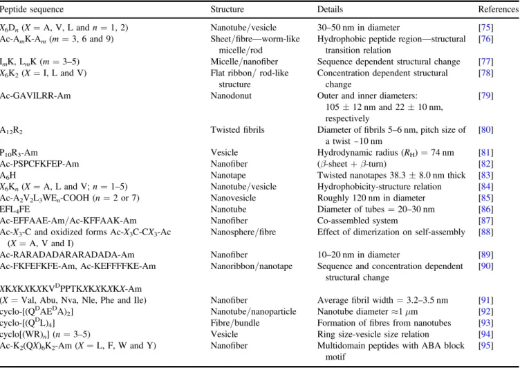
Peptides highlighted in this sectiontypically contain both hydrophobic and hydrophilic amino acid domains, as amphiphilic peptide designs readily self-assemble into well-organized nanostructures. The distribution andnumber ofhydrophobic and hydrophilic amino acids in the peptide sequence determine the final structure, including nanofibre, nanotape, nanorod, nanovesicle, nanotube and micelle, of the self-assembling peptides. Depending on the nature of the hydrophobic domain, theaggregation propensity and, simi-larly, thecritical aggregation concentration (CAC) of the peptides can be modulated, which is very important for the stability of the assembly in anaqueous environment [74].
Table1 summarizes the sequences of the peptides discussed in this section, the morphology of the nanostructures they form, and additional information regarding the structural features they exhibit.
The Zhang group has designed several short(7–8 amino acid) peptides and described them as a new class of surfac-tant-like peptides(SLPs) [75]. In their design, six consecutive
hydrophobic residues(A6, V6or L6) were attached to a head group with one or two aspartic acid residues, and charges on the N-termini were blocked by acetyl groups to prevent positive charge formation in the hydrophobic tail. The self-assembly of these peptides resulted in the formation of nanotubes or vesicles measuring~30–50 nm in diameter. The type of nanostructure formed was affected by the hydro-phobicity of the tail but not by the number of integrated charged residues. They also reported that similar peptides with two different positively charged(K and H) head groups exhibited analogous structures in transition electron micro-scopy(TEM) [96]. By using asimilar design, the influence of
the hydrophobic tail length on the formed nanostructure was investigated by Lu’s group with A3K, A6K, and A9K peptides [76]. These peptides exhibited structural transition as their
hydrophobic tail extended due to the change in packing within the nanostructure. While A3K self-assembled into unstable peptide sheet stacks, A6K and A9K formed long
fibrillarworm-like micelles with uniform diameters of 8±1 nm and short nanorods with smaller diameters ofaround 3±1 nm, respectively. The morphological diver-sity of peptide nanostructures stems from the equally diverse nature of the interactions between their constituent amino acids, with both hydrophobic and hydrophilic residues imparting characteristic properties to the overall structure. To gain insight into this cooperative effect, Han et al synthesized avariety of peptides containing three to five consecutive leucine or isoleucine residues attached to one polar lysine residue[77]. They systematically studied the role of hydrogen
bonding and its cooperative effect with hydrophobic inter-actions by changing the number and the type of the hydro-phobic residues. As expected, anincrease in the number of hydrophobic residues gave rise to adecrease in the CAC values of both LmK and ImK. Since isoleucine had a higher propensity to form β-sheet conformation, the ImK series formed long nanofibers through the combination of hydrogen bonding and hydrophobic interactions, whereas L3K self-assembled into spherical micelles. Moreover, the rest of the LmK series exhibited nanofiber morphology due to the dom-ination of intermolecular hydrogen bonding by additional leucine residues. In thecase of ImK assemblies, the diameter of the nanofibers gradually decreased as the number of iso-leucine residues increased, stemming from the change in
chain packing. In another comparative study conducted by Baumann and coworkers, I6K2, L6K2and V6K2peptides were prepared in anaqueous salt solution (2 mM NaCl) to inves-tigate the concentration dependence of the aggregation [78].
Although the secondary structures of all three peptides remained steady over the wide range of concentrations, morphological inspection of theself-assembled peptide structures revealed that peptides showed variance in their nanostructures as their concentrations changed. While pep-tides prepared at lower concentrations tended to form rod- and sheet-like structures with different cross sections, theaverage rod length and ribbon/sheet area couldbe tuned by var-yingthe concentration. The effect of the molecular geometry on the nanostructure formation and its relation to the concept of surfactant packing parameter were further analysed by Khoe et al [79]. A cone-shaped molecular structure
(Ac-GAVILRR-Am) was synthesized usingfive subunits of increasing size and hydrophobicity, starting with small gly-cine and alanine residues and ending with two bulky arginine residues that function as a cationic head group. Above its CAC, it self-assembled into both vesicular and donut-shaped nanostructures. Experimental results, together with model fittings, demonstrated that donut-shaped nanostructures had outer and inner diameters of 110 and 25 nm, respectively. Additionally, the diameter of the spherical nanovesicles Table 1.Recently reported peptide designs composed of only amino acids.
Peptide sequence Structure Details References X6Dn(X=A, V, L and n=1, 2) Nanotube/vesicle 30–50 nm in diameter [75]
Ac-AmK-Am(m=3, 6and 9) Sheet/fibre—worm-like
micelle/rod
Hydrophobic peptide region—structural transition relation
[76]
ImK, LmK(m=3–5) Micelle/nanofiber Sequence dependent structural change [77]
X6K2(X=I, L and V) Flat ribbon/ rod-like
structure
Concentration dependent structural change
[78]
Ac-GAVILRR-Am Nanodonut Outer and inner diameters: 105±12 nm and 22±10 nm, respectively
[79]
A12R2 Twistedfibrils Diameter offibrils 5–6 nm, pitch size of
a twist~10 nm
[80]
P10R3-Am Vesicle Hydrodynamic radius(RH)=74 nm [81]
Ac-PSPCFKFEP-Am Nanofiber (β-sheet+β-turn) [82]
A6H Nanotape Twisted nanotapes 38.3±8.0 nm thick [83]
X6Kn(X=A, L and V; n=1–5) Nanotube/vesicle Hydrophobicity-structure relation [84]
Ac-A2V2L3WEn-COOH(n=2 or 7) Nanovesicle Roughly 120 nm in diameter [85]
EFL4FE Nanotube Diameter of tubes=20–30 nm [86]
Ac-EFFAAE-Am/Ac-KFFAAK-Am Nanofiber Co-assembled system [87]
Ac-X3-C and oxidized forms Ac-X3C-CX3-Ac
(X=A, V and I)
Nanosphere/fibre Effect of dimerization on self-assembly [88]
Ac-RARADADARARADADA-Am Nanofiber 10–20 nm in diameter [89]
Ac-FKFEFKFE-Am, Ac-KEFFFFKE-Am Nanoribbon/nanotape Sequence and concentration dependent structural change
[90]
XKXKXKXKVDPPTKXKXKXKX-Am
(X=Val, Abu, Nva, Nle, Phe and Ile) Nanofiber Averagefibril width =3.2–3.5 nm [91]
cyclo-[(QD
AEDA)2] Nanotube/nanoparticle Nanotube diameter≈1 μm [92]
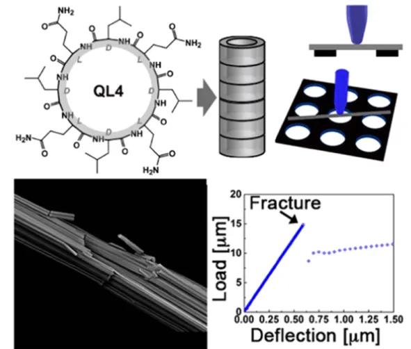
cyclo-[(QDL)
4] Fibre/bundle Formation offibres from nanotubes [93]
cyclo[(WR)n] (n=3–5) Vesicle Ring size-vesicle size relation [94]
Ac-K2(QX)6K2-Am(X=L, F, W and Y) Nanofiber Multidomain peptides with ABA block
motif
wereestimated to be around 41.5 nm, thesame as the dia-meter of the average thickness of the donut-shaped structures. The cationic arginine head group was also used in another SLPdesign to integrate twelve alanine residues [80]. This
oligomeric peptide was synthesized via theNCA poly-merization method rather than thestandard solid phase pep-tide synthesis method to prevent the aggregation problem encountered in the couplings of multiple identical amino acids. 2wt% samples were analysed in cryo-TEM and it was revealed that electrostatic repulsion between terminal arginine residues, together with hydrogen bonding of alanine residues, resulted in the formation of twistedfibrils. In another study, a proline-rich sequence was used instead of alanine-rich domains and one additional arginine residue was conjugated to the hydrophilic segment [81]. The change in the type of
amino acid completely changed the self-assembly mech-anism, leading to the formation of quite different supramo-lecular nanostructures. Owing to the fact that apyrrolidine ring in proline is responsible for the conformational con-straints, this block polypeptide formed stiff helical rod structures, known as polyproline type II (PPII) helices, in aqueous solution. In contrast, arginine residues provided flexibility to the structure. These two components created a rod-coil system that is capable of self-assembling into vesi-cular nanoaggregates with a unimodal distribution of hydro-dynamic radii. Proline residues were used in another study at both N- and C-termini of a nine-residual peptide to reduce the hydrogen bonding network in the β-sheet structure at the strand ends, and to bend the peptide chain by forming turns, favouring the formation of interfibrillar structure [82].
Addi-tionally, thehierarchical arrangement of these supramolecular aggregates exhibited a concentration dependent manner.
Unlike theabove-mentioned SLPs, Castelletto et al introduced for thefirst time ahistidine attached hexaalanine sequence, which had the ability to chelate to transition metal ions, particularly zinc cations, through its imidazole side chain [83]. They compared the structural differences
betweenA6H assemblies prepared in water and aZnCl2 containing solution at neutral and acidic pHs,and observed that while aqueous assemblies self-assembled into short sheets at pH 7, A6H dissolved in ZnCl2 solution formed pseudo-crystalline particles containing plate/tape-like sheets. This workexamined the effect of the number and type of integrated hydrophobic residues on the resulting supramole-cular nanostructure but not the length of the head group. Liu et al extended the hydrophilic amino acid chain length of X6, where X was alanine, valine or leucine, by introducing one to five lysine residues [84]. Extendingthe hydrophilic chain
length changed the surfactant-like character of peptides to amore block-type arrangement. Morphological analysis pointed out that alteration in the hydrophilicity varied the morphology of the peptide assemblies. Peptides with a longer hydrophilic segment but identical hydrophobic tail had a higher CAC, leading to the formation of vesicles rather than nanotubes. Accordingly, peptides having good solubility as well as very high CAC above the working concentration range failed to form any regular self-assembled structures. In another study done by van Hell et al, an approach previously
used by theZhang group [79] was adopted to synthesize
cone-shaped amphiphilic peptides[85]. A conical shape was
provided by the addition of amino acids having bulkier side chains near the charged glutamate residues at the C-terminus. However, here, two different peptides were synthesized with different lengths of hydrophilic domains(Ac-A2V2L3WE2/7 -COOH). They formed nanovesicles ofcomparable size with a diameter of approximately 120 nm, irrespective of the number of glutamate residues found in the head group.
By contrast to the classical design of traditional amphi-philic peptides, thedouble-headed architecture of bolaam-phiphiles, composed of ahydrophobic core and two hydrophilic moieties flankingboth ends of the core, haver-eceivedgrowing interest for their ability to form different self-assembled nanostructures[97–99]. Recently, theHamley
group reported that theydesigned alinear octapeptide (EFL4FE) self-assembled into nanotubes with diameters of 20–30 nm at high concentrations (1wt%), whereas they ten-ded to form sheet-like structures at lower concentrations[86].
In another study conducted by our group, co-assembly of oppositely charged Ac-EFFAAE-Am and Ac-KFFAAK-Am peptides at physiological pH resulted in the formation of nanofibers [87]. Recently, theCui group devised several
phenylalanine containing bolaamphiphiles, (EFFFFE, KFFFFKand EFFFFK), whose terminal charges were modulated to investigate the effect of electrostatic interactions on the resulting polymorphic nanostructure[100]. Thisled to
the twisting of the β-sheet tapes and accordingly the forma-tion of fibrils, twisted ribbons and belts.
Self-assembly can also be induced by utilizing specific chemical functionalities found in the peptide backbone. For instance, amphiphilic gemini peptides, formed as a result of linking two cysteine containing single chain peptides with disulphide bond through oxidation, were designed, and its self-assembly behaviour was compared with that of the corresponding single chain counterparts [88]. Furthermore,
three different hydrophobic amino acids(alanine, valineand isoleucine) were chosen to tune the molecular hydrophobicity of the peptides and to study its effect on the self-assembly together withdimerization. As the hydrophobicity of the single chain peptides increased, spherical aggregates became larger by as much as 5.5±2.2 nm,and even short fibrils with heights of 6.0±1.5 nm wereinitiated in the case of the strongly hydrophobic I3C peptide. After oxidation, additional constraints in molecular conformation were observed due to the gemini geometry, further enhancing intra- and inter-molecular interactions and supporting the self-assembly pro-cess. All three gemini peptides formed thinfibres with heights of approximately 1 nm and length of submicrometers. Hauser et alpreviously demonstrated the synthesis of ultrashort (tri-to heptamer) natural peptides and detailed their self-assembly mechanisms[101,102]. This group has also used cysteine in
a heptapeptide design (Ac-LIVAGKC-Am) to crosslink the system through disulphide bridging in addition to non-covalent interactions[103].
Amphiphatic peptides arranged in an alternating fashion were firstly developed by theZhang group [89, 104, 105].
and β-sheet structures thatfurther self-assembled into well-ordered nanofibres due toelectrostatic interactions. Although Zhang et al classified complementary ionic sides into various moduli based on the alternation of the charges, the(XZXZ)n motif, where X and Z represented polar and nonpolar amino acids, respectively, is the most frequently exploited sequence to direct self-assembly[106,107]. Beta-sheets are generally
arranged into bilayer structures where the hydrophobic side chains are localized in the interior part and the hydrophilic side chains are exposed to the aqueous medium. Lee et al dissected the effect of sequence pattern variation on the self-assembly of amphiphatic peptides by using thefrequently studied Ac-(FKFE)2-Am peptide as a model [90]. They compared the self-assembly behaviour of all four related sequences to that of amodel peptide. The alteration in the amino acid groupings and the disruption of the alternating pattern not only decreased the self-assembly ability but also changed the morphology of the resulting materials. On the other hand, Ramachandran et al worked with two oppositely charged amphiphatic peptides (KVW10 and EVW10) pre-senting either cationic or anionic amino acids to create a co-assembled system driven by charge neutralization [108].
While the individual peptide solutions did not have any fibrillar structures, nanofibrous network formation was observed upon mixing of oppositely charged peptide solu-tions. Schneider et aldesigned a series of amphiphatic pep-tides consisting of two-sided alternating nonpolar valine and polar lysine residues flanking a type II β-turn promoter sequence(-VDPPT-) [109,110]. This turn-inducing sequence
elicited a slight kink in the peptide skeleton and directed the self-assembly of molecules into bilayer β-sheet fibrils toge-therwith acontribution of alternating amino acid residues. In some cases, when some alterations were made in the amino acids, fibril lamination was observed, which is the main consequence of the disruption of hydrogen bonding pattern as well as β-hairpin conformation. Recently, Micklitsch et alalso investigated the influence of nonpolar amino acid units in the hydrophobic face of the hairpin on self-assembly behaviour and fibril morphology [91]. Six different amino
acids were selected based on the varying content of hydro-phobicity,β-sheet propensity and aromaticity. Although the type of amino acid used in the peptide design did not show asignificant influence on the local morphology of the fibrils, the residue type became an important factor when fibrils underwent higher order assembly.
Alternating sequences have been also used in cyclic peptide design to form nanotubular structures. While the assembly ofcyclic peptides was firstproposed by De Santiset al [111], theGhadiri group first demonstrated that
controlled acidification of an even-numbered pH responsive heterochiral cyclic peptide resulted in the formation of nanotubes[112]. The assembly of extended tubular structures
is predominantly driven by intermolecular hydrogen bonding between cyclic peptide rings where carbonyl and amide bonds are perpendicularly oriented to the plane of the ring, while amino acid side chains generate the exterior walls of the nanotube. It was recentlyreported that the morphology of the self-assembled nanostructures of acyclo-[(QDAEDA)2]
peptide was controlled by changing pH, reaction and soni-cation time. Also, the size of resulting cyclic peptide nano-tubes and nanoparticles were shown to be tuned by monitoring multiple parameters such as peptide concentration and PEG modification [92]. Rubin and coworkers showed
that cyclo-[(QDL)4] peptide monomers assembled into large, rod-like structures with diameters up to 2μm and lengths of tens to hundreds of micrometres were composed of thousands of individual nanotubes with adiameter of 2 nm [93]. Unlike
heterochiral cyclic peptides, surfactant-like cyclic peptides firstdeveloped by theParang group can also form self-assembled nanostructures in anaqueous environment depending on the hydrophobicity/charge balance in the peptide sequence [94]. Among the cyclic peptides, cyclo
[WR]n peptides formed vesicle-like structures whose dia-meters increased as the ring size increased. For amore detailed background on the design principles of cyclic pep-tides and related examples, we direct readers to recently published reviews[15,113,114].
Another class of peptide assemblies described by Hart-gerink group is multidomain peptides (MDPs) [115, 116].
The side chains of the peptides arearranged in an ABA block motif, where aB block composed of alternating hydrophilic and hydrophobic amino acid residues isthe major motif driving the self-assembly, and peripheral A blocks employ -charged amino acids to control the self-assembly through electrostatic interactions. The extendedβ-sheet conformation of the peptides leads to creating a facial amphiphile. Packing of two of the hydrophobic faces against each other formsa ‘hydrophobic sandwich’, which further elongatesthrough antiparallelβ-sheet hydrogen bonding. Although the length of the fibres depended on the design of theABA motif, the width and height of the fibres were 6 nm and 2 nm, respec-tively. The self-assembly of MDPs was further explored by introducing aromatic amino acid residues into the core and it was concluded that hydrogen bonding pattern changed depending on the type of substituted aromatic amino acid without affecting the basic nanofiber morphology [95].
4.2. Peptide amphiphiles
The second class of self-assembling peptide molecules reviewed in this section is peptide amphiphiles (PAs),also called lipidated peptides or lipopeptides, which are composed of two main regions, ahydrophobic alkyl tail and ahy-drophilic peptide sequence. These molecules are naturally present in living organisms, which have important roles asinitiators in the signal transduction pathways [117, 118]
and in the host defence mechanisms of bacteria [119]. The
lipidic parts of these molecules are believed to take part in protein–protein and protein–lipid interactions and provide a link to the cellular membrane[118].
Amphiphilicity is the main triggering factor for the self-assembly ofPA monomers into well-defined supramolecular nanostructures and the stability of the resulting assemblies can be further improved by enhancing the amphiphaticity of thePAs. Self-assembly is mediated by a variety of different noncovalent interactions, such as electrostatic interactions
between charged amino acids, hydrogen bonding, π–π stacking and hydrophobic interactions. These interactions may lead to the formation of nanostructures with diverse morphologies, including fibres, micelles, nanotapes, nano-tubes and nanosheets[70]. As described by theIsraelachvili
group, the balance between hydrophobic and electrostatic interactions needs to be taken into account whendetermining the geometry of amphiphiles with minimum free energy [120]. Since an alteration in the hydrophilic segment can lead
to a change in the critical packing parameter[121], it can
affect the morphology of the overall assembly [122, 123].
Also, Velichko et al investigated the contribution of hydro-phobic interactions and hydrogen bonding to the morphology of the resulting PA assemblies by molecular simulation techniques and created a phase diagram displaying distinct morphologies with respect to the corresponding variables [124]. Experimental results were also in good agreement with
those obtained by computational analysis. While ahigh content of intermolecular hydrogen bonds between peptide blocks favoured the formation of cylindrical PA nanofibers in
solution, PA molecules lackinghydrogen bonds had a ten-dency to form micellar structures[125,126]. Tsonchev et al
performed Monte Carlo simulations to demonstrate the role of electrostatic interactions in the self-assembly ofPA nanofi-bers[127]. Later, the proposed self-assembly mechanism was
further studied by Lee et al and Fu et al through molecular dynamic simulations and it was revealed that individual PAs initially formed spherical micelles, and they transformed into long thin fibres by merging with one another due to hydro-phobic interactions [128, 129]. The PA assemblies can be
engineered to display various morphologies and fulfil various functions by modifying the peptide building blocks. The type of hydrophobic group, the choice of amino acids and the secondary structure formed by the system affect the morph-ology of the final PA assembly. Recently published studies presenting the design of PAs are discussed in this section (table 2).
The Stupp groupdemonstrated that aC16H31O-V3A3E3 PA, in which hydrophilicity of the peptide domain increased towards the C-terminus, self-assembled into elongatedfibres Table 2.Recently published studies using peptide amphiphile architectures.
Peptide amphiphile Structure Details References C16H31O-V3A3E3 Nanofiber Salt-induced nanofiber formation [130,131]
C16H31O-F3E3 Twisted/helical ribbon Time/sequence dependent
mor-phological transformation
[132]
C16H31O-(VE)2, C16H31O-V2E2, C16H31O-(EV)2,
C16H31O-E2V2
Nanobelt/nanofiber/ nanoribbon
Structural variation in isomeric peptide amphiphiles
[133]
C16H31O-(VE)n(n=2, 4 and 6) Nanobelt Dimensions of belt structure versus
number of amino acid
[134]
C16H31O-WA4KA4KA4KA Worm-like micelle/
nanofibers Time dependent morphologicaltransition
[135]
C16H31O-A4LSQETFSDLWKLLPEN Worm-like micelle Secondary
structure-supramole-cular structure relation
[136]
C16H31O-KTTKS Flat tape-like/twisted
structure/spherical micelle pH tuned morphology [35] C12H23O-GAGAGAGY Nanofiber/twisted nanoribbon pH tuned morphology [137]
C16H31O-IAAAEEEE-Am Nanofiber pH and concentration dependent
self-assembly
[138]
C16H31O-KKFFVLK Nanotube-helical ribbon/
twisted tapes
Thermo-reversible transition [45]
C12H23O-VFDNFVLK-Am and C12H23O-VVAGE
(mixture)
Nanofiber Nanofiber diameter ≈10–20 nm [139]
C12H23O-P4R4-Am, C12H23O-P4K2R8-Am Nanosphere Diameter≈15–45 nm [140]
KLWVLPKCK2A2V2K(−OC12H23)-Am
KLWVLPKCK2K(−OC22H41)-Am
Nanofiber/nanosphere Importance ofβ-sheet forming region on self-assembly
[141]
CnH(2n−1)O-VRGDV(n=10, 12, 14 and 16) Nanofiber Tail length versus self-assembly at
different pH
[142]
C16H31O-H6-(OEG)4-Am,(OEG)4-H6K(-OC12H23)-Am Nanofiber/nanosphere Position of aliphatic tail-control on
morphology
[39]
C24H47O-GANPNAAG(diacetylene units on C24chain:
4,6- 10,12- or 16-18-positions)
Nanofiber Temperature versusfibre stability [143]
diC16H31O-EQLESIINFEKLTWE-Am Cylindrical micelle 8.0±2.3 nm in diameter,
poly-disperse in length
[144]
qC8-Tat, dC8-Tat, mC8-Tat Nanofiber Mean diameter of
nanofi-ber≈15 nm
after neutralization by pH adjustment or by the salt-mediated screening of charged glutamic residues. Although both for-mulations exhibited similar morphologies, salt-induced PA nanofibers formed stronger intra- and interfiber crosslinks through calcium mediated ionic bridges[130]. Additionally,
highly alignedfibrils were obtained usingthe same design by either injecting aheated PA solution into a saline solution or dragging the same solution through a film of calcium salt solution[131]. The lamellar plaque structure generated during
the heat treatment transformed into bundles of nanofibers upon cooling. Theyalso observed an unexpected morpholo-gical transformation from twisted ribbons into energetically more stable helical ribbons in C16H31O-F3E3PAs after aging them at room temperature for a month[132]. To clarify the
reason behind this phenomenon, asimilar peptide was syn-thesized by replacing phenylalanine residues with alanine residues and it was observed that thenewly designed peptide formed 7–9 nm diametercylindrical fibrils and did not exhibit any change after being aged, indicating that this conforma-tional change is due to the aromatic stacking rearrangement and stems from the existence of the phenyalanine residues. Another critical parameter causing the morphological change in the nanostructure is the concentration. Cui et al designed an alternating hydrophobic and hydrophilic amino acids con-taining PA, C16H31O-VEVE [146]. In the customized mole-cular design, laterally grown PAs self-assembled into one-dimensional ultralong and wide nanobelts, with widths ofthe order of 150 nm and height between 10 and20 nm, presenting peptide epitopes at the surfaces. With a decrease in con-centration, a morphological transition from large nanobelts to twisted nanoribbons was observed by TEM. The same peptide domain was also used in another study, together with its isomeric counterparts, to investigate the importance of pep-tide side chain interactions in the morphology of theresulting supramolecular assembly [133]. Each peptide amphiphile
isomer displayed a different one-dimensional nanostructure, such as nanobelts, nanofibres, twisted and helical ribbonsand nanotubes, due to the switch in the amino acid order. While alternating sequence bearing molecules, VEVE and EVEV, exhibitedflat morphologies as nanobelts and twisted ribbons, respectively, VVEE and EEVV peptide segments led to the formation of two distinct types of nanofibers. In addition to the amino acid order, the effect of the number of valine-glutamic acid(VE) dimeric repeats on the shape and dimen-sion of the supramolecular nanostructures was also system-atically studied[134]. As the length of the peptide sequence
increased, a shift from flat to cylindrical structures was observed. This effect was attributed to a higher tendency to form twistedβ-sheets by longer sequences.
In addition tothe discussed nanostructures, theTirrell group demonstratedtwo different peptides self-assembling into worm-like micelles due to their ɑ-helix propensities [135,136]. In the first design, lysine residues were
symme-trically distributed around the helical structure to provide individual helices in the micellar state [135]. The results
acquired from cryo-TEM, IR andcircular dichroism(CD) revealed that time dependent morphological transition from spherical to worm-like micelles was observed after days, and
they wereeventuallytransformed into long nanofibrillar structures with anouter diameter of ∼10 nm. In the second design, they explored the influence of hydrophobic amino acid residues on the self-assembly of PAs by conjugating four alanine residues between the palmitic acid and oligopeptide sequence [136]. The inclusion of hydrophobic alanine
resi-dues has resulted in major changes in the morphology of the PA structures, which formed worm-like micelles rather than nanoribbons, and were rich inβ-sheets rather than α-helices. Nanostructured features of the PAs can be manipulated by varying the external parameters such as pH, temperature or ionic strength. The effect of pH and temperature on the self-assembly behaviour of C16H31O-KTTKS was investigated in two separate studies [35, 147]. Whilst an increase in the
temperature gave rise to the formation of micellar structures rather than extended tape structure due to the disruption of hydrogen bonding, a gradual decrease in pH transformed nanotapes into twistedfibrils at pH 4 and spherical micelles at pH 2. Similarly, pH dependent morphological alteration was reported for C12H23O-GAGAGAGY PAs[137]. Cylindrical nanofibers observed at pH 9 transformed into twisted ribbons at pH 4, most likely due to the neutralization of carboxylic acid, and subsequently the weakening of electrostatic inter-actions, leading to the stacking of the β-sheet laminates. Furthermore, Ghosh et al demonstrated that the tendency of PAs to respond to pH change can be programmed by chan-ging the position of single hydrophobic amino acid residue in a short amphiphilic peptide[138]. As isoleucine moved away
from the alkyl tail, nanofiber formation was favoured over spherical micelles due to the enhanced propensity forβ-sheet formation. In another study carried out by theHamley group, thermo-responsive structural change was reported in PAs decorated with a KLVFF core motif that induced self-assembly throughπ–π stacking and hydrophobic interactions [45]. A reversible unwinding transition was observed between
twisted tapes and nanotubes/ribbons in a temperature dependent manner.
Co-assembly of two oppositely charged PAs can also lead to some modifications in the self-assembled nanos-tructures due to the electrostatic interactions occurring between charged amino acid residues. Cylindrical nanofibers were observed by theStupp group upon mixing oppositely charged palmitoylated peptides [148, 149]. Our group have
also reported that lauric acid conjugated lysine and glutamic acid bearing peptides self-assembled into high aspect ratio, one-dimensional fibrillar nanostructures [54]. We
alsoob-served that short bioactive sequences exploited in the PA design did not alter the shape of theresulting supramolecular assemblies [139, 150, 151]. TheHamley group studied the
co-assembly of oppositely charged PAs by mixing them at different ratios [152]. Co-assembly driven mainly by
elec-trostatic interactions brought about enhanced β-sheet forma-tion compared to samples prepared with a single component and resulted in the formation of nanotapes.
The shape of the nanostructure can be tuned by either varying or omitting the β-sheet forming domain [74,140,153]. Guler et al studied the importance of hydrogen
assemblies by areplacing trileucine sequence with triproline [74]. Due to the β-sheet breaking nature of proline residues,
spherical aggregates were observed instead of the cylindrical nanofibers obtained with the former system. Mammadov et al [154]and Mumcuogluet al [140] also used proline
residues to form micellar structures by disrupting theβ-sheet secondary structure to study the importance of nanostructural features in tuning theimmune response and to develop an efficient delivery vehicle for the transfection of oligonucleo-tides, respectively. Recently, Moyer et al demonstrated that nanofibre formation was disrupted when a β-sheet forming region(A2V2) was removed from the peptide sequence. This truncated sequence self-assembled into nanospheres with diameters in the range5–10 nm depending on the length of the tail region[141].
The conjugation of an alkyl group to a peptide segment was shown to enhance the thermal stability of the corresp-onding structure[14,118]. The influence of alkyl tail length,
its position and number on the self-assembly of PAs was investigated in detail by several research groups. The van Hest group examined the effect of alkyl tail length on the stability of β-sheet assemblies of GANPNAAG [155] and
KTVIIE[156] peptides. As the length of the tail increased, the
thermal stability of the PAs improved and the transition from β-sheet to random coil was observed at higher temperatures. Furthermore, Xu et al synthesized four PAs with different tail lengths by using the VRGDV sequence as the peptide domain and showed that PAs with shorter tail lengths did not main-tainstability at higher pH due to weaker hydrophobic inter-actions[142]. On the other hand, therelationship between the
sitewhere thehydrophobic segment is attached to the peptide domain and the resulting nanostructure shape was investi-gated by incorporating an alkyl tail on either theN- or C-terminus of an oligo-histidine peptide sequence,and it was revealed that cylindrical and spherical morphologies were obtained at physiological conditions, respectively [39]. He
et al reported that while two aliphatic tails attached to one side of the peptide sequence formed long fibrils, pepti-desconjugated from either side self-assembled into short twistedfibrils [157]. In addition, van del Heuvel et al showed
that thestability of the self-assembled nanofibres can be controlled by changing the position of the diacetylene moiety on the hydrophobic tail without varyingthe length of the tail [143]. TheTirrell group introduced adialkyl chain containing
peptides [158] and investigated the effect of the length of
double tail on the thermal stability of thecollagen-like structure in the peptide amphiphile [159]. They further
stu-died the effects of both the number and the length of alkyl tail on the nanostructured features of model collagen peptide amphiphiles by using a combination of small-angle neutron scattering and cryo-TEM[160]. In their design,dialkyl
con-jugatedT-cell epitope bearing apeptide self-assembled into 8 nm diameter of cylindrical micelles[144]. In addition to the
dialkyl tail bearing peptide amphiphiles, theCui group designed three Tat-peptide conjugates with different numbers of octanoic acid tails[145]. It was reported that while single
and double tail containing peptides could not form any well-defined nanostructures, four aliphatic tails attached one
self-assembled into afibrillar structure with adiameter of 15 nm, most likely due to enhanced intermolecular hydrogen bonding.
4.3. Peptides containing other hybrid systems
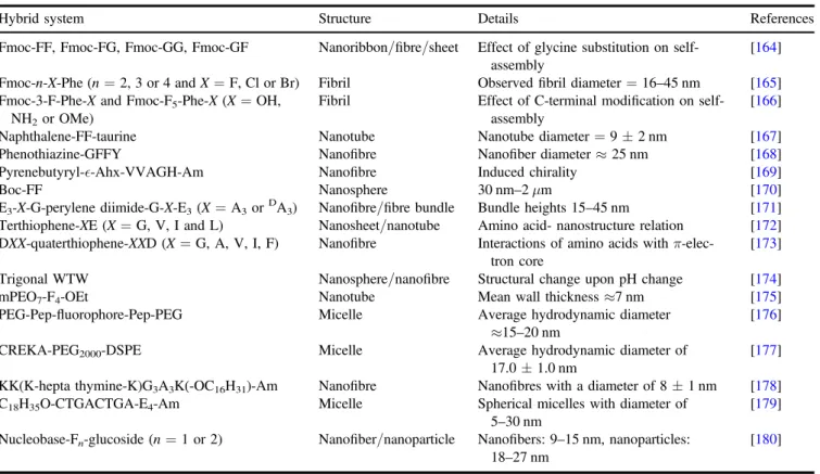
In this section, peptidic hybrid systems composed of various chemical groups besides peptides such as lipids, polymers, nucleobases, saccharides, aromatic groups, halogen elements, etc are elaborated to understand the key factors affecting the self-assembly behaviour of the related molecules. Table 3
depicts the self-assembled forms of recently reported hybrid peptide systems and gives additional informationregarding their assembly properties. Peptides were modified not only by changing their amino acid sequences, but also by using cap-ping molecules at the N- or C-termini or by inserting a linker between peptide domains. These modifications have been used both to investigate the mechanisms involved in self-assembly behaviour and to control the structural features of peptide assemblies for practical purposes[161–163].
Using aromatic moieties at the N-terminus of the peptide is another strategy fordrivingself-assembly by providing amphiphilicity to the structure. Unlike the assembly mech-anism of aliphatic peptide amphiphiles, here aromatic moiety dominantly directs the self-assembly by its planar structure and resulting geometric restrictions due to the preferred stacking arrangements [181]. Vegners et al synthesized an
aromatic peptide amphiphile, Fmoc-LD, which folded into filamentous micelles [182]. Various dipeptide combinations
have beenexploited togetherwith theFmoc (9-fluor-enylmethoxycarbonyl) unit since then [67, 183–186]. Tang
et al examined the self-assembly behaviour of Fmoc-dipep-tides composed of acombination of phenylalanine and gly-cine residues and revealed that the flexibility of the overall structure as well as the resultant conformation were affected bythe amino acid type and sequence, leading to the formation of structurally different assemblies[164]. In
addition,Fmoc-dipeptides, Fmoc-tripeptides, andtetra- and pentapeptide derivatives were also presented, which exhibited nanofibrous or nanotubular structures[187–190]. In addition, the effect of
ahalogen substituent and the position of its substitution on the aromatic side chain of Fmoc-F were systematically stu-died by theNilsson group [165]. Since the identity of halogen
and its position on the benzyl ring affected the self-assembly process and the morphology of the self-assembled fibrillar structures, it was hypothesized that the mentioned variations lead to the perturbation in the energetics of the aromaticπ–π interactions that driveself-assembly. The influence of the C-terminal modifications on fluorinated Fmoc-F derivatives (Fmoc-F5-Phe-OH and Fmoc-3-F-Phe-OH) was further investigated by the same group [166]. The hydrophobicity
and hydrogen bonding capacity of the C-terminus were adjusted by altering thecarboxylic acid group of theC-terminal with the amide and methyl ester derivatives. Their assembly kinetics and resultant structural features were stu-died either one by one or in co-assembled form. They recently reported that theelectronic nature of the substituent anchored
to the benzyl ring of Fmoc-F also exerted influence on the self-assembly[191].
Other than the Fmoc group, naphthalene [167, 192],
phenothiazine [168], pyrene [169], carboxybenzyl [193],
azobenzene [194, 195], naproxen [196] and benzimidazole
[197] moieties were also utilized as aromatic capping at the
N-terminus facilitating the self-assembly. TheXu group demonstrated that the incorporation of anon-proteinaceous amino acid, taurineand naphthalene into the peptide back-bone resulted in the formation of nanotubes, nanofibers or nanoribbons depending on the assembly conditions, such as temperature, sonication and pH [167]. In another study, Ou
et al showed that phenothiazine conjugated tetrapeptide (-GFFY) prepared at 0.2wt% formed uniform nanofibres with diameters of 25 nm [168]. On the other hand, pyrene was
conjugated to the peptide sequence (VVAGH) with an ò-aminohexanoic acid linker by our group. The formation of the nanofibres, observed in TEM, was mainly driven by the solvophobic effect [169]. The assembly also led to helical
organization of pyrene within thehydrophobic core and toacquiring chirality, which was verified by fluorescence spectroscopy and CD measurements.
Unlike aromatic capping groups, thetert-butoxycarbonyl (Boc) moiety was also anchored to short peptide sequences [170, 187]. TheGazit group studied the structural and
mechanical properties of theBoc-FF peptide and a morpho-logical characterization conducted by SEM and AFM revealed that Boc-FF peptide dissolved in organic solvent self-assembled into spherical structures whose diameters showed variation from 30 nm to 2μm. The same sequence
was also investigated in terms of the effect of solvent on the resultant morphology and a structural change was observed from spheres to nanotubes when water was used instead of ethanol as adiluent for the peptides prepared in HFIP [198].
Peptide π-electron rich systems are another well-studied class of hybrid peptide systems, and their self-assembled architectures exhibitdifferent photophysical, electricaland mechanical properties. The π-electron rich systems can be integrated into peptide backbone in many different ways: as a side chain [199], as a linker between two peptide sequences
[200] or as a capping molecule at the N-terminus [201]. While
designing a peptide and π-electron construct, anamino acid sequence/π-electron system pair should be selected carefully in terms of itsenergetic contributions to the self-assembly in order to form supramolecular structures with improved elec-tron transport properties [202, 203]. TheHodgkiss group
examined the thermodynamic factors affecting the self-assembly of peptide–perylene diimide conjugates by altering the peptide hydrophobicity, charge density, length, stereo-centre inversion and amphiphilic substitution[171]. The
self-assembly of alanine-rich conjugates resulted in the formation of bundles with heights of 15–45 nm. Moreover, changes in peptide hydrophobicity and the insertion of an imide hexyl group resulted in the most prominent effect with respect to changes in thermodynamic properties. As another π-con-jugated system, aterthiophene moiety was coupled to four different dipeptides by theStupp group, which assembled into one-dimensional nanostructures [172]. The main
con-sideration in their design was the selection of hydrophobic amino acids, based on van der Waals volume and Table 3.Recently reported hybrid peptide systems.
Hybrid system Structure Details References
Fmoc-FF, Fmoc-FG, Fmoc-GG, Fmoc-GF Nanoribbon/fibre/sheet Effect of glycine substitution on self-assembly
[164]
Fmoc-n-X-Phe(n=2, 3 or 4 and X=F, Cl or Br) Fibril Observedfibril diameter =16–45 nm [165]
Fmoc-3-F-Phe-X and Fmoc-F5-Phe-X(X=OH,
NH2or OMe)
Fibril Effect of C-terminal modification on self-assembly
[166]
Naphthalene-FF-taurine Nanotube Nanotube diameter=9±2 nm [167]
Phenothiazine-GFFY Nanofibre Nanofiber diameter ≈ 25 nm [168]
Pyrenebutyryl-ò-Ahx-VVAGH-Am Nanofibre Induced chirality [169]
Boc-FF Nanosphere 30 nm–2 μm [170]
E3-X-G-perylene diimide-G-X-E3(X=A3orDA3) Nanofibre/fibre bundle Bundle heights 15–45 nm [171]
Terthiophene-XE(X=G, V, I and L) Nanosheet/nanotube Amino acid- nanostructure relation [172]
DXX-quaterthiophene-XXD(X=G, A, V, I, F) Nanofibre Interactions of amino acids with π-elec-tron core
[173]
Trigonal WTW Nanosphere/nanofibre Structural change upon pH change [174]
mPEO7-F4-OEt Nanotube Mean wall thickness≈7 nm [175]
PEG-Pep-fluorophore-Pep-PEG Micelle Average hydrodynamic diameter
≈15–20 nm [
176]
CREKA-PEG2000-DSPE Micelle Average hydrodynamic diameter of
17.0±1.0 nm
[177]
KK(K-hepta thymine-K)G3A3K(-OC16H31)-Am Nanofibre Nanofibres with a diameter of 8±1 nm [178]
C18H35O-CTGACTGA-E4-Am Micelle Spherical micelles with diameter of
5–30 nm [
179]
Nucleobase-Fn-glucoside(n=1 or 2) Nanofiber/nanoparticle Nanofibers: 9–15 nm, nanoparticles:
18–27 nm [
hydrophobicity values, to study the impact of steric hindrance and hydrophobic van der Waals interactions on the supra-molecular structure. On the other hand, theTovar group worked with peptide–π–peptide triblock molecules [204], and
demonstrated that acidic solutions of quaterthiophene-peptide (DXX) conjugates prepared at 0.1wt% exhibited one-dimen-sional nanostructures[173]. Some variations in the width or
persistence length of the nanostructure were obtained depending on the type of amino acid residue used adjacent to theπ-core.
Peptides can alsobe functionalized on an organic tem-plate to provide symmetry in the structure and yield an ordered architecture. Different molecules such as tris (2-ami-noethyl)amine (TREN) [205],
1,3,5-tris(aminomethyl)-2,4,6-triethylbenzene [206], and pentaazacyclopentadecane [207],
were used to conjugate different peptides to these trigonal or pentagonal templates to afford nanoassemblies with different structures. TheKimizuka group designed aC3-symmetric peptide conjugate(Trigonal WTW) where an iodoacetoami-dated core was used for the coupling of an8-mer tryptophane zipper-forming peptide, and its self-assembly resulted in the formation of nanospheres and nanofibres depending on the pH[174].
Amphiphilicity in the hybrid structure can alternatively be achieved by anchoring hydrophilic polymers to hydro-phobic peptide segments, or vice versa, through different chemistries to afford copolymer conjugates of varied struc-tures[208]. The nature of the polymer, the chemical
hetero-geneity of the peptide, the conjugation site, and the reaction medium can affect the structure, dynamics and function of the corresponding hybrid system. A number of polymer–peptide conjugates have been reported, where polymers with different composition, number andlength ofside chains were used to create different forms of nanostructures[209,210]. Tzokova
et al showed amPEO7–F4–OEt conjugate via click chemistry and its self-assembly afforded nanotubes with a mean internal diameter of 3 nm and a mean wall thickness of roughly 7 nm [175]. While tetrapeptide domain provided the antiparallel
β-sheet structure, PEO chains assisted to the stabilization of the structure. It is also possible to synthesize amphiphilic triblock polymer–peptide conjugates. TheMandal group designed anABA type triblock conjugate composed of a hydrophobic peptide part and ahydrophilic PEG moiety at both ends of the peptide domain[176]. Micelle formation was observed when
thehybrid system underwent aggregation in water and the diameter of resultant spherical micelles was found to be 15–20 nm based on TEM imaging. In addition to the polymer conjugation, phospholipids have been used to create peptide hybrids [177, 211–213]. Tirrell and coworkers reported
amulticomponent system (peptide-PEG-DSPE) that self-assembled into micellar structures with an average hydro-dynamic diameter of 17±1 nm in anaqueous environment [177]. While the peptide domain served as the polar head
group, PEG and DSPE were used as the spacer and tail, respectively.
Similar to lipids, nucleobases or saccharides can also be conjugated to the peptides [186]. A peptide nucleic acid
(PNA)/peptide amphiphile conjugate, designed by theStupp
group, was constructed on a solid support and apoly-thymine PNA heptamer was built on the peptide amphiphile whose self-assembly resulted in the formation of uniform nanofibers with a diameter of 8±1 nm [178]. Similar approach was
later used by Zhang group to synthesize a series of peptide nucleic acid amphiphiles (PNAA) containing different hydrophobic and hydrophilic moieties [179]. The
self-assembly driven mainly by the base pair stacking of PNAA duplexes and intermolecular noncovalent interactions led to the formation of spherical micelles with adiameter of about 5−30 nm. On the other hand, Xu et al preferred utilizing simple building blocks, basically a nucleobase, amino acid and saccharide, rather than using peptide nucleic acid deri-vatives or glyco amino acid units to generate molecular architectures with defined structures [180]. Supramolecular
assemblies obtained by pH increment exhibited differences in morphology due to the number and type of presenting amino acid and nucleobase residues. Saccharide incorporated pep-tide conjugates were also developed, which can form uniform self-assembled nanostructures. They can be synthesized through a number of synthetic approaches, including the use of glycosylated amino acids [214], direct conjugation of the
sugar to the peptide backbone or amino acid side chains through amino bonds [215], or the use of a linker molecule
for indirect conjugation[216,217].
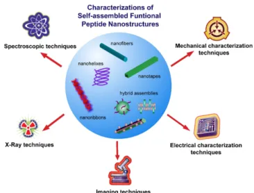
5. Characterization of self-assembled peptide nanostructures
Characterization of the self-assembled nanoscale architectures can be done usingsophisticated techniques due to their complex and dynamic nanoscale nature. Advances in mate-rials characterization methods, in conjunction with recent developments in nanotechnology, have resulted in the development of advanced tools and approaches for the investigation of chemical, physical, electrical and mechanical properties. These techniques can be classified as spectro-scopic, x-ray, microspectro-scopic, electrical and mechanical char-acterizations with various examples leading the material developments in different fields (figure2).
5.1. Spectroscopic analysis
Spectroscopic methods are used to understand the chemical and physical characteristics of nanoscale peptide organiza-tions, such as their bond properties, vibrational modes and covalent and noncovalent interactions. Although the basic principles of the spectroscopic techniques rely on the detec-tion of the transidetec-tions on the molecules such as nuclear spin, molecular vibrations or electronic states,the detection meth-ods for these transitions become different depending on the radiation source. The common spectroscopic techniques such as nuclear magnetic resonance (NMR), Fourier transform infrared spectroscopy (FTIR), Raman and CD spectroscopy have been widely used for the analysis of different self-assembled peptide nanostructures designed for various applications. NMR is a powerful technique enabling the