Electrospinning of nanofibers from non-polymeric systems: Electrospun
nanofibers from native cyclodextrins
Asli Celebioglu, Tamer Uyar
⇑UNAM-Institute of Materials Science & Nanotechnology, Bilkent University, Ankara 06800, Turkey
a r t i c l e
i n f o
Article history: Received 7 January 2013 Accepted 7 April 2013 Available online 6 May 2013 Keywords:
Cyclodextrin Electrospinning Nanofiber
a b s t r a c t
Electrospinning of nanofibers from non-polymeric systems is rather challenging, yet in this study, we have successfully performed electrospinning of nanofibers from two of the native cyclodextrins (CDs);
a-CD and b-CD. Electrospinning was carried out for highly concentrated solutions ofa-CD (120% up to 160%, w/v) and b-CD (120% up to 150%, w/v) in basic aqueous system. At optimal concentration level, the electrospinning of CD solutions yielded bead-free uniform CD nanofibers without using carrier poly-meric matrix. Similar to polypoly-meric systems, the electrospinning of CD solutions resulted in different mor-phologies and average fiber diameters depending on the CD type and CD concentration. The dynamic light scattering (DLS) and rheology measurements were performed in order to examine the electrospin-nability of CD solutions. The existence of CD aggregates via hydrogen bonding and very high solution vis-cosity and viscoelastic solid-like behavior of CD solutions were found to be the key factors for obtaining bead-free nanofibers from CDs. The addition of urea disrupted CD aggregates and lowered the viscosity significantly, and therefore, the urea-added CD solutions yielded beaded fibers and/or beads. Although the as-received CDs in powder form are crystalline, the structural analyses by XRD and HR-TEM indicated that electrospun CD nanofibers have amorphous characteristic without showing any particular orienta-tion or crystalline aggregaorienta-tion of CD molecules.
Ó 2013 Elsevier Inc. All rights reserved.
1. Introduction
Electrospun nanofibers have received great attention due to their unique properties including extremely high surface area, very light-weight, nano-porous features, and design flexibility for spe-cific physical and chemical functionalizations[1–3]. Unlike other nanofiber fabrication techniques, electrospinning is straightfor-ward, versatile, and very cost-effective for producing nanofibers from variety of materials such as polymers, polymer blends, sol– gels, emulsions, suspensions, and composite structures [1–7]. Due to their unique properties, electrospun nanofibers and their nanowebs are very promising candidates to be used in various fields such as membranes/filters, biotechnology, textiles, sensors, energy, electronics, and environment [1–3,7–15]. In electrospin-ning, high molecular weight polymer and high solution concentra-tions are generally used since entanglements and overlapping between polymer chains play crucial role for producing bead-free and uniform nanofibers [16–18]. For the electrospinnability of solutions, the importance of elasticity and relaxation time rather than molecular entanglements has also been reported [19,20]. The electrospinning of low molecular weight molecules is quite challenging. Yet, few examples have been recently reported about
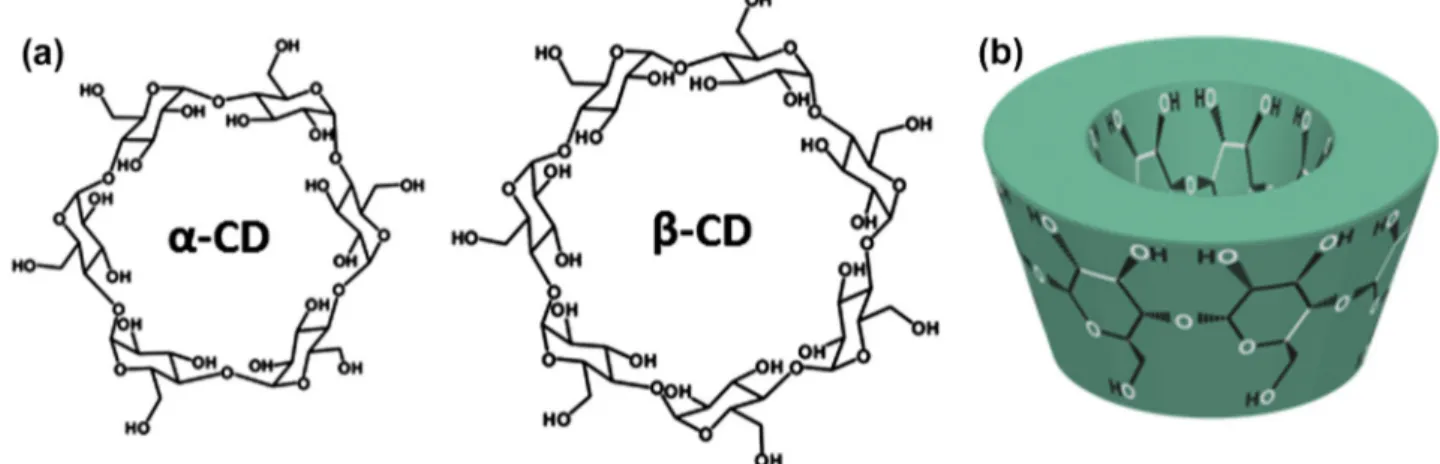
the electrospinning of non-polymeric systems such as phospholip-ids[21], diphenylalanine[22], gemini surfactant[23], heteroditop-ic monomer[24], and chemically modified cyclodextrins[25–29]. Besides solution electrospinning of low molecular weight com-pounds[21–29], melt electrospinning of small molecules such as 1,3,5-cyclohexane and 1,3,5-benzenetrisamides into fibers was also reported very recently [30]. These studies showed that the key factor for the electrospinnability of these low molecular weight molecules is the self-assembly and self-aggregation of the mole-cules in their concentrated solutions[21–29]or in their melt state [30]. In short, electrospinning of nanofibers from supramolecular structures is quite interesting and needs further investigation since electrospun supramolecular nanofibers can be used for designing and constructing new advanced functional nanofibrous materials. Electrospinning of nanofibers from polymers, which usually re-quire organic solvents, is very common; however, electrospinning of nanofibers from renewable resources is always attractive and on demand. Cyclodextrins (CDs) are naturally occurring water-sol-uble oligosaccharides, and they are produced from the enzymatic conversion of starch. CDs are one of the most studied supramolec-ular systems, and therefore, electrospinning of nanofibers from CDs would be quite fascinating due to their non-covalent host–guest inclusion complexation capability with other molecules. CDs are cyclic oligosaccharides having a toroid-shaped molecular structure (Fig. 1) which can form intriguing supramolecular structures by
0021-9797/$ - see front matter Ó 2013 Elsevier Inc. All rights reserved. http://dx.doi.org/10.1016/j.jcis.2013.04.034
⇑ Corresponding author. Fax: +90 312266 4365. E-mail address:[email protected](T. Uyar).
Contents lists available atSciVerse ScienceDirect
Journal of Colloid and Interface Science
w w w . e l s e v i e r . c o m / l o c a t e / j c i sforming host–guest inclusion complexes (ICs) with variety of molecules[31–33]. The native CDs have either six, seven, or eight glucopyranose units in their molecular structure and are named as
a
-CD, b-CD, andc
-CD, respectively (Fig. 1)[34]. Due to their non-covalent host–guest complexation ability, CDs are quite appli-cable in various field such as pharmaceuticals, functional foods, cosmetics, home/personal care, textiles, and filtration/separation/ purification systems[31,32,35–37].CDs can self-assemble and form considerable aggregates via intermolecular hydrogen bonding in their concentrated solutions [38,39]. So, this makes it possible for the electrospinning of nanof-ibers from highly concentrated CD solutions. In fact, in our very re-cent studies, we have successfully performed electrospinning of nanofibers from three different chemically modified CDs: hydroxy-propyl-beta-cyclodextrin (HPbCD), hydroxypropyl-gamma-cyclo-dextrin (HP
c
CD), and methyl-beta-cyclodextrin (MbCD)[25–27]. The chemically modified CDs have very high solubility when com-pared to native CDs (a
-CD, b-CD, andc
-CD); therefore, the prepa-ration of highly concentrated modified CD solutions in water or polar organic solvents was possible, and the electrospinning of nanofibers from these solutions were successful[25–27]. Follow-ing our studies, the electrospinnFollow-ing of nanofibers from HPbCD was also reported by two other research groups[28,29]. However, up to date, the electrospinning of native CD still remains a chal-lenge due to their low solubility when compared to that of chem-ically modified CDs. Native CDs are soluble in water, yet, their solubility is rather limited due to the presence of intramolecular hydrogen bonding within the CD molecule, which prevents the for-mation of hydrogen bond with surrounding water molecules [38,40]. Nevertheless, in this study, we were able to obtain highly concentrated homogeneous solutions ofa
-CD and b-CD by using 10% (w/v) NaOH aqueous solvent system. By using these highly concentrated CD solutions, our efforts on electrospinning of nanof-ibers from native CDs,a
-CD and b-CD, without using any carrier polymer matrix were quite successful. As anticipated, electrospin-ning of nanofibers from CDs would be very attractive since nanofi-brous materials with unique properties can be produced by combining the very large surface area of nanofibers with specific functionality of the CDs.2. Experimental 2.1. Materials
The
a
-CD and b-CD were obtained from Wacker Chemie AG (Germany) commercially. Sodium hydroxide (NaOH) (Fluka,P98%, small beads) was purchased. The de-ionized water was from the Millipore Milli-Q Ultrapure Water System. All the materi-als were used without any purification.
2.2. Preparation of CD solutions and electrospinning of CD nanofibers The electrospinning of CD (
a
-CD and b-CD) solutions was determined according to their solubility limits by trying different solvent types. Electrospinning requires highly concentrated CD solutions; therefore, in order to obtain highly concentrated homo-geneous CD solutions, various solvent types including water, N,N-dimethylformamide (DMF), dimethylacetamide (DMAc), and dimethyl sulfoxide (DMSO) as well as their mixtures were tested, and finally, we were able to get highly concentrated homogeneousa
-CD and b-CD solutions by using 10% NaOH aqueous solution. Therefore,a
-CD and b-CD nanofibers were electrospun from their 10% NaOH aqueous solution. The solutions were started to be pre-pared from 120% (w/v) CD concentration, and they were increased to the optimized level until the bead-free uniform nanofibers were produced. The homogenous nanofibers ofa
-CD and b-CD were ob-tained at 160% and 150% (w/v) concentrations, respectively. The ef-fect of urea on the fiber formation was investigated by adding 20% urea (w/w, with respect to CD) into the optimized concentration of CD solutions. For electrospinning, each CD solution was loaded into a syringe having metallic needle with 0.45 mm inner diameter, and it was positioned onto the syringe pump (Model: SP 101IZ, WPI) in order to control the flow of the solution. One of the electrodes of the high voltage power supply (Matsusada Precision, AU Series) was clamped to the metal needle tip, and the other was clamped to the grounded cylindrical aluminum collector. The electrospin-ning parameters were optimized as follows: applied voltage: 15 kV, tip-to-collector distance: 10 cm, and the solution flow rate: 1 mL/h. The electrospinning was performed in a horizontal position in an enclosed Plexiglass box at 25 °C and 25% relative humidity conditions.2.3. Measurements and characterization
The rheological properties of the CD solutions were studied by using rheometer (Anton Paar Physica MCR 301) equipped with cone-plate configuration (spindle type CP40-2). While the shear rate sweep tests were carried out at the range of 0–100 s 1, the frequency sweep oscillatory tests were done at the 0–10 Hz fre-quency range. For viscoelastic property measurements, the linear viscoelastic region was determined as 0.01% strain value. The aggregate size of CD solutions was measured by using Nano-ZS
Zetasizer dynamic light scattering (DLS) system (Malvern Instru-ments). Before the measurement, the temperature of cell was equilibrated at 25 °C for 2 min. The conductivity of the CD solu-tions was measured with a Multiparameter meter InoLab-Multi 720 (WTW) at room temperature. The scanning electron micro-scope (SEM) (Quanta 200 FEG, FEI) was used for the morphological characterization of nanofibers. Samples were sputtered with 5 nm Au/Pd (PECS-682) and the average fiber diameter (AFD) was calcu-lated from the SEM images by analyzing at least 100 fibers. X-ray
diffractometer (XRD) (X’Pert powder diffractometer, PANalytical) was used to determine the X-ray diffraction pattern of as-received CD powders and CD nanofibers with Cu K
a
radiation in the 2h = 5– 30° range. Transmission electron microscope (TEM) and high res-olution transmission electron microscope (HR-TEM) (FEI – Tecnai G2F30) were also used for the detailed structural characterization of CD nanofibers. For TEM imaging, HC200 TEM grids were at-tached on the aluminum foil and the samples were collected on the grids.Table 1
The characteristics of CD (a-CD and b-CD) 10% (w/v) NaOH aqueous solutions (DLS measurements of CD solutions at 25 °C summarizing the average diameter (nm) and polydispersity index (PDI) of CD aggregates), average fiber diameter, fiber diameter range, and fiber morphology of the electrospun CD nanofibers.
Solutions Viscosity (Pa s) Conductivity (mS cm 1) Intensity-average diameter/d (nm) – PDI
Average fiber diameter (fiber diameter range) (nm)
Fiber morphology
120%a-CD 0.16 14.33 5.50/0.55 – Bead structures
130%a-CD 0.20 11.22 6.04/0.47 – Bead structures with few fiber
formation
140%a-CD 0.26 9.06 6.38/0.40 – Bead structures with fiber
formation
150%a-CD 0.32 7.42 7.30/0.70 180 ± 80 (50–560) Beaded nanofibers
160%a-CD 0.43 5.93 8.29/0.48 375 ± 150 (80–940) Bead-free nanofibers
160%a-CD + 20% urea
0.33 4.42 6.04/0.81 – Bead structures with few fiber
formation
120% b-CD 0.13 12.05 4.98/0.36 – Bead structures
130% b-CD 0.16 9.77 5.14/0.53 – Bead structures with fiber
formation 140% b-CD 0.24 8.21 5.27/0.46 175 ± 70 (90–420) Beaded nanofibers 150% b-CD 0.33 7.25 5.96/0.43 220 ± 90 (90–460) Bead-free nanofibers 150% b-CD + 20% urea 0.15 5.53 4.91/0.92 – Bead structures
Fig. 2. The representative SEM images ofa-CD nanofibers obtained from 10% (w/v) NaOH aqueous solution at (a) 120% (w/v), (b) 130% (w/v), (c) 140% (w/v), (d) 150% (w/v), (e) 160% (w/v)a-CD concentrations and (f) the SEM image of bead structures with few fiber formation as a results of adding 20% (w/w) urea to the 160% (w/v)a-CD solution. (g) Fiber diameter distribution of electrospun nanofibers produced from 160% (w/v)a-CD concentration.
3. Results and discussion
3.1. Electrospinning of nanofibers from native cyclodextrins (
a
-CD and b-CD)Highly concentrated
a
-CD (120% up to 160%, w/v) and b-CD (120% up to 150%, w/v) solutions were prepared by dissolving the CDs in 10% (w/v) NaOH aqueous solution. The solution prepa-ration, electrospinning parameters/conditions, and the details of the characterization techniques are summarized in the experimen-tal section. The characteristics of the CD solutions, the morphology and the average fiber diameters of the electrospun CD nanofibers are summarized inTable 1. The representative scanning electron microscope (SEM) images of the electrospun CD (a
-CD and b-CD) nanofibers produced from different solution concentrations are de-picted inFigs. 2 and 3. The electrospinning of CD solutions resulted in different average fiber diameters and morphologies depending on the CD type and CD solution concentrations.We observed that the electrospinning of native CDs is quite similar to polymeric systems in which the solvent type, the solu-tion concentrasolu-tion/viscosity, and the solusolu-tion conductivity played a key role for the electrospinnability of CD nanofibers. That is, only the CD solutions having optimal concentration/viscosity and con-ductivity values were able to be electrospun into uniform nanofi-bers without any bead structure. In addition, we observed that the morphology and the diameter of the resulting electrospun nanofibers significantly vary with the type of CDs since the viscos-ity and conductivviscos-ity of the solutions were different from each other. In our very recent studies, similar findings were also ob-served in the case of electrospinning of chemically modified CDs (HPbCD, HP
c
CD, and MbCD)[27].For
a
-CD, vastly beaded structure along with very few nanofiber structures was obtained when 120% (w/v)a
-CD solution was elec-trospun (Fig. 2a). The bead structures were gradually eliminated as thea
-CD concentration was increased from 120% through 140% (w/v) (Fig. 2b and c), and nanofibers having very few beads were obtained at 150% (w/v) concentration (Fig. 2d). Finally, bead-free and uniforma
-CD nanofibers (Fig. 2e) having fiber diameter in the range of 80–940 nm (Fig. 2g) with an average fiber diameter (AFD) of 375 ± 150 nm were produced from the electrospinning of 160% (w/v)a
-CD solution. Likewise, we have also optimizedthe electrospinning of modified CDs (HPbCD, HP
c
CD, and MbCD) in water at 160% (w/v) CD concentration for obtaining bead-free nanofibers in our previously study[27].The bead structures at lower concentrations were due to the low solution viscosity and the presence of inadequate amount of
a
-CD aggregates, and therefore, mostly beads were formed instead of fully stretched fibers. This is because of the destabilization of the electrified jet during the electrospinning process. Bead-free nanof-ibers were obtained from 160% (w/v)a
-CD, suggesting that this is the optimal concentration where the solution viscosity and the amount and the size of the CD aggregates were sufficient for the electrospinning of bead-free and uniform nanofiber. This finding is very similar to electrospinning of polymeric systems in which low concentration of polymer solution yielded beaded fiber struc-ture due to the lack of polymer chain entanglements and overlap-ping, and mostly higher polymer concentrations are required for uniform fiber formation[1,2,41]. The rheology and DLS measure-ments (Figs. 4 and 5) indicated that as the concentration ofa
-CD increased from 120% through 160% (w/v), the solution became more viscous and considerable amount of CD aggregates was formed, and therefore, bead-freea
-CD nanofibers were success-fully obtained.Similarly, the electrospinning of b-CD solutions at low concen-tration (120% through 140%, w/v) yielded beaded nanofibers (Fig. 3a–c), but bead-free uniform b-CD nanofibers were achieved from the electrospinning of 150% (w/v) b-CD solution (Fig. 3d). The nanofiber electrospun from 150% (w/v) b-CD solution has AFD of 220 ± 90 nm and the fiber diameter was in the range of 90–460 nm (Fig. 3f).
In our previous study dealing with electrospinning of chemi-cally modified CDs (HPbCD, HP
c
CD, and MbCD) [27], we have shown that the morphologies and the diameters of the electrospun CD fibers were very much dependent on the CD type, CD concen-tration, and solvent type since the aggregation, viscosity, viscoelas-tic properties, and the solution conductivity were varied for each CD system. Here, it was also evident that electrospinning of native CDs was similar to chemically modified CDs where the fiber mor-phologies and the diameters were vary depending on the CD type and CD concentration. The optimal concentration for obtaining bead-free nanofibers was found as 160% and 150% fora
-CD and b-CD, respectively. As discussed in the later section, the rheologicalFig. 3. The representative SEM images of CD nanofibers obtained from 10% (w/v) NaOH aqueous solution at (a) 120% (w/v), (b) 130% (w/v), (c) 140% (w/v), (d) 150% (w/v) b-CD concentrations, and (e) the SEM image of bead structures as a results of adding 20% (w/w) urea to the 150% (w/v) b-b-CD solution. (f) Fiber diameter distribution of electrospun nanofibers produced from 150% (w/v) b-CD concentration.
properties of the
a
-CD and b-CD solutions were very similar, and therefore, fiber diameters close to each other were obtained, yet b-CD nanofibers were somewhat thinner than thea
-CD nanofibers since b-CD solution has lower concentration/viscosity and high conductivity values, and therefore, the electrified jet was subjected to more stretching during the electrospinning process[1,2]. These findings elucidated that electrospinning of CDs is quite similar to polymeric systems where the high solution concentration/viscosity is crucial for producing bead-free nanofibers from CDs.The addition of urea reduces the intermolecular interaction be-tween CD molecules by interrupting hydrogen bonding, and this resulted in disruption of self-association and aggregation of CDs in the solution[42,43]. The addition of urea (20% (w/w) with re-spect to CD) to the optimized concentrations of CDs caused signif-icant decrease in the viscosity and aggregate size (Table 1) since the urea breaks the hydrogen bonds among the CD molecules and disrupts the CD aggregates; therefore, electrospinning of CD solutions containing urea yielded beaded fibers or beads instead of uniform fibers (Figs. 2f and3e).
3.2. Cyclodextrin aggregation in solutions
The DLS measurements were performed in order to investigate the aggregation of CDs in their highly concentrated solutions. The size distribution of CD aggregates measured by DLS for
a
-CD and b-CD solutions is given inFig. 4, and the data are summarized in Table 1. The DLS measurements elucidated the presence of self-aggregateda
-CD molecules in their concentrated solutions (Fig. 4a andTable 1). The size of thea
-CD aggregates increased from 5.5 nm to 8.29 nm as the concentration was increased from 120% to 160% (w/v). The size of the aggregates decreased from 8.29 nm to 6.04 nm for the urea-added 160% (w/v)a
-CD solution confirming the depletion of thea
-CD aggregates with the addition of urea. In the case of b-CD, the aggregate size was increased from 4.98 nm to 5.96 nm as the b-CD concentration was increased from 120% to 150% (w/v) (Table 1). The size of thea
-CD aggregates wasslightly bigger than the size of the b-CD aggregates in the same solution concentration. The DLS and viscosity data are in good agreement with each other, and slight higher viscosity values were recorded for
a
-CD solutions when compared to b-CD solutions ow-ing to the higher amount of CD aggregates and their growow-ing sizes as the concentration of the CD increased in the solution. In brief, the DLS measurements confirmed that the CD molecules form sub-stantial amount of aggregates in their high concentrations, and this resulted in full stretching of electrified jet yielding bead-free uni-form fiber uni-formation without using any polymeric carrier. 3.3. Rheology of cyclodextrin solutionsThe rheological properties of CD solutions were examined by performing shear rate sweep viscosity and frequency sweep oscil-lation tests. Both of these tests were run for CD solutions having 120% (w/v) concentration up to the optimal concentration level (160% and 150% (w/v) for
a
-CD and b-CD, respectively) in which the bead-free nanofibers were obtained. The viscosity of CD solu-tions as a function of shear rate was displayed inFig. 5. It was ob-served that the viscosity of CD solution for the same concentration was being independent of shear rate indicating that the CD sys-tems show characteristic of a Newtonian fluid. In addition, the solution viscosity of CDs increases with the increasing CD concen-tration due to the presence of higher number of aggregates and their growing sizes (seeTable 1 andFig. 4). While the viscosity of 120% (w/v)a
-CD solution was measured as 0.16 Pa s, it reached to 0.43 Pa s for the 160% (w/v)a
-CD. The viscosity of b-CD solution was increased from 0.13 Pa s (120%, w/v) to 0.33 Pa s (150%, w/v). As mentioned previously, the addition of urea reduces hydrogen bonding between CD molecules and therefore disturbs the CD aggregation[42,43]. Here, the addition of 20% (w/w) urea (with re-spect to CD) to the optimized concentrations of CDs caused signif-icant decline in their viscosity levels. The viscosity of the CD solutions was decreased from 0.43 Pa s to 0.33 Pa s and 0.33 Pa s to 0.15 Pa s fora
-CD and b-CD, respectively. Similarly, the size ofFig. 4. Size distribution of (a)a-CD aggregates for 120%, 130%, 140%, 150%, 160% (w/v)a-CD and 20% (w/w) urea-added 160% (w/v)a-CD in 10% (w/v) NaOH aqueous solution; (b) 120%, 130%, 140%, 150% (w/v) b-CD, and 20% (w/w) urea-added 150% (w/v) b-CD in 10% (w/v) NaOH aqueous solution.
Fig. 5. Viscosity versus shear rate graphs of (a) 120% (j), 130% ( ), 140% ( ), 150% ( ), 160% ( ) and 20% (w/w) urea-added 160% (w/v) ( )a-CD solutions; (b) 120% (j), 130% ( ), 140% ( ), 150% ( ) ,and 20% (w/w) urea-added 150% (w/v) ( ) b-CD solutions.
the CD aggregates measured by DLS was decreased from 8.29 nm to 6.04 nm and 5.96 nm to 4.91 nm for
a
-CD and b-CD solutions containing urea, respectively. SEM images (Figs.2f and3e) clearly showed that the electrospinnability of CD solutions was signicantly affected with the addition of urea. Beads along with few fi-ber structures were obtained from the electrospinning of urea-containinga
-CD and b-CD solutions (Figs.2f and3e). It was also worth mentioning that the electrospinning of fibers from urea-con-taining CD solutions became much difficult since the jet breakup observed very frequently and much less fibers were deposited on the collector.The viscoelastic properties of the CD solutions were studied by the frequency sweep oscillation measurements.Fig. 6shows the storage and loss modulus of CD solutions as a function of fre-quency. For all CD solutions at given concentration, the storage modulus was higher than that of the loss modulus in the whole fre-quency range indicating that these highly concentrated CD solu-tions behave as a viscoelastic solid [44]. At a fixed CD concentration, both storage and loss modulus were steady under the applied frequency range, but increasing the concentration of CD solutions resulted in higher storage and loss modulus values. We have observed no crossover point between storage and loss modulus. In addition, the storage modulus was always higher than the loss modulus elucidated that CD solutions show predominantly solid-like behavior for CD systems in all studied concentrations. Moreover, at higher CD concentrations, the gap between storage and loss modulus became larger. This is mostly because the so-lid-like part becomes more pronounced as the concentration in-creases due to the increasing aggregation. Fig. 6a and b also indicates that the viscoelastic properties of
a
-CD and b-CD solu-tions are almost same with the similar modulus value for the same concentration. The oscillation tests were also applied to urea-added CD solutions, and the significant decrease for the modulus values was observed for the optimized concentrations of both CD types. As mentioned before, the addition of urea disturbs the hydrogen bonding among the CD molecules, so the size of the aggregates get smaller which results in solid density depression in the solution that supplies the elastic property. The modulus of urea-added 160% (w/v)a
-CD and 150% (w/v) b-CD solutions was decreased significantly and reached to the modulus value similar to their 120% (w/v) concentration level. As expected, the electros-pinning of the urea-added 160% (w/v)a
-CD and 150% (w/v) b-CD solutions resulted in similar fiber morphologies when compared to their 120% (w/v) solutions. In brief, the electrospinning of the urea-added CD solution did not result in uniform and bead-free nanofiber formation simply because of the break up of the electri-fied jet due to the presence of insufficient CD aggregates (Figs.2fand3e). Therefore, it is evident that the presence of substantial amount of CD aggregates via hydrogen bonding plays a key role for the electrospinning of uniform and bead-free nanofibers from CD solutions.
3.4. Structural characterization of the electrospun CD nanofibers The
a
-CD and b-CD are crystalline material having crystal struc-tures referred to as cage or channel type[34]. In the cage-type pack-ing, the CD molecules are in arrangement in which the cavity of each CD molecule is blocked by neighboring molecules. In the case of channel-type packing, the CD molecules are aligned and stacked on top of each other forming long cylindrical channels, and this channel-type packing is commonly observed when CDs form inclu-sion complexes with guest molecules [34]. Here, we have per-formed the structural analyses of CD nanofibers by XRD. The as-receiveda
-CD and b-CD in powder form are crystalline having a cage-type packing[34]. However, the XRD data of electrospuna
-CD and b--CD nanofibers showed that the diffraction peaks were ab-sent and only halo XRD pattern was observed indicating that these CD nanofibers have amorphous structure without any particular crystal formation (Fig. 7). The HR-TEM further proved that CD mol-ecules were randomly distributed without showing any presence of particular orientation or crystalline aggregation in the fibers (Fig. 8). It is likely that CD molecules could not pack into crystal structure because of the rapid evaporation of solvent along with the very fast and continuous stretching of the jet during the elec-trospinning process. We have also performed the XRD study for these CD nanofibers after 1 year of their production, and we ob-served that the amorphous structure was protected fora
-CD andFig. 6. Frequency depended storage modulus G’ (filled symbols) and loss modulus G’’ (open symbols) graphs of, (a)120%, 130%, 140%, 150%, 160%, and 20% (w/w) urea-added 160% (w/v)a-CD solutions; (b) 120%, 130%, 140%, 150%, and 20% (w/w) urea-added 150% (w/v) b-CD solutions.
Fig. 7. XRD patterns of as-receiveda-CD and b-CD powders and,a-CD and b-CD nanofibers.
b-CD nanofibers, and no transformation to cage-type packing oc-curred which is the most stable crystalline form for native CDs [34].
4. Conclusions
We have successfully produced nanofibers from native CDs of
a
-CD and b-CD via electrospinning technique without using any carrier polymeric matrix. At lower CD concentrations, beaded nanofibers were obtained, but, as the CD concentrations were in-creased, the transformation from beaded to bead-free nanofibers was observed. The optimal concentrations for producing bead-free nanofibers were 160% and 150% (w/v) fora
-CD and b-CD, respec-tively. The DLS and rheology measurements indicated the presence of self-associated CD aggregates in the solutions. It was found that the high solution viscosity and viscoelastic solid-like behavior of CD solutions played a key role for the electrospinning of bead-free nanofibers from these two native CD types. The size of CD aggre-gates got smaller, and the viscosity of the CD solutions decreased significantly with the addition of urea. This situation affected the electrospinnability of CD solutions and beaded fibers and/or beads were obtained. The XRD and HR-TEM studies revealed that electro-spun CD nanofibers were in amorphous state.CDs are naturally occurring non-toxic cyclic oligosaccharides having host–guest inclusion complexation capability with other molecules. So, electrospinning of CD nanofibers would have unique properties by combining the very large surface area of nanofibers with specific functionality of the CD. For instance, native CDs have different cavity size which can allow selective inclusion complexa-tion with various molecules of different sizes. In addicomplexa-tion, CDs are already being used in various fields including pharmaceutical, food, textiles, biotechnology, and filtration/separation systems. Hence,
CDs in the form of nanofibrous web may extend the use of CDs in the aforementioned areas.
Acknowledgments
State Planning Organization (DPT) of Turkey is acknowledged for the support of UNAM-Institute of Materials Science & Nano-technology. T. Uyar acknowledges EU FP7-PEOPLE-2009-RG Marie Curie-IRG for funding NANOWEB (PIRG06-GA-2009-256428) pro-ject. A. Celebioglu acknowledges TUBITAK-BIDEB for the national PhD study scholarship.
References
[1]S. Ramakrishna, K. Fujihara, W. Teo, T. Lim, Z. Ma, An Introduction to Electrospinning and Nanofibers, World Scientific Publishing Company, 2005. [2]J.H. Wendorff, S. Agarwal, A. Greiner, Electrospinning: Materials, Processing,
and Applications, Wiley-VCH, Germany, 2012.
[3]S. Agarwal, J.H. Wendorff, A. Greiner, Macromol. Rapid. Commun. 31 (2010) 1317.
[4]A. Greiner, J.H. Wendorff, Angew. Chem. Int. Ed. 46 (2007) 5670. [5]S. Agarwal, A. Greiner, Polym. Adv. Technol. 22 (2011) 372.
[6]S. Zhan, D. Chen, X. Jiao, S. Liu, J. Colloid Interface Sci. 308 (2007) 265. [7]C. Shin, J. Colloid Interface Sci. 302 (2006) 267.
[8]X. Li, M. Cao, H. Zhang, L. Zhou, S. Cheng, J. Colloid Interface Sci. 382 (2012) 28. [9]C. Su, C. Shao, Y. Liu, J. Colloid Interface Sci. 359 (2011) 220.
[10] S. Ramakrishna, R. Jose, P. Archana, A. Nair, R. Balamurugan, J. Venugopal, W. Teo, J. Mater. Sci. 45 (2010) 6283.
[11]S. Agarwal, A. Greiner, J.H. Wendorff, Adv. Funct. Mater. 19 (2009) 2863. [12]J. Xie, X. Li, Y. Xia, Macromol. Rapid Commun. 29 (2008) 1775.
[13]R. Rahul, S. Kumar, R. Sridhar, J. Sundaramurthy, V.J. Reddy, S. Ramakrishna, J. Mater. Chem. 22 (2012) 12953.
[14]V. Thavasi, G. Singh, S. Ramakrishna, Energy Environ. Sci. 1 (2008) 205. [15]K. Yoon, B.S. Hsiao, B. Chu, J. Mater. Chem. 18 (2008) 5326.
[16]M.G. McKee, G.L. Wilkes, R.H. Colby, T.E. Long, Macromolecules 37 (2004) 1760.
[17]P. Gupta, C. Elkins, T.E. Long, G.L. Wilkes, Polymer 46 (2005) 4799. [18]S.L. Shenoy, W.D. Bates, H.L. Frisch, G.E. Wnek, Polymer 46 (2005) 3372. [19]J.H. Yu, S.V. Fridrikh, G.C. Rutledge, Polymer 47 (2006) 4789.
[20] S. Talwar, A.S. Krishnan, J.P. Hinestroza, B. Pourdeyhimi, S.A. Khan, Macromolecules 43 (2010) 7650.
[21]M.G. McKee, J.M. Layman, M.P. Cashion, T.E. Long, Science 311 (2006) 353. [22]G. Singh, A.M. Bittner, S. Loscher, N. Malinowski, K. Kern, Adv. Mater. 20 (2008)
2332.
[23]M.P. Cashion, X. Li, Y. Geng, M.T. Hunley, T.E. Long, Langmuir 26 (2010) 678. [24]X. Yan, M. Zhou, J. Chen, X. Chi, S. Dong, M. Zhang, X. Ding, Y. Yu, S. Shaod, F.
Huang, Chem. Commun. 47 (2011) 7086.
[25]A. Celebioglu, T. Uyar, Chem. Commun. 46 (2010) 6903. [26]A. Celebioglu, T. Uyar, Langmuir 27 (2011) 6218. [27]A. Celebioglu, T. Uyar, Nanoscale 4 (2012) 621.
[28]J.L. Manasco, C.D. Saquing, C. Tang, S.A. Khan, RSC Adv. 2 (2012) 3778. [29]W. Zhang, M. Chen, B. Zha, G. Diao, Phys. Chem. Chem. Phys. 14 (2012) 9729. [30] J.C. Singer, R. Giesa, H.W. Schmidt, Soft Matter 8 (2012) 9972.
[31]J. Szejtli, Chem. Rev. 98 (1998) 1743. [32]A.R. Hedges, Chem. Rev. 98 (1998) 2035.
[33]L.X. Song, L. Bai, X.M. Xu, J. He, S.Z. Pan, Chem. Rev. 253 (2009) 1276. [34]W. Saenger, J. Jacob, K. Gessler, T. Steiner, D. Hoffmann, H. Sanbe, K. Koizumi,
S.M. Smith, T. Takaha, Chem. Rev. 98 (1998) 1787.
[35]Í.X. García-Zubiri, G. González-Gaitano, J.R. Isasi, J. Colloid Interface Sci. 307 (2007) 64.
[36]H.M.C. Marques, Flavour Frag. J. 25 (2010) 313. [37]N. Morin-Crini, G. Crini, Prog. Polym. Sci. 38 (2013) 344.
[38]M. Messner, S. Kurkov, P. Jansook, T. Loftsson, Int. J. Pharm. 387 (2010) 199. [39]M. Bonini, S. Rossi, G. Karlsson, M. Almgren, P. Nostro, P. Baglioni, Langmuir 22
(2006) 1478.
[40] J. Szejtli, J. Mater. Chem. 7 (1997) 575. [41]T. Uyar, F. Besenbacher, Polymer 49 (2008) 5336. [42]L. Szente, J. Szejtli, G. Kis, J. Pharm. Sci. 87 (1998) 778. [43]W. Hinze, D. Pharr, Z. Fu, W. Burkert, Anal. Chem. 61 (1989) 422. [44]O. Jazkewitsch, H. Ritter, Macromolecules 44 (2011) 375. Fig. 8. TEM and HR-TEM images of electrospun (a-i, ii)a-CD and (b-i, ii) b-CD