Original Article / Özgün Makale
Effect of mitomycin-C applied through different approaches
following tracheal surgery on development of granulation tissue
and level of nephrotoxicity in rats
Sıçanlarda trakea cerrahisi sonrası farklı yaklaşımlarla uygulanan mitomisin-C’nin granülasyon dokusu gelişimi ve nefrotoksisite düzeyi üzerine etkisi
Mustafa Kuzucuoğlu1, Ruhan Deniz Topuz2, Eren Altun3
Received: April 09, 2018 Accepted: July 25, 2018
Institution where the research was done:
Balıkesir University Health Application and Research Hospital, Balıkesir, Turkey Medicine Faculty of Trakya University, Edirne, Turkey
Author Affiliations:
1Department of Thoracic Surgery, Balıkesir University Health Application and Research Hospital, Balıkesir, Turkey 2Department of Thoracic Surgery, Medicine Faculty of Trakya University, Edirne, Turkey
3Department of Pathology, Balıkesir University Health Application and Research Hospital, Balıkesir, Turkey
Correspondence: Mustafa Kuzucuoğlu, MD. Balıkesir Üniversitesi Sağlık Uygulama ve Araştırma Hastanesi Göğüs Cerrahisi Kliniği, 10145 Balıkesir, Turkey.
Tel: +90 266 - 612 10 10 e-mail: [email protected]
©2019 All right reserved by the Turkish Society of Cardiovascular Surgery.
Kuzucuoğlu M, Topuz RD, Altun E. Effect of mitomycin-C applied through different approaches following tracheal surgery on development of granulation tissue and level of nephrotoxicity in rats. Turk Gogus Kalp Dama 2019;27(1):73-79
Cite this article as:
ÖZ
Amaç: Bu çalışmada trakea cerrahisi uygulanan sıçanlarda farklı ilaç uygulama yaklaşımları ile uygulanan mitomisin-C’nin cerrahi bölgede granülasyon dokusu gelişimi ve böbrek fonksiyonları üzerine etkisi değerlendirildi.
Çalışma planı: Elli erkek erişkin Sprague Dawley sıçan (ağırlık ort. 200 g-300 g) beş gruba ayrıldı. Tüm gruplara anestezi altında trakeanın beşinci ve altıncı kıkırdak halkası arasından kesi yapıldı ve kesi 6/0 monofilaman emilebilir dikiş ile primer olarak onarıldı. Deney hayvanlarına atandıkları topikal, intraperitoneal enjeksiyon, yara kenarlarına enjeksiyon ve inhalasyon yolu gruplarına uygun olarak bir doz 0.5 mg mitomisin-C uygulandı. Kontrol grubu kabul edilen bir gruba mitomisin-C uygulanmadı. Cerrahiden dört hafta sonra sıçanlar sakrifiye edildi ve takiben trakeaları çıkarıldı. Trakea dokuları histopatolojik olarak epitelizasyon, fibrozis, fibroblast miktarı, angiogenezis ve enflamatuar yanıt açısından değerlendirildi. Trakea çapı ve duvar kalınlığı ölçüldü. Kan üre ve kreatinin değerleri nefrotoksisite açısından değerlendirildi ve sıçanlar immunhistokimyasal olarak glomeruler patoloji açısından incelendi.
Bul gu lar: Kontrol grubuna kıyasla topikal mitomisin-C uygulanan grupta epitelizasyon istatistiksel olarak anlamlı derecede yavaşladı (p<0.01), trakea çapı istatistiksel olarak anlamlı derecede daha genişti (p<0.05) ve trakea duvar kalınlığı anlamlı olarak daha kalındı (p<0.01).
Sonuç:Trakea cerrahisi sonrası topikal olarak uygulanan mitomisin-C epitelizasyonu yavaşlatarak granülasyon dokusu gelişimini azaltmakta ve trakea çapının daha geniş kalmasını sağlamaktadır.
Anahtarsözcükler: Granülasyon dokusu; mitomisin-C; cerrahi, trakea.
ABSTRACT
Background: This study aims to evaluate the effect of mitomycin-C applied through different drug administration approaches on the development of granulation tissue in the field of surgery and renal functions in rats which underwent tracheal surgery.
Methods: Fifty male adult Sprague Dawley rats (weighing mean 200 g to 300 g) were divided into five groups. An incision was performed between the fifth and sixth cartilage ring of the trachea in all groups under anesthesia and the incision was primarily repaired with a 6/0 monofilament absorbable suture. A single dose of mitomycin-C 0.5 mg was applied in the experimental animals appropriate with their assigned groups as topical, intraperitoneal injection, injection to the wound edges, and through inhalation. No mitomycin-C was administered in one group which was accepted as the control group. Rats were sacrificed four weeks after surgery and their tracheas were excised subsequently. Tracheal tissue samples were histopathologically evaluated in terms of epithelization, fibrosis, amount of fibroblasts, angiogenesis, and inflammatory response. Diameter and wall thickness of the tracheas were measured. Blood urea and creatinine levels were evaluated for nephrotoxicity, and the rats were immunohistochemically examined for glomerular pathology.
Results: Epithelization was statistically significantly decelerated (p<0.01), diameter of the trachea was statistically significantly larger (p<0.05), and wall thickness of the trachea was significantly thicker in the group with topical mitomycin-C application compared to the control group (p<0.01).
Conclusion:Topically applied mitomycin-C following tracheal surgery slows down epithelization and, thus, decreases the development of granulation tissue and maintains a wider diameter of the trachea.
Tracheal stenosis is defined as congenital or acquired stenosis of the trachea. While congenital causes include tracheomalacia, cardiovascular anomalies and congenital tumors, post-intubation tracheal stenosis (PITS) is the leading cause for acquired disease.[1] Post-intubation tracheal stenosis
was first described by Cooper and Grillo.[2] Excessive
pressure applied to the trachea, mucosal damage, segmental ischemia and necrosis in the concerned segment and granulation tissue formation in an ischemic region are responsible for intubation-related tracheal stenosis. Likelihood of PITS development varies between 0.6-21%.[1,3]
Post-intubation tracheal stenosis is still a serious problem despite the developments in protective measures such as measurement of the intubation tube cuff pressure, developments in patient monitorization, and early tracheostomy procedure in patients who need longer term intensive care unit care. No current algorithm or standard approach is available for the treatment of PITS. Dilation using rigid bronchoscopy, tracheal stents, interventional bronchoscopic procedures and surgical resection are utilized.[3-5] The
most important problem in the surgical procedures is granulation tissue, which develops after surgery and leads to repeat procedures.
Various studies have been conducted and various chemical agents have been tried for prevention of postoperative granulation tissue formation. Mitomycin-C (MMC), which is an anti-neoplastic agent, has an important place among these. Studies have revealed that MMC reduces postoperative granulation tissue; however, the agent has been administered locally in all of the studies. Therefore, in this study, we aimed to evaluate the effect of MMC applied through different drug administration approaches on the development of granulation tissue in the field of surgery and renal functions in rats which underwent tracheal surgery.
MATERIALS AND METHODS
The study was conducted at Balıkesir University, Faculty of Medicine in December 2016. Fifty male adult Sprague Dawley rats (weighing mean 200 g to 300 g) were used after having obtained approval from The Animals Ethics Committee. The rats were divided into five equal groups according to the MMC administration method. All rats were administered ketamine (100 mg/kg) and xylazine hydrochloride (10 mg/kg) via the intra-muscular route before the surgical procedure. After anesthesia induction, the animals were placed in the supine position on the
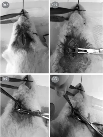
operating table after the neck region had been shaved. The neck was placed in hyper-extension in order to provide airway patency. The thyroid tissue was lateralized with a vertical incision in the neck and the trachea was reached through the sternocleidomastoid muscle. The dissected trachea was suspended through a dissector. A horizontal incision was performed so as to include 60% of the tracheal ring between the fifth and sixth tracheal cartilages with scalpel and the trachea was repaired with separated stitches using 6/0 mono-filament absorbable sutures (Figure 1).
The first group was the control group and no medications were administered. Group 2 was administered 0.5 mg MMC via the intra-peritoneal route after surgery. Group 3 was injected a total of 0.5 mg MMC to the incision lines before repair after trachea dissection. Group 4 was administered 0.5 mg MMC topically onto the repaired tracheal tissue after surgery through being absorbed in cotton. The rats in group 5 inhaled 0.5 mg nebulized MMC by dilution
Figure 1. (a) Neck was placed in hyper-extension in order to provide airway patency. (b) Dissected trachea was suspended through a dissector. (c) A horizontal incision was performed so as to include 60% of tracheal ring. (d) Trachea was repaired using 6/0 mono-filament absorbable sutures.
(a)
(c)
(b)
with 3 mL distilled water after the animals had completely recovered from anesthesia and respiration had returned to normal after surgery.
The rats were fed with pellet feed and water ad libitum at proper humidity and 24°C room temperature, 12 hours night and day cycle for four weeks. At the end of four weeks, blood samples were drawn from the hearts under anesthesia and the animals were sacrificed thereafter. Urea and creatinine were examined in order to investigate the nephrotoxic effect of MMC. The tracheas and kidneys of rats were removed for histopathological and immunohistochemical analyses.
All tissues were soaked in 10% formaldehyde for 24 hours and embedded in paraffin blocks by a pathologist blinded to the study. Thin sections of 5 mm were obtained from each block. Epithelization, inflammation and fibrosis were evaluated with hematoxylin-eosin and Masson trichrome staining. Epithelial coating and epithelial regeneration at the trauma site were evaluated. The scoring system and subjective scales were used for detection of fibrosis, epithelization and collagen accumulation, amount of fibroblast, inflammatory response and degree of angiogenesis. The wall thickness and diameter of the
tracheas were calculated using the D4.30 program with NIS element under Nikon Eclipse Ci bio-microscope (Nikon, Amsterdam, Netherlands).
Statistical analysis
Each MMC group was compared with the control group for statistical analyses. The Mann-Whitney U test was used for the statistical analyses and a p level of <0.05 was accepted as statistically significant.
RESULTS
The study was begun with 50 rats in five groups. One rat in the control group died due to pulmonary edema during the surgical procedure. While urea and creatinine levels were found to be non-elevated in 49 rats that were scarified after drawing blood from the heart, no glomerular pathology was determined in the immunohistochemical analyses of the rats.
The excised surgical region was evaluated using the immunohistochemical method. The amount of fibroblasts, level of fibrosis, angiogenesis, collagen structure and inflammatory response were evaluated. No difference was determined between the control group and the study groups. When epithelization
3 4 * 3 3 2 2 1 1 0 0 2 1 0 0.5 1.5 2.5 0.0 Control Control Control Control Intraperitoneal Inhalation Incisional Local D eg re e D eg re e D eg re e D eg re e 1.0 2.0 (a) (c) (b) (d)
Figure 2. Epithelization. (a) Inhalation group compared to control group (p=0.17), (b) local group compared to control group (p=0.0266), (c) intraperitoneal group compared to control group (p=0.3289), (d) incisional group compared to control group (p=0.2335). Mann-Whitney U test.
Groups
Groups
Groups
was evaluated histopathologically, epithelization was determined to have slowed down significantly in the topical MMC group compared to the control group (p<0.01) (Figure 2) and no significant difference was determined in the other groups.
The diameter of the trachea was measured microscopically from epithelium to epithelium and wall thickness was measured from inward to outward. The diameter of the trachea (p<0.05) (Figure 3) and the wall thickness of the trachea (p<0.01) (Figure 4) were determined to have significantly increased in the topical MMC group compared to the control group and no significant difference was determined in the other groups. When cartilage overlapping was evaluated, no significant difference was determined in any groups compared to the control group.
DISCUSSION
Rigid bronchoscopy and dilation, bronchoscopic laser, graft inter-position, tracheal stent insertion and
tracheal resections are applied for the treatment of tracheal stenosis.[6] Tracheal resection is accepted as
the definitive treatment method in benign tracheal stenosis.[3,6,7] Grillo et al.[3] performed tracheal
resection on 503 patients due to benign tracheal resection. Of these, 324 underwent anastomosis from trachea to trachea, 117 patients underwent crico-tracheal anastomosis and 62 patients underwent anastomosis from the trachea to the thyroid or the crico-thyroid cartilage. The authors have reported favorable or satisfactory outcomes in 93.7% of the patients.
El-Fattah et al.[8] reported favorable or satisfactory
outcomes in 85.7% of the patients in their study in which they performed crico-tracheal resection in 35 patients with Grade 3 and 4 benign tracheal resection. Likewise, Nandakumar et al.[6] yielded
successful outcomes in 92.85% of 14 patients who had undergone tracheal stenosis. On the other hand, Ulusan et al.[9] reported the success rate as 68.2% in 22 patients
2000 2000 2000 2000 1500 1500 1500 1500 1000 1000 1000 1000 500 500 500 500 0 0 0 0 Control Control Control Control Inhalation Groups Groups Groups Groups Local * Intraperitoneal T ra ch ea d ia m et er ( µ m) T ra ch ea d ia m et er ( µ m) T ra ch ea d ia m et er ( µ m) T ra ch ea d ia m et er ( µ m) Incisional (d) (b) (c) (a)
Figure 3. Diameter of trachea (a) Inhalation group compared to control group (p=0.3920), (b) local group compared to control group (p=0.0433), (c) incisional group compared to control group (p=0.9438), (d) intraperitoneal group compared to control group (p>0.9999). Mann-Whitney U test.
who had undergone tracheal stenosis due to PITS, while Abbasidezfouli et al.[10] demonstrated a success
rate of 61.5% in their study conducted with 26 patients. Altuntas et al.[11] found a significant relationship
between the duration of mechanical ventilation and the length of trachea resected in patients with benign tracheal stenosis treated with tracheal resection. They showed that patients who underwent mechanical ventilation for over 10 days required longer tracheal resection in their treatment.
Granulation tissue at anastomosis line is among the most common complications seen in patients who undergo tracheal resection and the likelihood of this complication is higher when non-absorbable sutures are used.[12] While granulation tissue developed in
49 out of 503 patients in the study of Grillo et al.,[3]
44 of them were included in the 186 patients who had undergone the operation until 1978. The authors reported that granulation tissue formation decreased after Vicryl polyglactin had been introduced instead of Tevdek polyester.
Granulation tissue and re-stenosis developed postoperatively in five out of 22 patients (22.7%) in the study of Ulusan et al.[9] and these patients were treated
with tracheal silicon stent after dilation with rigid bronchoscopy.
The rate of granulation tissue formation was determined as 31.4% in the study of El-Fattah et al.[8] Eleven patients who had developed granulation
tissue were treated with repeated rigid bronchoscopic dilations under general anesthesia and administered 2 mg/mL topical MMC. Nandakumar et al.[6]
administered 1 mg/mL MMC topically onto the surgical line and did not detect granulation tissue formation in their postoperative follow-up in their study including 14 patients who had undergone tracheal resection.
Mitomycin-C is an antibiotic and anti-neoplastic agent obtained from streptomyces caespitosus. MMC, which shows its anti-neoplastic effect through impairment of deoxyribonucleic acid synthesis, was shown to impair fibroblast proliferation and activity when used locally.[13,14] Khaw et al.[15] revealed that a
Figure 4. Wall thickness of trachea (a) Inhalation group compared to control group (p=0.0649), (b) local group compared to control group (p=0.0041), (c) incisional group compared to control group (p=0.3493), (d) intraperitoneal group compared to control group (p=0.6447). Mann-Whitney U test.
Control Inhalation Groups Control Groups W al l t hic kn es s ( µ m) W al l t hic kn es s ( µ m) W al l t hic kn es s ( µ m) W al l t hic kn es s ( µ m) Incisional 800 800 800 800 1000 1000 600 600 600 600 400 400 400 400 200 200 200 200 0 0 0 0 Control Groups Intraperitoneal Control Groups Local ** (a) (c) (b) (d)
single dose of locally administered MMC eradicated epithelization during 36 hours in glaucoma surgery.
Moreover, Arbag et al.[13] investigated granulation
tissue formation after tracheostomy in their study conducted with 16 New Zealand rabbits. The investigators divided the rabbits into two groups; while they only opened tracheostomy in one group, they administered 0.4 mg/mL MMC topically for five minutes during surgery in the other group. The fibroblast level and the subepithelial tissue thickness were shown to be significantly lower in the MMC group; however, the inflammatory cell count remained unchanged.
Lampus et al.[16] investigated 12 Wistar rats
undergoing anoplasty in two groups, one of which was the control group. While one group underwent the anoplasty procedure only, the other group was administered 0.5 mg/mL MMC topically for five minutes following the anoplasty procedure. The authors reported that the fibroblast amount was significantly lower, and that epithelization and collagen accumulation were higher in the MMC group.
Furthermore, Iñiguez-Cuadra et al.[17] divided
18 rabbits into three groups and performed tracheal resection and end-to-end anastomosis in all. Group 1 received saline solution. Group 2 received a low dose of MMC (0.2 mg/mL) and group 3 received a high dose of MMC (0.5 mg/mL) for five minutes. Rabbits that were followed-up for one year underwent 54 surgical procedures, 35 bronchoscopies and 18 biopsies. No difference was determined between the saline solution group and the low dose MMC group. The airway diameter was found to be narrower and fibrosis development was found to be at a higher extent in the high dose MMC group compared to the other group.
Ward and April[18] carried out laser and rigid
bronchoscopy in five patients who had developed granulation and scar tissue after surgery. Three of the patients were administered low dose MMC together with dilation due to recurrent granulation tissue formation and they were seen not to require dilation in their two-year follow-up after MMC administration.
Until today, MMC has not been used topically for prevention of granulation tissue after tracheal resection. We aimed to investigate the effect of MMC administered through different routes on the development of granulation tissue and found that only topical MMC could prevent granulation tissue, and that the airway diameter was larger in patients who were administered topical MMC. We have also
demonstrated that a single dose of MMC administered through different routes does not lead to renal toxicity.
In conclusion, topical mitomycin-C administration after trachea surgery reduces granulation tissue development through slowing down the epithelization and enables the tracheal diameter to remain larger. It may be concluded that short-standing, single and low-dose mitomycin-C administration is effective for the prevention of granulation tissue formation following trachea surgery. In line with our findings, we may suggest topical application of low dose mitomycin-C in the development of resistant granulation tissue after tracheostomy or trachea surgery. However, there is a need for both additional animal studies and experimental clinical trials with respect to the amount of mitomycin-C, mode of application and timing of the application.
Declaration of conflicting interests
The authors declared no conflicts of interest with respect to the authorship and/or publication of this article.
Funding
The authors received no financial support for the research and/or authorship of this article.
REFERENCES
1. Gökçe Ş, Koç F, Aydın D, Akşit S. A case report: An acquired tracheal stenosis. J Pediatr Res 2015;2:164-6. 2. Cooper JD, Grillo HC. The evolution of tracheal injury due
to ventilatory assistance through cuffed tubes: a pathologic study. Ann Surg 1969;169:334-48.
3. Grillo HC, Donahue DM, Mathisen DJ, Wain JC, Wright CD. Postintubation tracheal stenosis. Treatment and results. J Thorac Cardiovasc Surg 1995;109:486-92.
4. Türkyılmaz A, Aydın Y, Ermancık M, Erdem AF, Eroğlu A. The treatment of post-intubation tracheal stenosis. EAJM 2007;39:189-93.
5. Wain JC. Postintubation tracheal stenosis. Chest Surg Clin N Am 2003;13:231-46.
6. Nandakumar R, Jagdish C, Prathibha CB, Shilpa C, Sreenivas V, Balasubramanya AM, et al. Tracheal resection with end-to-end anastomosis for post-intubation cervical tracheal stenosis: study of 14 cases. J Laryngol Otol 2011;125:958-61. 7. Anand VK, Alemar G, Warren ET. Surgical considerations in
tracheal stenosis. Laryngoscope 1992;102:237-43.
8. El-Fattah AMA, Ebada HA, Amer HE, Abosamra MM, Tawfik A. Partial cricotracheal resection for severe upper tracheal stenosis: Potential impacts on the outcome. Auris Nasus Larynx 2018;45:116-22.
9. Ulusan A, Sanli M, Isik AF, Celik İA, Tuncozgur B, Elbeyli L. Surgical treatment of postintubation tracheal stenosis: A retrospective 22-patient series from a single center. Asian J Surg 2018;41:356-62.
10. Abbasidezfouli A, Shadmehr MB, Arab M, Javaherzadeh M, Pejhan S, Daneshvar A, et al. Postintubation multisegmental
tracheal stenosis: treatment and results. Ann Thorac Surg 2007;84:211-4.
11. Altuntas B, Aydın Y, Eroglu A. Does duration of mechanical ventilation affect the resection lenght in benign tracheal stenosis? Turk Gogus Kalp Dama 2016;24:711-6.
12. Wynn R, Har-El G, Lim JW. Tracheal resection with end-to-end anastomosis for benign tracheal stenosis. Ann Otol Rhinol Laryngol 2004;113: 613-7.
13. Arbag H, Avunduk MC, Ozer B, Ozturk K, Ulku CH. Increased expression of epidermal growth factor receptors in the tracheal epithelia after topical mitomycin-C in rabbits. Auris Nasus Larynx 2005;32:65-70.
14. Hartnick CJ, Hartley BE, Lacy PD, Liu J, Bean JA, Willging JP, et al. Topical mitomycin application after laryngotracheal reconstruction: a randomized, double-blind, placebo-controlled trial. Arch Otolaryngol Head Neck Surg
2001;127:1260-4.
15. Khaw PT, Doyle JW, Sherwood MB, Grierson I, Schultz G, McGorray S. Prolonged localized tissue effects from 5-minute exposures to fluorouracil and mitomycin C. Arch Ophthalmol 1993;111:263-7.
16. Lampus HF, Kusmayadi DD, Nawas BA. The influence of topical mitomycin-C on total fibroblasts, epithelialization, and collagenization in anoplasty wound healing in Wistar rats. J Pediatr Surg 2015;50:1347-51.
17. Iñiguez-Cuadra R, San Martín Prieto J, Iñiguez-Cuadra M, Zúñiga Erranz S, Jofré Pavez D, González Bombardiere S, et al. Effect of mitomycin in the surgical treatment of tracheal stenosis. Arch Otolaryngol Head Neck Surg 2008;134:709-14. 18. Ward RF, April MM. Mitomycin-C in the treatment of
tracheal cicatrix after tracheal reconstruction. Int J Pediatr Otorhinolaryngol 1998;44:221-6.