Evaluation of corneal thickness with spectral-domain optical coherence tomography
following keraring implantation for keratoconus: five year follow-up
Mustafa Eliaçık,, MDa, Ahmet Kırgız,, MDb, and Beril Tülü Aygün, MD b
aSchool of Medicine, Department of Ophthalmology, Medipol University, Istanbul, Turkey;bOphthalmology Department, Beyoglu Eye Training and Research Hospital, University of Health Sciences, Istanbul, Turkey
ABSTRACT
To evaluate the change in the position of intrastromal corneal ring segments (ICRS) implanted in keratoconus patients within five years of implantation. Participants in this study included keratoconus patients who received Keraring 10 ICRS implantation and had a follow-up time of at least 5 years. The distances from apex to anterior corneal surface (AA), from outer basal corner to posterior corneal surface (BP), and from inner basal corner to posterior corneal surface (CP) were measured at every postoperative visit (6 months, 1 year, 3 years, and 5 years) and compared to each other. Thirty eyes of 22 patients were included. The CP showed a statistically significant decrease at all 15 time points (p < .001); however, no statistically significant difference was found at 5 years regarding AA or BP (p > .05 for all). Triangular ICRS implanted in keratoconus patients remained stable for five years without any complications, which is an extremely important aspect of ICRS surgery. The only difference was a slight posterior movement of the inner basal corner, although without anterior chamber perforation.
ARTICLE HISTORY
Received 21 December 2019 Revised 10 March 2020 Accepted 23 March 2020
Introduction
Intrastromal corneal ring segments (ICRS) is commonly used to rehabilitate vision and to improve corneal irregularity in kera-toconus (KC),1 pellucid marginal degeneration,2 and corneal ectasia following refractive surgery.3ICRS acts as a new corneal surface with a smaller diameter by flattening the central cornea and reducing the asymmetry with their arc-shortening effect.4
One of the most commonly used poly(methyl methacry-late) (PMMA) types of ICRS today, Keraring (Mediphacos, Belo Horizonte, Brazil), has a triangular cross-section and is implanted into the corneal tunnel, with the apex closer to the anterior and the base closer to the posterior corneal surface.5 The triangular design has a prismatic effect, which aims to decrease the photic phenomena.6
The depth of implantation is crucial to preventing compli-cations, which may occur due to incorrect placement in one way or another. Superficial implantation may cause complica-tions like ring segment extrusion, whereas deep implantation may result in anterior chamber perforation.7 For this reason, accurately evaluating the depth of the ICRS in the cornea is critical during follow-up visits.
In order to determine the depth of the ICRS, anterior segment optic coherence tomography (AS-OCT) has been used as a valuable tool, as it shows the distances from the ICRS to the corneal surfaces and creates images of higher resolution than the topography.8 Several studies have used AS-OCT to report the depth of the ICRS within the cornea in the early postoperative period,8-10but the long-term posi-tion of the ICRS was not studied. The aim of this study was to
evaluate the change in the position of ICRS implanted in KC patients within five years of implantation.
Materials and Methods
This retrospective study included patients with KC who had undergone Keraring (Mediphacos, Belo Horizonte, Brazil) ICRS implantation in the Ophthalmology Department of Istanbul Medipol University in the years 2011–2013, and who had at least 5 years of follow-up visits. The study was approved by the Clinical Research Ethics Committee of Istanbul Medipol University (Project # 10840098 –604.01.01-E.12593) and followed the tenets of the Declaration of Helsinki. Informed consent was obtained from all patients.
All patients had KC with at least 350μm corneal thickness at the thinnest point, and ICRS implantation was recom-mended due to intolerance of or dissatisfaction with glasses and contact lenses. Two ring segments were implanted in all cases; eyes with a central cone received two symmetrical ring segments, and the thicker ring segment was placed on the inferotemporal region, where the cone is more prominent in slightly asymmetrical corneas.
Patients were excluded from the study if they had a history of corneal cross-linking in the previous year, a history of any ocular surgery other than corneal cross-linking and ring implantation, any type of corneal scarring and/or corneal disease other than KC, and who had experienced any perio-perative complications. All patients had a detailed preopera-tive ophthalmological examination.
CONTACTBeril Tülü Aygün [email protected] Beyoglu Eye Training and Research Hospital, Ophthalmology Department, Istanbul Health Sciences University, Istanbul, Turkey 34421
2020, VOL. 45, NO. 11, 1359–1363
https://doi.org/10.1080/02713683.2020.1749667
Surgical Technique
All surgical procedures were performed by a single surgeon (ME), followed the same steps under sterile conditions, and used topical anesthesia. After the center of the pupil was marked with a methylene blue-dyed Sinskey hook to determine corneal vertex, the intrastromal tunnel was created using the femtose-cond laser (IntraLase Corp, Irvine, California, USA). The tunnel depth was targeted at 75-80% of the thinnest point in the optical zone correspondent to the ICRS, and the energy was set to 1.5 mJ to create a 1.4-mm incision site on the steepest axis. The two Keraring segments (selected according to the Keraring Calculation Guidelines 2009) were placed into the tunnels through the incision sites by forceps and spatulas that were provided by the manufacturers. After the ICRS was implanted, a bandage contact lens was placed on all eyes and was removed after 24 hours. Patients were instructed to use dexamethasone 0.1% and moxifloxacin 0.5% eye drops 4 times a day for 1 month. Postoperative examinations were scheduled at 1 day, 1 week, 1 month, 6 months, 1 year, 3 years, and 5 years.
OCT Examination
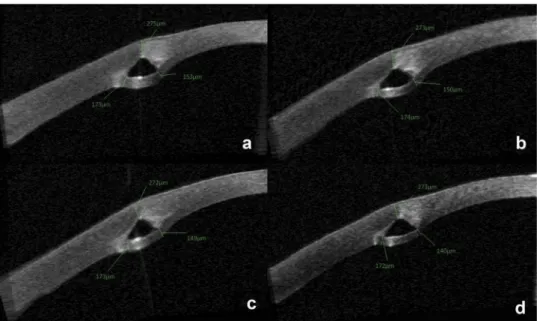
AS-OCT (Visante OCT Model 1000, axial and transverse resolutions 18 and 60 μm, Carl Zeiss, Germany) imaging was obtained with every patient at every visit. One of the corneal rings was randomly selected, and the cross-section of the segment’s midpoint was evaluated. Following Gorgun et al.,9distances were recorded using the software’s calipers by the same investigator (ME): from apex to anterior corneal surface as AA, from outer basal corner to posterior corneal surface as BP, and from inner basal corner to posterior cor-neal surface as CP. The measured distances were recorded for the same segment at each postoperative visit for every patient and were compared accordingly.
Statistical Analysis
Statistical analysis was performed using Statistical Package for Social Sciences 22.0 (SPSS, Inc., Chicago, USA). A Shapiro-Wilk test was used to determine whether the data were appropriate for normal distribution. One-way repeated measures analysis of var-iance (ANOVA) was applied to compare the distances of AA, CP, and BP, with Bonferroni correction for post-hoc analysis.P values below 0.05 were accepted as statistically significant.
Results
Thirty eyes of 22 patients (14 male, 8 female) were included in this study. The mean age of the patients was 36.4 ± 7.2 years, with a range of 23–49 years. Two segments were implanted in all eyes; the thickness of ring segments varied between 150 and 300μm, and the arc lengths varied between 90 and 210 degrees, with an inner diameter of 5 mm.
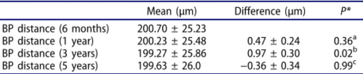
The change in distances over 6 months, 1 year, 3 years, and 5 years of AA, BP, and CP are shown inTables 1-3, respec-tively. The distance defined as CP showed a statistically sig-nificant decrease at all time points (p < .001), whereas no
statistically significant difference was found at 5 years for AA or BP (p > .05 for all).
The AS-OCT images of a representative case through the time points are shown inFigure 1. No postoperative compli-cations were observed during the 5-year period in any patient. Discussion
AS-OCT has been a valuable tool in assessing the position of ICRS and therefore might detect any potential complications related to depth. Many studies have reported that the post-operative evaluations of patients found that implanted ICRS showed shallower tunnel depth than intended – especially with manual surgery, which causes the ICRS to be closer to the anterior corneal surface over time.10–12 However, over time and with the availability of AS-OCT, both the distance to the anterior corneal surface and the distance to the poster-ior corneal surface have gained importance.9 Since the implanted ICRS remains in the cornea of the patients for many years, it is crucial to observe the ICRS position over a long period of time with AS-OCT.
This study evaluated the three distances from the triangu-lar Keraring segments to the anterior and posterior corneal
Table 1.The change in the AA distance over time.
Mean (µm) Difference (µm) P* AA distance (6 months) 283.43 ± 28.36 AA distance (1 year) 282.80 ± 27.85 0.63 ± 0.40 0.743a AA distance (3 years) 282.40 ± 27.84 0.40 ± 0.27 0.891b AA distance (5 years) 282.63 ± 27.33 −0.23 ± 0.48 0.999c AA: from apex to anterior corneal surface
*Pairwise comparison with Bonferroni post hoc analysis acompared to 6 months
b
compared to 1 year ccompared to 3 years
Table 2.The change in the BP distance over time.
Mean (µm) Difference (µm) P* BP distance (6 months) 200.70 ± 25.23
BP distance (1 year) 200.23 ± 25.48 0.47 ± 0.24 0.36a BP distance (3 years) 199.27 ± 25.86 0.97 ± 0.30 0.02b BP distance (5 years) 199.63 ± 26.0 −0.36 ± 0.34 0.99c BP: from outer basal corner to the posterior corneal surface
*Pairwise comparison with Bonferroni post hoc analysis a
compared to 6 months bcompared to 1 year c
compared to 3 years
Table 3.The change in the CP distance over time.
Mean(µm) Difference (µm) P* CP distance (6 months) 142.96 ± 4.33
CP distance (1 year) 140.40 ± 4.38 2.56 ± 0.27 0.001a CP distance (3 years) 138.06 ± 4.38 2.33 ± 0.34 0.001b CP distance (5 years) 133.40 ± 4.37 4.66 ± 0.62 0.001c CP: from inner basal corner to the posterior corneal surface
*Pairwise comparison with Bonferroni post hoc analysis a compared to 6 months b compared to 1 year ccompared to 3 years 1360 M. ELIAÇIK ET AL.
surfaces over the long term, and it found that the distance between the inner basal corner and the posterior corneal surface statistically significantly decreased at five years post-operation. In their study comparing the intended and achieved distances, Sanctis et al.8 found that the outer basal corner was farther from the posterior cornea than the inner basal corner, which is consistent with the findings of Gorgun et al.9as well as this study. The current study showed that this oblique positioning lasts for five years and that the imbalance increases between the two corners, bringing the inner corner closer to the posterior surface over time. This finding might indicate possible endothelial cell damage in long-term follow-ups of ICRS patients.
Kling et al.13have suggested that the oblique positioning of a triangular ICRS (like Keraring) might be attributed to the incompatibility of the rigid triangular shape with the curved soft cornea, contrasting previous studies proposing that the ICRS had rotated inside the tunnel.14This incompatibility is also evident when the smooth surface of the tunnel created by the femtosecond laser is compared with the edges of the triangle-shaped ring segment, which might prevent the segment from fitting properly into the tunnel. Coskunseven et -al.15reported that segment displacement occurred in 0.8% of Keraring cases, which might also support this idea. Additionally, other studies have reported cell accumulation and increased cell density in cornea adjacent to the ICRS, and which might have contributed to the ring segment’s change of position over the years.16,17It is also necessary to consider the idea that KC is a disease characterized by a progressive thin-ning of the cornea; increased collagen and keratocyte destruc-tion, and reduced cross-links within the keratoconic cornea, have been previously seen.18
AS-OCT has been used to assess the depth and position of the triangular ICRS in the short term in several studies;9,10,19 however in this study, the five-year outcomes are presented, with the distances to both the anterior and the posterior corneal surfaces, which might give surgeons valuable insight
about the final results and possible complications of their patients. This insight especially matters in a chronic disease like KC, because the patients will most likely require a refractive aid like ICRS, possibly for life. Similarly, a few important studies in the literature reporting on the long-term outcomes of ICRS concluded that the method is safe and stable over time.20–23
Another interesting outcome of this study is that the inner base corner of the ICRS moved more posteriorly at every time point; the CP distance’s change was statistically significant in the comparison of 1 year to 3 years, and of 3 years to 5 years (p < .001). This trend might indicate a probable risk of ante-rior chamber perforation in the long term. The cases reported in the literature show that anterior chamber perforation is more likely to occur intraoperatively7,24 or immediately postoperation.25To our knowledge, the only late-onset ICRS intrusion into the anterior chamber was described by Moshirfar et al., and it occurred 7 years post-operation;26 however, the ICRS of that specific patient was an INTACS segment, and they reported that the patient engaged in habi-tual eye-rubbing, which caused progressive corneal thinning. The endothelial break into the anterior chamber was docu-mented with AS-OCT. Therefore, a late-onset perforation to the anterior chamber from the inner base corner remains a possible complication, and the distances from the segments to both the anterior and posterior corneal surfaces need to be evaluated with AS-OCT during follow-ups to foresee any potential intrusions.
Even though it was not the primary objective of this study, it is noteworthy to mention that none of the patients had any post-operative complications (neither extrusion nor anterior chamber perforation) in the five years of the study, in accordance with the study of Barbara et al.11 Nonetheless, rare cases have been reported in the literature that showed late extrusion, even as late as 20 years post-implantation.27
This study has its limitations. For one, the design of the study is retrospective, which is also one of the main reasons for the small
sample size. As it was not the object of this study, the clinical characteristics of the patients– such as refractive error and visual acuity– were not analyzed during the time points, and connec-tions between them and the differences in distances could not have been examined. Additionally, the endothelial cell density was not counted at postoperative visits and compared to the preopera-tive data in order to reveal any possible endothelium damage. However, due to the retrospective design and the lack of aware-ness of this outcome of ICRS being closer to the posterior corneal surface, the researchers failed to gather sufficient postoperative data in this matter.
In conclusion, AS-OCT enables more precise imaging of ICRS and depicts the area not only above but also below the segment. This study provides enlightening information about the long-term stability of ring segments. During the five years post-implantation, the ICRS moved significantly closer to the posterior corneal sur-face at the inner base corner, which might help to predict any complications and/or clinical signs in the future.
Declaration of Interest
Authors report no conflict of interest.
Funding
Authors did not receive any funding. No funder is present.
ORCID
Beril Tülü Aygün http://orcid.org/0000-0003-4091-7094
References
1. Ertan A, Colin J. Intracorneal rings for keratoconus and keratectasia. J Cataract Refract Surg.2007;33(7):1303–14. doi:10.1016/j.jcrs.2007.02.048. 2. Moshirfar M, Edmonds JN, Behunin NL, Christiansen SM. Current Options in the Management of Pellucid Marginal Degeneration. J Refract Surg. 2014;30(7):474–85. doi:10.3928/ 1081597X-20140429-02.
3. Alió J, Salem T, Artola A, Osman A. Intracorneal rings to correct corneal ectasia after laser in situ keratomileusis. J Cataract Refract Surg.2002;28(9):1568–74. doi:10.1016/s0886-3350(01)01275-5. 4. Burris TE, Ayer CT, Evensen DA, Davenport JM. Effects of
intrastromal corneal ring size and thickness on corneal flatten-ing in human eyes. Refract Corneal Surg. 7(1):46–50. http:// www.ncbi.nlm.nih.gov/pubmed/2043543. Accessed September 9, 2019.
5. Piñero DP, Alio JL. Intracorneal ring segments in ectatic corneal disease - a review. Clin Experiment Ophthalmol. 2010;38 (2):154–67. doi:10.1111/j.1442-9071.2010.02197.x.
6. Siganos D, Ferrara P, Chatzinikolas K, Bessis N, Papastergiou G. Ferrara intrastromal corneal rings for the correction of keratoconus. J Cataract Refract Surg.2002;28(11):1947–51. doi:10.1016/s0886-3350(02)01495-5. 7. Kanellopoulos AJ, Pe LH, Perry HD, Donnenfeld ED. Modified
intracorneal ring segment implantations (INTACS) for the management of moderate to advanced keratoconus: efficacy and complications. Cornea. 2006; 25(1):29–33. [Accessed September 17, 2019]. http://www.ncbi.nlm.nih.gov/pubmed/ 16331037.
8. de Sanctis U, Lavia C, Nassisi M, D’Amelio S. Keraring Intrastromal Segment Depth Measured by Spectral-Domain
Optical Coherence Tomography in Eyes with Keratoconus. J Ophthalmol.2017;2017:1–9. doi:10.1155/2017/4313784. 9. Gorgun E, Kucumen RB, Yenerel NM, Ciftci F. Assessment of
Intrastromal Corneal Ring Segment Position With Anterior Segment Optical Coherence Tomography. Ophthalmic Surgery, Lasers, and Imaging. 2012;43(3):214–21. doi: 10.3928/15428877-20120301-01.
10. Lai MM, Tang M, Andrade EMM, Li Y, Khurana R, Song JC, Huang D. Optical coherence tomography to assess intrastromal corneal ring segment depth in keratoconic eyes. J Cataract Refract Surg.2006;32(11):1860–65. doi:10.1016/j.jcrs.2006.05.030. 11. Barbara R, Barbara A, Naftali M. Depth evaluation of intended vs
actual intacs intrastromal ring segments using optical coherence tomography. Eye.2016;30(1):102–10. doi:10.1038/eye.2015.202. 12. Monteiro T, Alfonso JF, Franqueira N, Faria-Correia F,
Ambrósio R, Madrid-Costa D. Predictability of tunnel depth for intrastromal corneal ring segments implantation between manual and femtosecond laser techniques. J Refract Surg. 2018;34 (3):188–94. doi:10.3928/1081597X-20180108-01.
13. Kling S, Marcos S. Finite-element modeling of intrastromal ring seg-ment implantation into a hyperelastic cornea. Invest Ophthalmol Vis Sci.2013;54(1):881–89. doi:10.1167/iovs.12-10852.
14. Cochener B, Savary-LeFloch G, Colin J. Effect of intrastromal corneal ring segment shift on clinical outcome: one year results for low myopia. J Cataract Refract Surg. 2000;26(7):978–86. doi:10.1016/s0886-3350(00)00537-x.
15. Coskunseven E, Kymionis GD, Tsiklis NS, Atun S, Arslan E, Charalambos SS, Jankov M, Pallikaris IG. Complications of intrastro-mal corneal ring segment implantation using a femtosecond laser for channel creation: a survey of 850 eyes with keratoconus. Acta Ophthalmol.2011;89(1):54–57. doi:10.1111/j.1755-3768.2009.01605.x. 16. Pérez-Merino P, Parra F, Ibares-Frías L, Gallegoa P, Vázquez-Lasab
B, Benitob L, San Románb J, Martínez-Garcíac C, Merayo-Llovesa J. Clinical and pathological effects of different acrylic intracorneal ring segments in corneal additive surgery. Acta Biomater. 2010;6 (7):2572–79. doi:10.1016/j.actbio.2010.01.014.
17. Twa MD, Ruckhofer J, Kash RL, Costello M, Schanzlin DJ. Histologic Evaluation of Corneal Stroma in Rabbits After Intrastromal Corneal Ring Implantation. Cornea. 2003;22 (2):146–52. doi:10.1097/00003226-200303000-00014.
18. Andreassen TT, Simonsen AH, Oxlund H. Biomechanical proper-ties of keratoconus and normal corneas. Exp Eye Res. 1980;31 (4):435–41. doi:10.1016/s0014-4835(80)80027-3.
19. Ramos JLB, Li Y, Huang D. Clinical and research applications of anterior segment optical coherence tomography - a review. Clin Experiment Ophthalmol. 2009;37(1):81–89. doi: 10.1111/j.1442-9071.2008.01823.x.
20. Torquetti L, Berbel RF, Ferrara P. Long-term follow-up of intras-tromal corneal ring segments in keratoconus. J Cataract Refract Surg.2009;35(10):1768–73. doi:10.1016/j.jcrs.2009.05.036. 21. Torquetti L, Ferrara G, Almeida F, Cunha L, Araujo LPN,
Machado AP, Lyra JM, Merayo-Lloves J, Ferrara P. Intrastromal corneal ring segments implantation in patients with keratoconus: 10-year follow-up. J Refract Surg.2014;30(1):22–26. doi:10.3928/ 1081597X-20131217-02.
22. Vega-Estrada A, Alió JL, Brenner LF, Burguera N. Outcomes of intrastromal corneal ring segments for treatment of keratoconus: five-year follow-up analysis. J Cataract Refract Surg. 2013;39 (8):1234–40. doi:10.1016/j.jcrs.2013.03.019.
23. Alfonso JF, Fernández-Vega-Cueto L, Lisa C, Monteiro T, Madrid-Costa D. Long-Term Follow-up of Intrastromal Corneal Ring Segment Implantation in Pediatric Keratoconus. Cornea.
2019;38(7):840–46. doi:10.1097/ICO.0000000000001945. 24. Ruckhofer J, Stoiber J, Alzner E, Grabner G. Multicenter European
Corneal Correction Assessment Study Group. One year results of European Multicenter Study of intrastromal corneal ring segments. Part 2: complications, visual symptoms, and patient satisfaction. J Cataract Refract Surg. 2001;27(2):287–96. doi: 10.1016/s0886-3350(00)00740-9.
25. Park S, Ramamurthi S, Ramaesh K. Late dislocation of intrastro-mal corneal ring segment into the anterior chamber. J Cataract Refract Surg.2010;36(11):2003–05. doi:10.1016/j.jcrs.2010.08.016. 26. Moshirfar M, Bean AE, Desautels JD, Birdsong OC. Corneal Hydrops Secondary to Intrastromal Corneal Ring Intrusion into the Anterior
Chamber 7 Years after Implantation: A Case Report. Ophthalmol Ther.2017;6(2):373–79. doi:10.1007/s40123-017-0105-7.
27. Oatts JT, Savar L, Hwang DG Late extrusion of intrastromal corneal ring segments: A report of two cases. Am J Ophthalmol Case Reports.2017;8:67–70. doi:10.1016/j.ajoc.2017.10.004