247 Indian Journal of Anaesthesia | Volume 64 | Issue 3 | March 2020
Letters to Editor
An alternative and novel
usage for ultrasound‑guided
erector spinae plane block:
Extracorporeal shock wave
lithotripsy in a paediatric patient
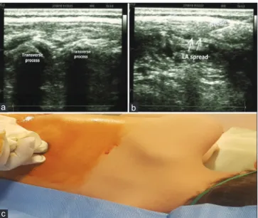
Sir,Erector spinae plane block is a novel paraspinal interfascial plane block.[1] It has been performed for several indications, and its usage for paediatric patients have been increased recently.[2,3] It may be used safely especially in paediatric analgesia management instead of central neuroaxial techniques because of its distance to the important anatomical structures.[2,3] Herein, we would like to share our experience using erector spinae plane block (ESPB) for management of interventional pain during extracorporeal shock wave lithotripsy (ESWL) in a 2-year-old paediatric patient. Written informed consent for the procedure and future publishing were obtained from parents of patient. Two-year-old boy was scheduled to undergo ESWL due to multiple renal stones. The patient was taken to the ESWL unit. Midazolam (0.1 mg·kg−1) and propofol (1 mg·kg−1) was administered IV for sedation. The patient was placed in the right lateral decubitus position. Under aseptic conditions; ultrasound-guided ESPB was performed from the level of the Th10 with a high frequency, sterile sheath covered, 12 MHz linear US probe. Following the visualisation of the erector spinae muscle above the transverse process, 22G, 50 mm block needle was inserted in a cranial-caudal direction using the in-plane technique into the deep fascia. 6 ml of 0.25% bupivacaine was injected into the interfacial area[2] [Figure 1]. ESWL was commenced 10 minutes after the block. There were no changes in arterial pressure, heart rate, respiratory rate, and SpO2 during ESWL. No additional analgesic drug or sedation was performed to the patient during the procedure. The patient was immobile during ESWL. Patient was comfortable at the postoperative period and started to walk 1 hour after the ESWL.
Anaesthesia and analgesia management is a very important issue during ESWL. ESWL is a daycare procedure, patients usually are discharged in the same day. Effective opioid-free techniques may be
used for post-procedural pain management.[4] The patient should be immobile during the procedure, and the sedation should be light for protection of the spontaneous respiration. So, regimens such opioid-free anaesthesia may be preferred. QLB block may be performed in paediatric patients for abdominal procedures like ESWL analgesia and provides at Th7-L1 dermatomes.[4] However, there have been some reports of complications related to QLB. QLB is administered in closer proximity to the intra-abdominal solid organs such as liver and kidney. Ahiskalioglu et al. reported the visualisation of hepatomegaly during the QLB block.[5] If this block is performed in less skilled hands, a number of complications such as solid organ injury may occur during block application.
ESPB is a novel block defined for use in both acute and chronic pain and sometimes as the main anaesthesia method in both paediatric and adult patients.[1-3] ESPB is performed distant to solid organs and vascular structures. The transverse process is a natural anatomical barrier in front of the needle and visualisation is simpler under ultrasound guidance, so it may be performed safely.[1,2]
To conclude, ESPB may provide effective interventional analgesia during paediatric ESWL safely for opioid-free analgesia treatment.
Declaration of patient consent
The authors certify that they have obtained all appropriate parent consent forms. In the form the parent(s) has/have given his/her/their consent Figure 1: (a) Sonographic anatomy of the block (b) Needle direction and
spread of local anaesthetic. LA indicates local anaesthetic (c) Patient
position and probe localisation during ESPB performing
c
b a
Page no. 87 [Downloaded free from http://www.ijaweb.org on Tuesday, April 14, 2020, IP: 212.154.57.44]
248 Indian Journal of Anaesthesia | Volume 64 | Issue 3 | March 2020 Letters to Editor
for his/her/their child’s images and other clinical information to be reported in the journal. The parents understand that the name and initials of their child will not be published and due efforts will be made to conceal the identity, but anonymity cannot be guaranteed.
Financial support and sponsorship Nil.
Conflicts of interest
There are no conflicts of interest.
Mursel Ekinci, Bahadir Ciftci, Selcuk Güven1, David T Thomas2
Departments of Anesthesiology and Reanimation and 1Urology,
Istanbul Medipol University, School of Medicine, Mega Medipol University Hospital, 2Department of Medical Education, Maltepe
University Faculty of Medicine, Istanbul, Turkey
Address for correspondence:
Dr. Bahadir Ciftci, Department of Anesthesiology and Reanimation, Mega Hospital Complex, Istanbul Medipol University School of Medicine, 34070, Bagcilar, Istanbul, Turkey. E‑mail: [email protected]; [email protected]
Submitted: 07‑Nov‑2019 Revised: 14‑Dec‑2019 Accepted: 05‑Jan‑2020 Published: 11‑Mar‑2020 REFERENCES
1. Tulgar S, Ahiskalioglu A, De Cassai A, Gurkan Y. Efficacy of bilateral erector spinae plane block in the management of
pain: Current insights [Internet]. J Pain Res 2019;12:2597-613. Available from: http://dx.doi.org/10.2147/jpr.s182128. 2. Aksu C, Gurkan Y. Defining the indications and levels of erector
spinae plane block in pediatric patients: A retrospective study of our current experience [Internet]. Cureus 2019;11:e5348. doi: 10.7759/cureus.5348.
3. De la Cuadra-Fontaine JC, Concha M, Vuletin F, Arancibia H. Continuous erector spinae plane block for thoracic surgery in a pediatric patient. Pediatric Anesthesia 2018;28:74-5. 4. Yayik AM, Celik EC, Ahiskalioglu A. An unusual usage for
ultrasound guided Quadratus lumborum block: Pediatric extracorporeal shock wave lithotripsy. J Clin Anesth 2018;46:47-8.
5. Ahiskalioglu A, Yayik AM, Aydin ME, Ahiskalioglu EO, Caglar O. Incidental hepatomegaly during quadratus lumborum block in pediatric patient. J ClinAnesth 2019;54:112-3.
How to cite this article: Ekinci M, Ciftci B, Güven S, Thomas DT.
An alternative and novel usage for ultrasound‑guided erector spinae plane block: Extracorporeal shock wave lithotripsy in a paediatric patient. Indian J Anaesth 2020;64:247‑8.
© 2020 Indian Journal of Anaesthesia | Published by Wolters Kluwer ‑ Medknow
This is an open access journal, and articles are distributed under the terms of the Creative Commons Attribution‑NonCommercial‑ShareAlike 4.0 License, which allows others to remix, tweak, and build upon the work non‑commercially, as long as appropriate credit is given and the new creations are licensed under the identical terms.
Access this article online
Quick response code
Website: www.ijaweb.org
DOI:
10.4103/ija.IJA_819_19
Inadvertent dural puncture: Is
excess flexion of spine a possible
cause?
The rate of inadvertent dural puncture during epidural catheter placement varies from 0.19% to 3.6%.[1,2] The performance of lumbar punctures and epidural catheterisations relies primarily on the palpation of anatomical landmarks, the angle of needle progression and the distance from the skin to the target space; hence, the skill of the operator is a major influencing factor. The 4 P’s (position, preparation, projection, and puncture) continue to remain the very essence of these procedures. Accidental dural puncture (ADP) during placement of an epidural catheter for anaesthesia is a well-known complication. Risk factors for this iatrogenic
complication include patient movement and repeated epidural trials.[3] Kuroda K et al investigated retrospectively factors related to accidental dural
puncture during epidural anaesthesia.[4] They
concluded that the chances of ADP were more in lower thoracic and lumbar region, and in elderly patients. In our clinical experience of inadvertent lumbar dural puncture, we feel that patient positioning could have (acute anterior flexion of lumbar spine) resulted in decreased posterior epidural space [Figure 1]. Anterior flexion of spine is required for proper insertion of Tuohy needle during epidural anaesthesia. This position of acute flexion can result in decreased posterior epidural space as dura comes closer to ligamentum flavum. This flexion that can help us pierce the dura in subarachnoid block, would also increase our chances of inadvertent dura puncture in case of epidural anaesthesia. Figure 1 shows sketch
Page no. 88 [Downloaded free from http://www.ijaweb.org on Tuesday, April 14, 2020, IP: 212.154.57.44]