DOI 10.1007/s00345-017-2058-9
ORIGINAL ARTICLE
A prospective randomized comparison of micropercutaneous
nephrolithotomy (Microperc) and retrograde intrarenal surgery
(RIRS) for the management of lower pole kidney stones
Abdulkadir Kandemir1 · Selcuk Guven2 · Mehmet Balasar1 ·
Mehmet Giray Sonmez1 · Hakan Taskapu1 · Recai Gurbuz1
Received: 21 February 2017 / Accepted: 31 May 2017 / Published online: 6 June 2017 © Springer-Verlag GmbH Germany 2017
stones up to 15 mm in diameter. However, prolonged hos-pital stay and scopy times are the main disadvantages of Microperc and further research is needed to evaluate the renal tubular damages caused by both of these methods. Keywords Microperc · Retrograde intrarenal surgery · Lower pole kidney stones · Flexible · Ureteroscopy · Percutaneous nephrolithotomy
Introduction
The incidence of renal calculi is rising and more patients are presenting with small renal calculi [1]. The main pur-pose of the treatment is to provide the least morbidity rate with full stone-free cases as minimal invasive. Choosing the most appropriate treatment option depends on many factors such as; size, placement and number of the stones, comorbidity of the patient, and urinary system anatomy. The treatment options for small renal calculi (<1.5 cm) are ESWL, retrograde intrarenal surgery (RIRS), and percuta-neous nephrolithotomy (PCNL) [2]. Low stone clearance rates and the need for multiple intervention sessions in the lower pole stones are disadvantages of ESWL [3]. Even though RIRS is one of the standard treatment options and has a better safety profile for small kidney stones, its cost much more than ESWL and stone removal rate is lower than PCNL [2, 4, 5]. On the other hand, high stone clear-ance rates of PCNL are associated with a significant risk of morbidity which can be decreased with reduction of tract size [6, 7].
To prevent the drawback of PCNL, Mini-PCNL [8] and Microperc [9] were developed, respectively. Microperc was described by Bader et al. Thanks to the instrument, it was defined as the ‘‘all-seeing needle’’. Differing from standard Abstract
Objective To make a comparison between the safety and efficacy of micropercutaneous nephrolithotomy (microp-erc) and retrograde intrarenal surgery (RIRS) for the man-agement of lower pole kidney stones up to 15 mm.
Patients and methods 60 patients presenting with solitary lower pole kidney stones up to 15 mm were included in the study between March 2013 and December 2015. Patients were randomized into Microperc or RIRS groups with computer-generated numbers.
Results The mean stone size was 10.6 (5–15) and 11.5 (7–15) mm for Microperc and RIRS groups, respectively (P = 0.213). In the Microperc group, the scopy time was 158.5 s, while in the RIRS group, the scopy time was 26.6 s (P = 0.001). The hospitalization period in the Microperc group was 542 h, while it was 19 h in the RIRS group (P = 0.001). No statistical differences were observed dur-ing the operatdur-ing time, pre-operative–post-operative hemo-globin (Hb), serum creatinine, and estimated glomerular filtration speed (e-GFR) values and stone-free rates. No intraoperative complications were observed in either of the groups, while post-operative complications were observed in six patients in Microperc Group and five patients belong-ing to the RIRS Group (P = 0.922).
Conclusions Both Microperc and RIRS are safe and effec-tive alternaeffec-tives, and have similar stone clearance and com-plication rates for the management of lower pole kidney
* Abdulkadir Kandemir [email protected]
1 Department of Urology, Meram Medical Faculty, Necmettin
Erbakan University, 42080 Akyokus¸, Konya, Turkey
2 Department of Urology, School of Medicine, Istanbul
methods and Mini-PCNL, stones are reachable by a single step and left to pass spontaneously after being fragmented and crumbled into smaller pieces. It was reported that in general, the increased rate of Microperc stone clearance is comparable to PCNL with decreased morbidity due to the smaller tract size, and lower costs compared to ESWL and RIRS [10, 11]. In the only prospective and randomized study which evaluates Microperc and RIRS for both renal stones, Sabnis et al. reported that the stone clearance rates were high and the complications were low for both tech-niques. They also concluded that Microperc was associated with higher hemoglobin loss, increased pain, and higher analgesic requirements, while RIRS was associated with a higher requirement for JJ stenting [12]. Regardless of either of these techniques, removal of lower pole kidney stones is more challenging and there is limited information in lit-erature about the prospective comparison of Microperc and RIRS which is controversial for both techniques. In a ret-rospective comparative clinical study, Armagan et al. sug-gested that Microperc is an alternative technique to flexible ureteroscopy for lower pole stones [13].
In this prospective, randomized, controlled study, we aimed to compare the safety and efficacy of Microperc and RIRS in lower pole kidney stones up to 15 mm dependent on different features.
Materials and methods Patients
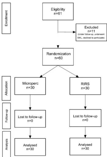
This is single center study. After the institutional ethics committee approval, a parallel-arm randomized controlled study was conducted in Necmettin Erbakan University Meram Medical School, Department of Urology between March 2013 and December 2015. All participants were informed in advance about the study and their consent was obtained individually. Patients who had a single lower pole kidney stone up to 15 mm without contraindications to microperc and RIRS were included. Exclusion criteria were patients with multiple stones, renal failure, bleeding disorders, stones in renal anomalies, and who had stones other than kidney such as bladder and ureteral stones, chil-dren cases, and pregnancy. Sixty patients presenting with a single lower pole kidney stone up to 15 mm in diameter were prospectively randomized into Microperc or RIRS group. Randomization was done with computer-generated numbers. Patients and authors assessing the outcomes were informed about the procedure. Figure 1 shows the flow of the patients in the study. Both Microperc and RIRS inter-ventions were performed by three different surgeons who had both Microperc and RIRS experience. A single <1.5 cm
stone was detected in the lower calyx of the kidney by non-contrast spiral CT scan.
The stone-free rate in the post-operative 3rd month was the primary outcome. The secondary outcomes of complications, blood transfusion, operative time, need for blood transfusion, and hemoglobin deficit were com-pared between the treatment groups. All the patients were examined with urine cultures before the operation. Both the Microperc and RIRS Group were compared in terms of stone clearance rates, scopy and operation time, intra-operative and post-intra-operative complications (according to the Clavien–Dindo classification system), pre-operative and post-operative hemoglobin (Hb), serum, creatinine and estimated glomerular filtration speed rates (e-GFR), and post-operative hospitalization periods. Fluoroscopic screening time is defined as the sum of instant and short time views during the process which was specified on the Ziehm Vision R C-Arm System panel (Germany). No patient needed nephrostomy. A DJ ureteral stent was placed in the case of high volume stone burden. The stone-free rate in the post-operative 3rd month was the primary out-come. The secondary outcomes of complications, blood
transfusion, operative time, need for blood transfusion, and hemoglobin deficit were compared between the treat-ment groups. The Chronic Kidney Disease Epidemiology Collaboration formula was used for the Glomerular filtra-tion speed calculafiltra-tion [14]. Residue stone present in kidney was examined 3 months after the operation with non- con-trast spiral CT. Clinically insignificant residual fragments (CIRFs) are defined as asymptomatic, non-obstructing residual fragments smaller than 4 mm on CT. For compli-cations which occurred during the operation, the Satava classification was used, whilst the Clavien–Dindo classifi-cation was used for post-operative compliclassifi-cations.
Microperc technique
A ureteral catheter was placed in a transurethral way under general anesthesia. Then, the patient was converted to the proper position, and percutaneous access was obtained by the placement of a 16 gauge all-seeing needle (PolyDiagnost, Pfaffenhofen, Germany) with optical guidance under fluoro-scopic or ultrasonographic guidance into the appropriate calix. Access with the all-seeing needle was successful in all patients. A surgeon controlled the irrigation system (IP 200, PolyDiagnost, Pfaffenhofen, Germany) or alternatively a 20 mL was used for irrigation. The inner needle was removed. A three-way connector was attached to the proximal end of the sheath. The telescope was passed through the connector side port, and the other side port was used for irrigation. The con-nector central port was used to pass the laser fiber. The calcu-lus was then fragmented by a holmium: yttrium–aluminum-garnet (YAG) laser using a 200 micron fiber. At the end of the procedure, we monitored the fragmentation by fluoroscopy. Operation time for Microperc includes the time used for the initial cystoscopy, retrograde catheter placement, and to turn the patient prone until the post-operative fluoroscopic control.
RIRS technique
Under general anesthesia, patients were in the lithotomy position during the procedure. A hydrophilic guide wire (0.035/0.038-inch) was inserted under fluoroscopic guid-ance during the semi-rigid ureteroscopic evaluation of the relevant ureter. After placement, the ureteral access sheath (UAS) was inserted into the ureter, the stone was accessed using a flexible video ureterorenoscope (F-URS) (URF-V: Olympus, Japan). Stone fragmentation was enabled via Hol-mium: YAG laser. During RIRS, the stones were fragmented in the lower calyx or in more accessible locations after repo-sitioned by a stone basket according to surgeons’ preference and especially in patients’ with difficult anatomy.
For spontaneous passage, all renal stones were frag-mented to the necessary size. After this procedure, a JJ
stent was placed in some patients. Operation time for RIRS includes the times used for the initial cystoscopy, retrograde guide wire, and ureteral access sheath place-ment until the post-operative fluoroscopic control.
Statistical analysis
Research data are created by the statistics package pro-gram, SPSS for Windows version 14.0. During process of data analyzing, χ2 (Chi-squared) test is used for categori-cal data, while t test and Mann–Whitney test are used for continuous data according to normality analyze. T test and General Linear Model analyzing method were used for the comparison in between pre- and post-values of paired groups. P < 0.05 was accepted as statistically significant.
Results
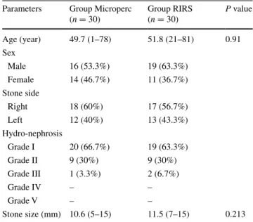
The patient demographics and stone characteristics were similar in both groups (Table 1). Regarding the Micro-perc group, lower kidney calyx stone was present in the right kidney of 18 patients (60%) and the left kidney of 12 patients (40%). The average age was 49.7 (1–78). In the RIRS group, kidney stone was present in right kidney of 17 patients (56.7%) and the left kidney of 13 patients (43.3%). The average age of RIRS group was 51, 8 (21– 81), and when comparing the Microperc group, the aver-age aver-age was not statistically significant (P = 0.9).
Operation and scopy time of the Microperc group was 59.04 min and 158.48 s, while in the RIRS Group, they
Table 1 Baseline demographic and clinical characteristics for each group
Parameters Group Microperc (n = 30) Group RIRS (n = 30) P value Age (year) 49.7 (1–78) 51.8 (21–81) 0.91 Sex Male 16 (53.3%) 19 (63.3%) Female 14 (46.7%) 11 (36.7%) Stone side Right 18 (60%) 17 (56.7%) Left 12 (40%) 13 (43.3%) Hydro-nephrosis Grade I 20 (66.7%) 19 (63.3%) Grade II 9 (30%) 9 (30%) Grade III 1 (3.3%) 2 (6.7%) Grade IV – – Grade V – – Stone size (mm) 10.6 (5–15) 11.5 (7–15) 0.213
were 51.05 min and 26.58 s. For the operation time, no statistical difference is observed between the two groups, while in scopy time, a statistical difference was observed (P = 0.126, P = 0.001). In 16 RIRS patients (53.3%), the ureteral access sheath insertion failed. After the dila-tation of the orifice or obstructed ureter, the F-URS was transferred by dragging over the guide. Access to the col-lecting system with the all-seeing needle was successful in the Microperc group. Post-operative discharge time for this was 54.2 h, while it was 19 h for the RIRS Group. This was statistically significant (P = 0.001). When the residue stone rates are considered; the comparison is 16.7–13.3% and no statistically significant difference was observed between the two groups (P = 0.158). A DJ ureteral stent was placed in nine cases in the Microperc group and 14 in the RIRS Group.
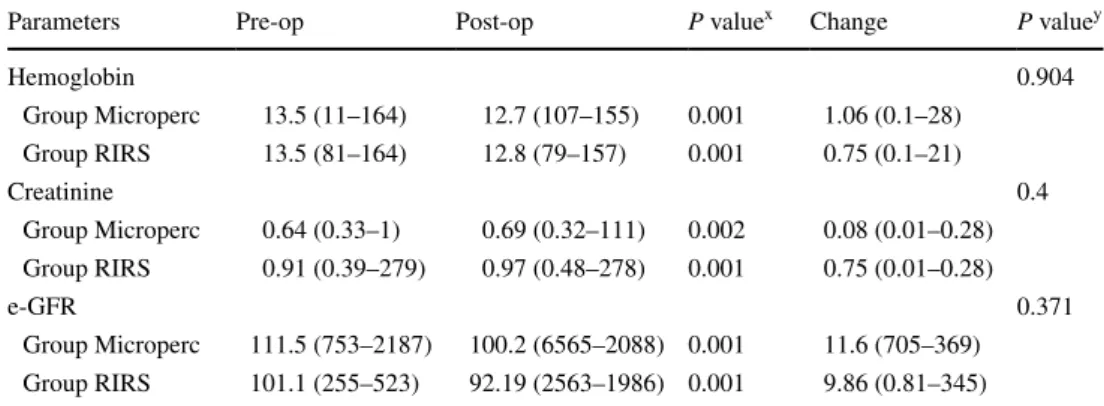
When pre- and post-operative hemoglobin, creatinine and e-GFR values were examined of the groups; in nei-ther of the groups, no statistically significant decrease in post-operative hemoglobin, creatinine, and e-GFR values was observed when compared to the pre-operative values (see Table 2 for P values. Further comparison of these values between the two groups was not statistically sig-nificant (P = 0.904, P = 0.371) (Table 2).
In post-operative 3rd month non-contrast urinary CT; in the Microperc group, 25/30 (83.3%) patients had final stone-free situation, 2/30 (6.6%) patients had clinically insignificant residual fragments (CIRFs), and residue stone was diagnosed in the last 3/30 (10%) patients. These patients were undergone re-microperc; the patients were stone-free after the second procedure. In the RIRS Group, 26/30 (86.7%) patients had final stone-free situation, while residual fragment was observed in 4/30 (13.3%) patients. Residual stones of patients are still being followed.
In the Microperc group, residual stones were observed in five cases in the post-operative 3rd month with CT imag-ing. Two of these were clinically insignificant residual
fragments. The remaining 3 were undergone re-microperc; the patients were stone-free after the second procedure.
In the Microperc Group, one patient required blood transfusion and an infection necessitating additional anti-biotics occurred in 1 (Grade II). Fever and renal colic occurred in four patients post-operatively which was resolved with antipyretic and analgesic therapy (Grade I). In the RIRS group, additional antibiotics needed and hos-pitalization periods were prolonged in two patients (Grade II) and fever and renal colic occurred in four patients post-operatively (Grade I). In terms of post-operative compli-cations, no statistical significance was observed between both groups according to Clavien–Dindo classification (P = 0.819). In neither of the groups, any urosepsis or adja-cent organ injury was observed. Emergent open surgery was not needed in any of these cases.
Discussion
Management of lower pole stones is harder than the other kidney stones due to their inherent anatomic challenges. Techniques such as ESWL, RIRS, and PCNL have their own advantages and disadvantages, and it is still contro-versial which one is the better option for the treatment of lower pole stones that are smaller than 15 mm in diam-eter [10]. ESWL has low success and higher retreatment rates in lower pole stone treatment. On the other hand, it enabled the intrarenal surgery and percutaneous inter-ventions to become a significant option with endourologi-cal instrumentation and technologiendourologi-cal advances in lower pole stone treatment. Even though percutaneous neph-rolithotomy has higher success rates, it is more invasive and has higher complication rates compared to RIRS [15, 16]. RIRS has deflection challenges under flexible ureteroscopy and higher cost of treatment as limitations. Donaldson et al. argued that in ESWL, RIRS, and PNL
Table 2 Comparison of the Groups’ pre- and post-operative laboratory tests and e-GFR levels
x Comparisons made in between Micro-PNL and RIRC groups’ pre- and post-operative laboratory tests y Comparisons made between groups’ post-operative period laboratory tests changes based on
pre-opera-tive tests
Parameters Pre-op Post-op P valuex Change P valuey
Hemoglobin 0.904 Group Microperc 13.5 (11–164) 12.7 (107–155) 0.001 1.06 (0.1–28) Group RIRS 13.5 (81–164) 12.8 (79–157) 0.001 0.75 (0.1–21) Creatinine 0.4 Group Microperc 0.64 (0.33–1) 0.69 (0.32–111) 0.002 0.08 (0.01–0.28) Group RIRS 0.91 (0.39–279) 0.97 (0.48–278) 0.001 0.75 (0.01–0.28) e-GFR 0.371 Group Microperc 111.5 (753–2187) 100.2 (6565–2088) 0.001 11.6 (705–369) Group RIRS 101.1 (255–523) 92.19 (2563–1986) 0.001 9.86 (0.81–345)
techniques’ meta-analysis, 2 cm or smaller stones are being assessed, PNL is the most successful technique in terms of stone-free rate and its recent modifications can reduce morbidity and convalescence, but it makes the process harder in decision making of the patients and informing of clinicians because of its limitations in well-designed studies [15]. De et al. suggested that PCNL provides overall significantly higher stone-free rates than RIRS, at the expense of higher complication rates, blood loss, and longer length of stay, with no differences in sur-gical time and secondary procedures [16].
Microperc was first presented in 2010 by Desai et al. to decrease PCNL’s morbidity rates caused by its wide tract sizes [17]. Later on, in many studies, Microperc was stated as safe and effective in small- and medium-sized renal stones. Our study is the first randomized one which compares Microperc and RIRS in the treatment of lower pole kidney stones less than 15 mm in diameter.
In our study, similar results of stone-free rates were obtained in Microperc and RIRS groups, 83.3 and 86.7%, respectively (P = 0.158). It was mentioned in the previ-ous meta-analysis that more hemoglobin decrease was observed in percutaneous interventions; however, in this study, no significant difference was observed in inter-group hemoglobin decrease (P = 0.904). Hospital stay is one of the critical limitations of PNL, even though stone-free rate is high. As in the standard PNL, in our study, hospital stay was found to be significantly longer in the Microperc group compared to the RIRS group (P = 0.001). While, the ureteral access sheath inser-tion failed in 16 patients (53.3%) who underwent RIRS, access to the collecting system with the all-seeing needle was successful in all patients of the Microperc group.
Complication rates were similar in the two groups studied. According to the Clavien–Dindo classifica-tion, no statistically significant difference was observed between the Microperc and RIRS groups in terms of post-operative complication (P = 0.819). No urosepsis or adjacent organ injury was observed in any of the groups, which composed of 60 patients in total. Emergent open surgery or nephrectomy was not needed in any of these cases. Scopy time which is one of the main limitations of the stone management methods for both the patients and the operation team was significantly longer in the Micro-perc group (P = 0.001). This finding is critical and needs to be confirmed with further studies. With the increasing experience, our scopy time has decreased during RIRS. Moreover, we have been using fluoroscopy only for resid-ual fragment control for most of the patients. However, we perform all-seeing needle access under fluoroscopic control and this increases the scopy time. Scopy times for both groups are comparable with the previous studies.
The change of blood creatinine level in both the Microperc and RIRS groups was 0.08 and 0.075, respec-tively. No significant difference in the change of blood creatinine level was observed (P = 0.4). In addition, the pre and post-operative e-GFR level changes in both groups were 11.6 and 9.86 mL\min\1.73 m2, respectively. This was not statistically significant when the two groups were compared to each other in terms of the changes in e-GFR levels (P = 0.371). Though serum creatinine and e-GFR levels show kidney functions, normal status of these values does not exactly inform about the renal tubu-lar damages. To evaluate kidney damages during both techniques, further research is needed [18].
The UAS insertion failure in 16 cases (53.3%) is a high failure rate compared to the previous studies. This may be due to a small number of patients in the cohort (UAS insertion failure in our clinic is normally slightly lower (35%), unpublished data). We do not think that it is an anatomic variation specific to the lower pole stones; however, further research is needed on this subject. On the other hand, in cases in which UAS could not be fur-ther advanced into the ureter even if the slightest diffi-culty, we did not insist on this issue, balloon dilation, passing the ureterorenoscope over a guidewire or passive dilation of the ureteral orifice was used according to the preference of the urologist.
One of the limitations of the study is the relatively low number of patients with a single center experience. For more accurate and realistic results, multicenter work is needed. However, this is the first study, which focuses directly on single lower pole kidney stones up to 15 mm. After the enrollment of the cases, the study period took more than 32 months that can be reasonable accepted for such kind of selective stone patient group.
In conclusion, for the treatment of lower pole single and <1.5 cm stones, the stone clearance rates in both Microp-erc and RIRS are high with a low complication rate and are comparable. However, the prolonged hospital stay, scopy times of Microperc, and ureteral access sheath placement failure of RIRS are technique disadvantages. Moreover, additional research and studies are needed to explore the renal tubular damages caused by both methods.
Author contributions AK was involved in project development and data analysis and wrote the manuscript; SG edited the manuscript; HT and AK was involved in data collection; MB was involved in data analysis; MGS was involved in project development; RG was involved in project development.
Compliance with ethical standards
Conflict of interest The authors declare that they have no conflict of interest with this work.
Ethical standards The study protocol is compliant with ethical stand-ards and was approved by the local ethical committee.
References
1. Menon M, Resnick MI (2007) Urinary lithiasis: etiology, epide-miology and pathogenesis. In: Wein AJ (ed) Campell’s Urology vol. 2, 2nd edn. Sounders, Philadelphia
2. Türk C, Knoll T, Petrik A, Sarica K, Seitz C, Straub M. EAU Guidelines on Urolithiasis. Uroweb 2012. Available at: http:// www.uroweb.org/gls/pdf/20_Urolithiasis_LR%20March%20 13%202012.pdf. Accessed 14 November 2012
3. Srisubat A, Potisat S, Lojanapiwat B, Setthawong V, Laopaiboon M (2009) Extracorporeal shock wave lithotripsy (ESWL) versus percutaneous nephrolithotomy (PCNL) or retrograde intrarenal surgery (RIRS) for kidney stones. Cochrane Database Syst Rev 4:CD007044
4. Koo V, Young M, Thompson T, Duggan B (2011) Cost-effec-tiveness and efficiency of shockwave lithotripsy vs flexible uret-eroscopic holmium:yttrium-aluminium-garnet laser lithotripsy in the treatment of lower pole renal calculi. BJU Int 108:1913–1916 5. Bryniarski P, Paradysz A, Zyczkowski M, Kupilas A, Nowa-kowski K, Bogacki R (2012) Randomized controlled study to analyze the safety and efficacy of percutaneous nephrolithotripsy and retrograde intrarenal surgery in the management of renal stones more than 2 cm in diameter. J Endourol 26:52–57 6. Goger E, Guven S, Gurbuz R, Yilmaz K, Kilinc M, Ozturk A
(2012) Management of a colon perforation during pediatric percutaneous nephrolithotomy. J Endourol 26(9):1118–1120. doi:10.1089/end.2011.0433
7. Guven S, Istanbulluoglu O, Gul U, Ozturk A, Celik H, Aygün C, Ozdemir U, Ozturk B, Ozkardes H, Kilinc M (2011) Success-ful percutaneous nephrolithotomy in children: multicenter study on current status of its use, efficacy and complications using Clavien classification. J Urol 185(4):1419–1424. doi:10.1016/j. juro.2010.11.055
8. Helal M, Black T, Lockhart J, Figueroa TE (1997) The Hickman peelaway sheath: alternative for pediatric percutaneous nephroli-thotomy. J Endourol 11:171–172
9. Bader M, Gratzke C, Schlenker B et al (2010) The “All-seeing needle”- an optical puncture system confirming percutaneous access in PNL. J Urol Suppl 183:e734 (abstract 1890)
10. Kiremit MC, Guven S, Sarica K, Ozturk A, Buldu I, Kafkasli A, Balasar M, Istanbulluoglu O, Horuz R, Cetinel CA, Kandemir A, Albayrak S (2015) Contemporary management of medium-sized (10–20 mm) renal stones: A Retrospective Multicenter Observational Study. J Endourol 29(7):838–843. doi:10.1089/ end.2014.0698
11. Resorlu B, Unsal A, Ziypak T, Diri A, Atis G, Guven S, Sancak-tutar AA, Tepeler A, Bozkurt OF, Oztuna D (2013) Comparison of retrograde intrarenal surgery, shockwave lithotripsy, and per-cutaneous nephrolithotomy for treatment of medium-sized radio-lucent renal stones. World J Urol 31(6):1581–1586. doi:10.1007/ s00345-012-0991-1
12. Sabnis RB, Ganesamoni R, Doshi A, Ganpule AP, Jagtap J, Desai MR (2013) Micropercutaneous nephrolithotomy (microp-erc) vs retrograde intrarenal surgery for the management of small renal calculi: a randomized controlled trial. BJU Int 112(3):355– 361. doi:10.1111/bju.12164 (PubMed PMID: 23826843) 13. Armagan A, Karatag T, Buldu I, Tosun M, Basibuyuk I,
Istan-bulluoglu MO, Tepeler A (2015) Comparison of flexible uret-erorenoscopy and micropercutaneous nephrolithotomy in the treatment for moderately size lower-pole stones. World J Urol 33(11):1827–1831. doi:10.1007/s00345-015-1503-x
14. Levey AS, Stevens LA, Schmid CH et al (2009) A new equa-tion to estimate glomerular filtraequa-tion rate. Ann Intern Med 150:604–612
15. Donaldson JF, Lardas M, Scrimgeour D et al (2015) Systematic review and meta-analysis of the clinical effectiveness of shock wave lithotripsy, retrograde intrarenal surgery, and percuta-neous nephrolithotomy for lower-pole renal stones. Eur Urol 67:612–616
16. De S, Autorino R, Kim FJ et al (2015) Percutaneous nephro-lithotomy versus retrograde intrarenal surgery: a systematic review and meta-analysis. Eur Urol 67:125–137
17. Desai MR, Sharma R, Mishra S, Sabnis RB, Stief C, Bader M (2011) Single-step percutaneous nephrolithotomy (microperc):the initial clinical report. J Urol 186:140–145 18. Balasar M, Pis¸kin MM, Topcu C, Demir LS, Gürbilek M,
Kan-demir A, Öztürk A (2016) Urinary kidney injury molecule-1 levels in renal stone patients. World J Urol. doi:10.1007/ s00345-016-1765-y