© 2018 Indian Chest Society | Published by Wolters Kluwer - Medknow 339
pulmonary radiograph, bilateral hilar fullness and right hilar calcification were determined. Evaluations of the laboratory test were within normal limits. In the respiratory function test, an obstruction was determined in the small airway. Thorax computed tomography (CT) was taken with an initial diagnosis of broncholithiasis. Cartilage calcifications were observed in the trachea and the walls of both main bronchi and segmentary bronchi. Right hilar calcified lymph nodes were seen, and in the right middle lobe, the soft‑tissue appearance was observed to be of calcifications, the largest of which was 20 mm × 24 mm, adjacent to an oblique fissure. There were also nodular calcifications within the soft tissue. Fiber‑optic bronchoscopy (FOB) was applied. An appearance of increased cartilage was seen in the trachea and under the right system mucosa. The right middle lobe entry was seen to have narrowed, and no endobronchial lesions or fistula was observed. Bronchial lavage cytology was reported as nonspecific and atypical cells were not determined. With a clinical and radiological diagnosis of broncholithiasis, the patient was followed up without medication.
Endobronchial tumors with calcification or tracheobronchial disease with mural calcification can mimic broncholithiasis.[2] Broncholithiasis, which is defined as the presence of a
calcified fragment of tissue within a bronchus,[1] is an uncommon condition which is difficult to diagnose. The most common cause of broncholithiasis is erosion by and extrusion of a calcified contiguous lymph node into the bronchial lumen, a finding usually associated with tuberculosis or histoplasmosis.[2,3] The clinical manifestation of broncholithiasis is when calcified stones erode or break loose into the airways. As these stones are composed of 85%–90% calcium phosphate and 10%–15% calcium carbonate, the composition closely resembles that of bone.[4] The most common symptoms are a nonproductive cough and hemoptysis. Fever leading to secondary pulmonary infections may develop together with chest pain and purulent phlegm.[5,6] Occasionally, broncholytic expectoration may be seen, which is known as lithoptysis. As this is not commonly seen, a case diagnosed with lithoptysis is here presented.
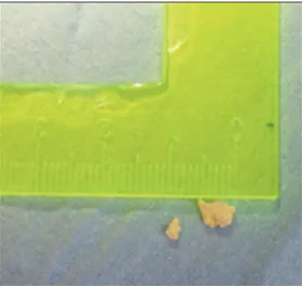
A 65‑year‑old male presented at the Chest Diseases Polyclinic with the complaint of two hard bodies expectorated after coughing [Figure 1]. The patient had a 50‑packet/year smoking history and continued to smoke. There was no history of tuberculosis or contact with. The physical examination was normal. On the posterior–anterior
Broncholithiasis presenting with lithoptysis
Berna Akinci Ozyurek, Serife Savas Bozbas1Department of Pulmonary Medicine, Ataturk Chest Diseases and Chest Surgery Education and Research Hospital, 1Department of Pulmonary Medicine, Baskent University Faculty of Medicine, Ankara, Turkey
ABSTRACT
Address for correspondence: Dr. Berna Akinci Ozyurek, Ataturk Chest Diseases and Chest Surgery Education and Research Hospital, Ankara, Turkey. E‑mail: [email protected]
Case Report
Broncholithiasis is defined as calcified or ossified material in the bronchial lumen. The most frequently seen symptoms are a non-productive cough and hemoptysis. Occasionally, broncholytic expectoration can be seen, which is known as lithoptysis. As this is not a common finding, a case diagnosed with lithoptysis is presented here.
KEY WORDS: Broncholithiasis, calcification, lithoptysis
Access this article online
Quick Response Code:
Website:
www.lungindia.com
DOI:
10.4103/lungindia.lungindia_304_17 How to cite this article: Ozyurek BA, Bozbas SS. Broncholithiasis presenting with lithoptysis. Lung India 2018;35:339-40. This is an open access journal, and articles are distributed under the terms of the Creative Commons Attribution-NonCommercial-ShareAlike 4.0 License, which allows others to remix, tweak, and build upon the work non-commercially, as long as appropriate credit is given and the new creations are licensed under the identical terms.
For reprints contact: [email protected]
Ozyurek and Bozbas: Case of broncholithiasis
340 Lung India • Volume 35 • Issue 4 • July-August 2018
Other causes include Cryptococcus, coccidioidomycosis, actinomycosis, aspergillosis, nocardiosis, silicosis, and malignancy.[7] Although uncommon, broncholithiasis has also been reported to be a cause of diffuse tracheobronchial calcification,[5] which is often associated with advanced age.[8] In a previous study using thorax CT, tracheobronchial calcification was determined in 26% of patients aged 40–59 years, and in 65% of males and in 40.5% of females aged 60–79 years.[9] The current patient was 65 years old. Broncholithiasis is a rarely seen condition, and the incidence is 0.1%–0.2% within respiratory system diseases.[10] In the diagnosis of broncholithiasis, bronchoscopy, thorax CT, and high‑resolution CT can be used. CT findings can be strongly suggestive of broncholithiasis when there is a calcified nodule that is either endobronchial or peribronchial and is associated with findings of bronchial obstruction such as atelectasis, obstructive pneumonitis, or bronchiectasis.[2] Due to the airway anatomy and lymph node distribution, it is most often seen in the proximal right‑side midlobe and the anterior segment bronchi of the upper lobes[6,11] Bronchoscopy combined with CT examination is helpful in diagnosing and typing broncholithiasis. An optimal treatment method is either bronchoscopic removal of broncholithiasis or thoracotomy. In the current case, subabdominal right hilar calcified lymph nodes were determined on thorax CT, and soft tissue was observed as calcifications, the widest of which was 30 mm × 24 mm, adjacent to an oblique fissure in
the right middle lobe. On FOB, the right middle lobe entry was observed to be narrowed.
Treatment is not required in asymptomatic cases. In symptomatic cases (when there is hemoptysis, recurrent pneumonia, or the development of bronchoesophageal fistula), bronchoscopy and surgical treatment are recommended.[10] As the current case was asymptomatic, follow‑up without treatment was planned and in a 5‑year period, no complications have developed. In cases that present with expectoration of foreign material together with a cough, broncholithiasis should be kept in mind. The coughing out of a broncholith and lithoptysis can confirm the diagnosis. When there are recurrent symptoms or the development of complications, treatment should be planned on a patient‑specific basis or monitoring for asymptomatic patients.
Financial support and sponsorship Nil.
Conflicts of interest
There are no conflicts of interest.
REFERENCES
1. Dixon GF, Donnerberg RL, Schonfeld SA, Whitcomb ME. Clinical commentary: Advances in the diagnosis and treatment of broncholithiasis. Am Rev Respir Dis 1984;129:1023‑30.
2. Seo JB, Song KS, Lee JS, Goo JM, Kim HY, Song JW, et al. Broncholithiasis: Review of the causes with radiologic‑pathologic correlation. Radiographics 2002;22:S199‑213.
3. Zhang H, Li L, Xiao H, Sun XW, Wang Z, Zhang CL, et al. Silicotuberculosis with oesophagobronchial fi stulas and broncholithiasis: A case report. J Int Med Res 2017; 1‑7:300060516680440.
4. Singer JP, Jones K, Lazarus SC. Bronchiolitis and Other Intrathoracic Airway Disorders. Murray and Nadel’s Textbook of Respiratory Medicine, 2016; 50,:897‑911.e5.
5. Fraser RS, Müller NL, Colman N, Paré PD. Fraser and Parés Diagnosis of Diseases of the Chest. 4th ed. Philadelphia, Pa: Saunders; 1999. p. 2287‑9.
6. Kelley WA. Broncholithiasis: Current concepts of an ancient disease. Postgrad Med 1979;66:81‑6, 88, 90.
7. Kwas H, Guermazi E, Zendah I, Khattab A, Ghédira H. Broncholithiasis: An uncommon cause of recurrent pneumonia. Tunis Med 2016;94:81. 8. Edge JR, Millard FJ, Reid L, Simon G. The radiographic appearances of
the chest in persons of advanced age. Br J Radiol 1964;37:769‑74. 9. Lloyd DC, Taylor PM. Calcification of the intrathoracic trachea
demonstrated by computed tomography. Br J Radiol 1990;63:31‑2. 10. Chujo M, Yamashita S, Kawano Y, Miyawaki M, Imakiire T, Kawahara K,
et al. Left sleeve basal segmentectomy for broncholithiasis. Ann Thorac Cardiovasc Surg 2008;14:101‑4.
11. Faber LP, Jensik RJ, Chawla SK, Kittle CF. The surgical implication of broncholithiasis. J Thorac Cardiovasc Surg 1975;70:779‑89.
Figure 1: Broncholithiasis (0.2 cm, 0.6 cm)