Kocatepe Tıp Dergisi Kocatepe Medical Journal 2014;15(3):322-5
CASE PRESENTATION / OLGU SUNUMU
A Case of Pulmonary Emphysema Presenting as Photopenia on
Myocardial Perfusion Scintigraphy
Miyokard Perfüzyon Sintigrafisinde Fotopeni Gösteren Pulmoner Amfizem Vakasi
Zeynep ERDOĞAN
1, Güler SİLOV
1, Ayşegül ÖZDAL
1, Özgül TURHAL
1,
Mehtap BAŞBUĞ
2, İlknur IŞIK
31
Kayseri Training and Research Hospital, Department of Nuclear Medicine, Kayseri
2
Haydarpaşa Training and Research Hospital,Department of Nuclear Medicine, İstanbul
3Dumlupinar University School of Medicine Kütahya Evliya Celebi Training and Research Hospital, Department of
Nuclear Medicine, Kütahya
Geliş Tarihi / Received: 20.08.2012 Kabul Tarihi / Accepted: 12.02.2013 ABSTRACT
Noncardiac findings are not common on routine myocard perfusion scintigraphy (MPS). In this case in which large photopenic area of left lung that corresponded to emphysema during the evaluation of unprocessed gated single photon emission computed tomography and raw data who received MPS and ventilation-perfusion scintigraphy for detecting the reason of chest pain was presented. We want to serve this case because of this incidental finding is not defined in the literature previously.
Keywords: Myocardial perfusion scintigraphy; extracardiac uptake; emphysema.
ÖZET
Miyokard perfüzyon sintigrafisinde kalp dışı bulgular yaygın değildir. Bu vaka çalışmasında göğüs ağrısı nedeniyle miyokard perfüzyon sintigrafisi ve akciğer ventilasyon-perfüzyon sintigrafisi yapılan hastada işlemlenmemiş gated SPECT görüntülerinde ve ham dataların değerlendirilmesinde amfizemle uyumlu olarak sol akciğerde geniş fotopenik alan izlenmiştir. Bu tesadüfi bulgu daha önce literatürde tanımlanma-dığı için sunulmak istenmiştir.
Anahtar Kelimeler: Miyokard perfüzyon sintigrafisi; ekstrakardiyak tutulum; amfizem.
INTRODUCTION
Myocard perfusion scintigraphy (MPS) is a non-invasive modality that is widely used to aid decision making in the investigation of suspected coronary artery disease and the radiopharmaceuticals used include 201Tl-thallous chloride and 99mTc-labeled methoxyisobutylisonitrile (MIBI) or tetrofosmin (1). Although 99mTc-MIBI was developed initially as an agent for MPS, several studies have shown that it accumulates in cardiac and extracardiac tissues, depending on the etiologic processes involved. Depending on the patient’s body size and the camera
Yazışma Adresi / Correspondence: İlknur IŞIK, M.D.
Dumlupinar University School of Medicine Kütahya Evliya Celebi Training and Research Hospital, Department of Nuclear Medicine, Kütahya [email protected]
field of view, the lower thorax and upper abdomen are visualized during cardiac acquisition of rest and stress studies. Thus, abnormal radiopharmaceutic localization in noncardiac areas such as parts of the lungs, liver, spleen, and kidneys can be observed. Noncardiac findings are unusual and the incidence of unusual extracardiac findings on MPS is about 1,2 % (2).
Diffuse pulmonary 99m Tc-MIBI uptake on MPS is usually bilateral and related to coronary artery disease (3, 4). Conversely, in this case report we present a patient who had decreased unilateral left pulmonary MIBI uptake that corresponded to emphysema and air trapping on the concurrent computer tomography (CT) of the chest.
Kocatepe Tıp Dergisi 2014;15(3):322-5
Kocatepe Tıp Dergisi, Cilt 12 No:3, Eylül 2011
323
Erdoğan et al. CASE PRESENTATION
A 74-years-old woman with chronic obstructive pulmonary disease (COPD) complained chest discomfort was underwent both ventilation-perfusion scintigraphy (VPS) and rest-stress gated MPS. The images compatible with hypertrophic left ventricle, enlarged right ventricle related to COPD and decreased pulmonary activity in the left lung (Figure I). The gated single-photon emission computed tomography (SPECT) images showed vigorous left ventricular systolic function with cavity obliteration. Left ventricle ejection fraction was calculated to be greater than 65 %, and systolic volume and
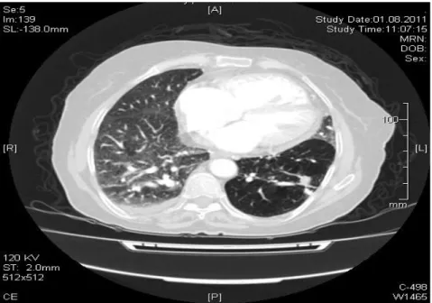
end-diastolic volume were calculated to be 1 ml and 18 ml respectively. In this patient, lung/ heart ratio was calculated 0,22 from the left lung and 0,38 from the right lung. Decreased activity in the left lung was also visualized on the raw data images. VPS showed multiple matched perfusion-ventilation defects and central airway deposition of the radiopharmaceutical consistent with COPD (Figure II). Thorax CT compatible with emphysema in the bilateral upper lobe and left lower basal segments. CT showed evidence of emphysema and air trapping (Figure III).
Figure I: MPS and SPECT images compatible with hypertrophic left ventricle, enlarged right ventricle and decreased pulmonary activity in the left lung.
Kocatepe Tıp Dergisi 2014;15(3): 322-5 324
Pulmonary Emphysema on Myocardial Perfusion Scintigraphy
Miyokard Perfüzyon Sintigrafisinde Pulmoner Amfizem
Figure III: Evidence of emphysema and air trapping on transverse section of CT images.
DISCUSSION
Myocardial perfusion studies with Tc-99m MIBI are routinely performed to evaluate patients with ischemic heart disease. Tc-99m MIBI is a cationic complex which diffuses passively through the capillary and cell membrane. Within the cell it is localized in the mitochondria, where it is trapped and retention is based on intact mitochondria, reflecting viable myocytes. In patients referred for evaluation of myocardial perfusion, extracardiac findings on MPS are uncommon, including increased focal uptake or decreased uptake (5). Abnormalities of extracardiac activity in the thorax include diffusely bilaterally increased pulmonary activity and increased activity in the sternum and/or vertebrae in patients with anemia or hypoxemia. Diffusely decreased to absent activity in the lung and/or the areas of absent to decrease activity in the chest is pleural effusion and pericardial effusion (6).
In our case, decreased pulmonary uptake of left lung in myocard perfusion scintigraphy was caused by emphysema in that pulmonary parenchyma could not take up tracer in the lungs. Pulmonary emphysema is characterized by irreversible destruction of lung parenchyma. Emphysema is defined as a condition of the lung characterized by abnormal, permanent enlargement of the air spaces distal to the terminal bronchiole, accompanied by destruction of alveolar
walls (7). Because emphysema decreases the elastic recoil force that drives air out of the lung and thereby reduces maximal expiratory airflow, the disease is clinically classified as a COPD (8).
Extracardiac activity can be an indirect but important indication for a variety of non-cardiac disorders that may occasionally mimic cardiac symptoms such as emphysema. The primary aim of MPS is the evaluation of myocardial perfusion, so processed tomographic slices include only the heart. Therefore, because the unprocessed data include the physiologic or pathologic radiopharmaceutical uptake in the rest of the imaged body visualized within the filed of view, it is important that the interpreting physician evaluate all the information available.
As conclusion, we presented a case who received MPS and VPS for detecting the reason of chest discomfort. Chest discomfort is a physically and emotionally distressing symptom, which often poses a diagnostic dilemma for the physician regarding the underlying etiology and extent of evaluation. The classical features of angina do not distinguish the origin of the pain. However, differentiation between cardiac and non-cardiac origins of discomfort is extremely important in the subsequent management of the patient. Large areas of photophenia in the
Kocatepe Tıp Dergisi 2014;15(3):322-5
Kocatepe Tıp Dergisi, Cilt 12 No:3, Eylül 2011
325
Erdoğan et al. thorax were noted from raw data of MPS and SPECT images. CT showed evidence of emphysema and air trapping. The interpretation of MPS should not be limited to the heart, careful inspection and interpretation of the raw data besides the routine evaluation of myocardial reconstructed SPECT slices on MPS is important for these extracardiac findings (9). In agreement with the conclusion of Jones et al, any noncardiac finding from an MPS that arouses suspicion of a serious disease warrants direct contact with the referring physician for further evaluation, as extracardiac findings and non-perfusion abnormalities may on occasion account for the patient’s symptoms (9).
REFERENCES
1. Ritchie JL, Bateman TM, Bonow RO, et al. Guidelines for clinical use of cardiac radionuclide imaging: report of the American College of Cardiology/American Heart Association Task Force on Assessment of Diagnostic and Therapeutic Cardiovascular Procedures (Committee on Radionuclide Imaging), developed in collaboration with the American Society of Nuclear Cardiology. J Nucl Cardiol 1995;2(2 Pt 1):172-92.
2. Gedik GK, Ergün EL, Aslan M, et al. Unusual extracardiac findings detected on myocardial perfusion single photon emission computed tomography studies with Tc-99m sestamibi. Clin Nucl Med 2007;32(12):920-6.
3. Choy JB, Leslie WD. Clinical correlates of 99mTc sestamibi lung uptake. J Nucl Cardiol 2001;8(6):639-44.
4. Giubbini R, Campini R, Milan E, et al. Evaluation of technetium-99m-sestamibi lung uptake: correlation with left ventricular function. J Nucl Med 1995;36(1):58-63.
5. Williams KA, Hill KA, Sheridan CM. Noncardiac findings on dual-isotope myocardial perfusion SPECT. J Nucl Cardiol 2003;10(4):395-402.
6. Shih WJ. Thoracic and abdominal abnormalities found on raw data ımages of cardiac SPECT. Ann Nucl Med Sci 2005;(18):99-110.
7. Snider GL, Kleinerman JL, Thurlbeck WM, et al. The definition of emphysema: report of a National Heart, Lung, and Blood Institute, Division of Lung Diasease Workshop. Am Rev Respir Dis 1985;132(1):182-5. 8. MacNee W. Pathogenesis of chronic obstructive pulmonary disease. Proc Am Thorac Soc 2005;2(4):258-66;discussion 290-1.
9. Jones SE, Aziz K, Yasuda T, et al. Importance of systematic review of rotating projection images from Tc99m-sestamibi cardiac perfusion imaging for non-cardiac findings. Nucl Med Commun 2008;29(7):607-13.