Histopathological findings and immunohistochemical distribution of
bacterial antigen in Balb/c mice experimentally infected with
Arcobacter butzleri*
Ahmet ATA
1, Latife ÇAKIR BAYRAM²
1Işık Veterinary Clinic, Kocasinan, Kayseri; ² Erciyes University, Faculty of Veterinary Medicine, Department of Pathology, Kayseri, Turkey.
Summary: The aim of this study was to determine the tissue colonization and pathogenicity of Arcobacter butzleri in
BALB/c mice. For this purpose, 60 adult mice were distributed into three experimental groups consisting of 16 animals in each and into three control groups consisting of 4 animals in each. One ml of A. butzleri suspension containing 1x10⁹ CFU/mL was inoculated by peroral (PO), intraperitoneal (IP) and intramammary (IMG) routes to BALB/c mice in experimental groups in order to determine the clinical and pathological findings. Animals in control groups received 1 mL of sterile saline solution. Duration of the experiment was limited for 15 days. The BALB/c mice were found to be susceptible to infection by the three ways of inoculation. In infected mice, diarrhea, weakness, decrease of body temperature (IP and IMG); hepatomegaly, splenomegaly (IP); swelling of mammary glands, exudation, abscess formation (IMG) were seen. Mortality was determined as 25% in PO inoculation group and as 95% in IP IMG inoculation groups. In histopathological examination, acute/chronic inflammation characterized with neutrophil and plasma cells and local necrosis or congestion (IP) were determined in the lungs, kidneys, regional lymph nodes, liver, spleen, stomach and colon. However, severe, diffuse and chronic pyogranulomatous and necrotizing inflammation and interstitial fibrosis were observed in mammary gland and its subcutaneous tissues (IMG). In all of inoculation groups, A. butzleri was reisolated until the 5th days of post inoculation. The results of this study have suggested that BALB/c mice might be an appropriate experimental animal model for studying A.butzleri infection.
Keywords: Arcobacter butzleri, BALB/c mouse, experimental infection.
Deneysel olarak Arcobacter butzleri ile infekte edilmiş Balb/c farelerde bakteriyel antijenin
immunohistokimyasal dağılımı ve histopatolojik bulgular
Özet: Bu çalışmada BALB/c farelerde Arcobacter butzleri’nin (A.butzleri) patojenitesini ve doku kolonizasyonunu ortaya
koymak amaçlanmıştır. Toplam 60 adet fare kullanıldı. Her birinin deney grubunda 16 adet, kontrollerinde ise 4’er adet fare kullanılan 3 grup oluşturuldu. Araştırma 15 günle sınırlandırıldı. A.butzleri izolatı ml'sinde 1x10⁹ bakteri içerecek şekilde BALB/c farelere peroral (p.o), intraperitoneal (i.p) ve meme bezi içine (m.b.i) verilerek klinik ve patolojik bulgular değerlendirildi. BALB/c fareler üç inokulasyon yoluyla da infeksiyona duyarlı bulundular. Mortalite, i.p ve m.b.i inokulasyon gruplarında %95, p.o grupta ise %25 olarak saptandı. Klinik ve patolojik bulgular i.p. ve m.b.i. inokulasyon gruplarında gözlendi. İnfekte gruplarda, ishal, halsizlik, vücut ısısının düşmesi (i.p ve m.b.i); hepatomegali, splenomegali (i.p); infekte meme bezlerinde şişkinlik, eksudasyon, apse formasyonu (m.b.i) gözlendi. Histopatolojik olarak, akciğer, karaciğer, böbrek, dalak, bölgesel lenf düğümleri, mide, kolon ve nötrofil ve plazma hücreleri ile karakterize akut/kronik yangı, lokal nekroz ve konjesyon (i.p), meme bezi ve deri altı dokusunda ise şiddetli, diffuz, kronik pyogranulomatoz-nekrotik yangı ve intersitisyel fibrosis (m.b.i) saptandı. Tüm inokulasyon gruplarında Arcobacter butzleri inokülasyon sonrası 5. güne kadar izole edildi. Sonuç olarak, BALB/c farelerin Arcobacter butzleri’nin deneysel infeksiyonu için uygun deney hayvanı olarak kullanılabileceği kanısına varıldı.
Anahtar sözcükler: Arcobacter butzleri, BALB/c fare, deneysel infeksiyon
Introduction
The genus Arcobacter is included in
Campylobacteraceae family, which also includes the Campylobacter genus. Arcobacter spp. are phenotypically similar to Campylobacter species, but they differ from
Campylobacter and other microorganisms in this family
with two important characteristics such as their ability to survive and propagate at low temperatures (15-25 °C) and under aerobic conditions (16). The Arcobacter genus currently includes 17 species (16). Among the Arcobacter
species, A. butzleri, Arcobacter cryaerophilus (A.
cryaerophilus) and Arcobacter skirrowii (A. skirrowii)
have been isolated from several diseases in human (22, 25, 29) and in animals (1, 15, 23, 24, 28). Although the pathogenicity of Arcobacter is not fully elucidated, today
Arcobacter spp. are known as potential water and
food-borne pathogens (1, 16). A. butzleri is the most commonly isolated Arcobacter species (1, 12, 22). In human, A. butzleri is considered to be a pathogen that can cause severe diarrhea and bacteremia (8). Bacteremia associated with A. butzleri and A. cryaerophilus have been reported in children, in the elderly and in the patients with chronic diseases (19, 24, 29). Common symptoms of Arcobacter infections in human are abdominal pain and cramps along with persistent diarrhea (18, 24). It has been reported that A. butzleri caused no clinical finding except the bleeding due to
placenta previa in mothers through vertical/
transplacental transport of the agent, whereas in newborns death was observed as the result of respiratory distress, intravascular coagulation and acute renal failure (26). In addition, some studies have shown that A.
butzleri is also the cause of diarrhea in non-human
primates (6). A.butzleri have been considered as the most serious agent in this genus (16) because of its isolation from the gastrointestinal infections (18-20, 24) as well as extra-intestinal invasive diseases (19, 30) in human.
Arcobacter species, except Arcobacter nitrofigilis and Arcobacter halophilus, have been incriminated with
various animal diseases including abortion, septicemia, mastitis, gastritis and enteritis (6, 11, 16, 18, 21). On et al. (23) reported reproductive disorders, chronic abortion problems as well as opportunistic pathogens that play a role in the disease process in Arcobacter infections in animals. The isolation of A. butzleri and Arcobacter
cryaerophilus from the preputial fluids of bulls (11, 23)
and isolation of Arcobacter skirrowii from the preputial fluids the pigs (24) have suggested that venereal route is a source of contamination for Arcobacter species in animals (10). Arcobacter species were also determined in the cases of diarrhea and enteritis behind the reproductive disorders in livestock (24). Arcobacter species has been isolated from aborted fetuses of pig (11, 15, 23), ovine (18, 24) and bovine (14, 24) with diarrhea (18, 27).
Very limited knowledge is available regarding the molecular mechanisms of the pathogenesis of Arcobacter
spp. that genetically differ from Campylobacter spp. (8,
16). However, the studies concerning the pathogenicity of Arcobacter have been based on the data of the
Campylobacter and Helicobacter species (16). One of the
studies on the pathogenicity of Arcobacter species was in piglets (27), and the other one was carried out by the administration of Arcobacter species from several origins to chickens and turkeys (28). A few studies were reported
in experimental animals such as rat, but in these studies infections were induced solely by oral route or through testicles. In the study conducted by Adesiji et al. (2), in which healthy adult male albino rats were infected with solely a single oral challenge of A. butzleri isolated from stool of healthy chicken, severe histopathological lesions in small intestine was reported. In another experimental study of Adesiji et al. (3), inoculation of A. butzleri that was isolated from pig into the testicles of albino rats resulted in degeneration in the testicles. Arcobacter could be a potential pathogen in albino rats while the selected haematological and electrolyte parameters may be good diagnostic indicators of the animal response to the bacterial infections (4).
To the author’s knowledge, no study is available regarding the infections caused by Arcobacter species, thus the histopathological changes in mice. Therefore, this study was performed to investigate the pathological findings and tissue colonization of A. butzleri in the infection induced with oral, intraperitoneal and intramammary routes in pregnant BALB/c mice. This
study is of importance for determining the
histopathological changes due to Arcobacter, and for creation of an experimental model animal in human studies as well as for contributing to the compensation of the lack in the literature.
Materials and Methods
Experimental animals: In this study, 60 adult,
healthy, female BALB/c mice mean body weight of (25-30 g), which had been provided by Erciyes University Experimental Clinics and Research Center, were used with the permission of Ethics Committee of Faculty of Veterinary Medicine, University of Erciyes (2004-042).
Bacterial strain and preparation of inoculum: The
strain of A. butzleri, which had been isolated from human gastrointestinal infection, and confirmed by Multiplex Polymerase Chain Reaction (m PCR) (5), was provided by University of Erciyes, Faculty of Veterinary Medicine, Department of Microbiology. For preparation of inoculum, A. butzleri strain was inoculated onto Mueller Hinton agar supplemented with 5% sheep blood,
and incubated at 30oC in microaerophilic conditions for
24-48 hours. The bacterial culture was suspended in 0.9% sterile saline and standardized by McFarland’s Nephelometry of 5 that approximately corresponding to
109 colony forming unit per mL (CFU/mL).
Experimental design: In the study, 60 animals were
allocated into six groups containing 16 mice in each of three infection groups [peroral (PO), intraperitoneal (IP) and intramammary (IMG)] and 12 mice in controls (4 animals in each control of infection groups). A 0.1 mL of
A. butzleri suspension containing 1x10⁹ CFU/mL was inoculated by oral, intraperitoneal and intramammary
routes to the mice in PO, IP and IMG groups respectively. The agent was given with a sterile nutrition syringe to mice in PO group and with injection to the mice in IP group. The mice in IMG group were infected
on 10th day of the gestation. The mice were anesthetized
with ether, then the inocula were injected into the nipple of left (L4) and right (R4) mammary glands. Intramammary injection of mice was carried out as described previously (9, 13). Normal saline solution (0.9% NaCl) was given with sterile nutrition syringe orally to the mice in control of PO group and with injection to the mice in IP and IMG groups. Animals were observed for clinical signs during the study.
Sample collection: Four animals (either dead or
sacrificed) from each of the infection groups and two animals from each of their controls were sacrificed on the 2nd, 5th, 10th and 15th days of post inoculation (pi), and tissue samples (the liver, intestine stomach, spleen, heart, lungs kidneys, pancreas, cerebrum, cerebellum and mammary glands) were collected for microbiological and histopathological analysis after the gross macroscopic examinations at necropsy.
Microbiological analysis - isolation and identification:
The collected tissue samples were homogenized with sterile saline for 2 minutes with Stomacher-Bag mixer, and then fortified in double-strength Arcobacter Enrichment Broth (AEB) (Oxoid CM965) prepared using Arcobacter enrichment basal medium supplemented with cefoperazone, amfoterisin and teicoplanin (CAT) under
microaerophilic condition at 30 oC for 48 hours. The
enriched samples were inoculated onto blood agar (Oxoid) containing 5% (v/v) defibrinated sheep blood with membrane filtration technique as described
elsewhere (7, 17), and incubated at 35-37oC under
microaerophilic conditions for 48-72 hours. The colony morphology and microscopic morphology and staining characteristics of the bacteria were examined for the identification. This procedure was applied until the isolation of the agent. Arcobacter butzleri 10828 strain obtained from Laboratorium voor Microbiologie en Microbielle Genetica (LMG), Ghent, Belgium was used as the control organism for the isolation and identification.
Preparation of tissue section for the histopathological examinations: The tissue samples (the liver, intestine
stomach, spleen, heart, lungs kidneys, pancreas, cerebrum, cerebellum and mammary glands) that were collected from either sacrificed or dead animals were fixed in 10 % buffered neutral formalin solution. After the routine alcohol-xylol process, tissue samples were embedded in paraffin and sectioned in 5-6 μm. Two of four sections taken from each blocks were mounted on normal slides and the other two were mounted on adhesive slides for HaematoxylinxEosin (HxE) and
Brown-Brenn Gram stainings. Unstained sections were used for immunohistochemical staining of A. butzleri antigen with Avidin-Biotin-Peroxidase Complex (ABC) staining technique with a commercially available kit (Ultravision HRP mouse, TM-125-HL Labvision)
according to manufacturer’s instructions. All
preparations were examined under a light microscope. The immunoreactivity were scored according to the severity of the stain (1= mild, 2= moderate, and 3= severe). Sera obtained from two adult New Zealand white rabbits that were hyperimmunized with multiple injections of killed whole A. butzleri strain served as the primary polyclonal antibody. New Zealand rabbits were inoculated 3 times with 0.25, 0.5, and 1 mL of
(approximately 1 × 10⁹ cfu/mL) via an ear vein at 5- day
intervals. Blood samples were collected from the rabbits on day 15, after the last injection, and sera were separated and stored at –20 °C. Serial dilutions (log2) of the primary antiserum—from 1/2 to 1/256— were made to obtain optimal primary antibody titers. The serum had an agglutinating titer of 1: 64. Primary antibody was used at a dilution of 1: 64.
Results
Clinical findings: In all infection groups, clinical
signs began to appear on day 2 pi. Eyes were closed in infected animals, and diarrhea, weakness, decreases in body temperature and marked tremors were observed. Conjunctivitis was developed in mice in IMG group as a different finding from the PO and IP groups. In PO group, clinical signs appeared in three mice, no clinical sign was observed in the remaining. The deaths occurred on day 3 after infection, and mortality was 25%. In IP and IMG groups, the deaths occurred on days 2-5 after infection. The mortality rates reached up to 95% in these two groups. Only two mice in each group had no clinical sign, and they survived until the end of the study, and these animals were sacrificed on the 15th day of pi.
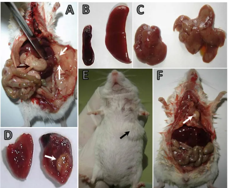
Macroscopic findings: In PO group, serosal vessels
were plump, especially, in the large intestine. Mesenterial lymph nodes were swollen, and their cut surfaces were wet. In IP group, a rather watery and foul-smelling feces were found in the small intestine. In one case, pinhead foci were present in duodenum, jejunal serosa and also in the fundus of glandular stomach. Splenomegaly and a few miliary foci were observed in the spleen (Figure 1A). In the most of the cases hepatomegaly was observed (Figure 1B). Liver was swollen and congested. In addition, in some cases grayish-white miliary foci of varying severity were observed. In one case, left and median lobes of the liver were bonded to the periton with an abscess of 0.5 cm in diameter (Figure 1C). In the cross-sectioned fluctuating mass, there was a creamy, yellowish-white content. The abscess was observed in the
myocardium in the same mouse (Figure 1D). In IMG group, in all infected mice, swelling, congestion, varying degrees of subcutaneous gelatinous-purulent exudate were observed in infected mammary glands (left cervical: LC, left thoracic: LT, left abdominal: LA, left inguinal: LI, right cervical: RC, right thoracic: RT, right abdominal: RA, right inguinal: RI), (Figure 1E). In some cases (case no:IMG-6,9), diffuse abscesses embracing the infected mammary gland and surrounding tissues were
present. It was observed that abscesses were
subcutaneous and in some cases penetrated to the chest (Figure 1F). When the fluctuating abscess sectioned, a creamy, yellowish-white content was seen. No lesion was found in any of the control animals.
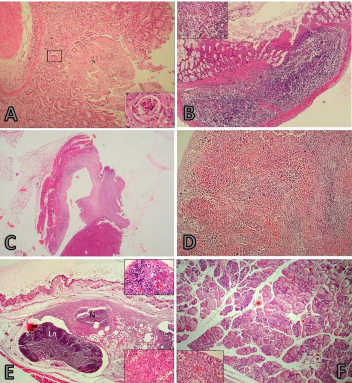
Microscopic findings: In PO group, severe colitis
was observed in large intestine. Crypt epithelium had been desquamated. Neutrophilic reaction and necrosis were present between the gland of colon, and it was much more intense, especially, in the submucosa that advanced to muscle layers, as well as the cellular increase was observed in lymphoid follicles in the submucosa (Figure 2A and 2B). Severe congestion was present in the mesenterial lymph nodes. The endothelium of vascular sinuses was plump. The lumens of vascular sinuses were expanded by dark colored lymphoblastic cells and by desquamated endothelial cells. The striking findings were hemorrhage in the white and red pulp of the spleen and as well as lymphoid depletion.
Figure 1. A-D) IP experimental group A) Adhesion of left and median lobes of liver to the skin with the abscess (white arrows). A pinhead sized focus (black arrow) on the fundus of glandulary stomach. B) Spleen of a control animal, Splenomegaly (right). C) Hepatomegaly (right), liver of a control animal (left). D) Myocardium of a control animal (left), an abscess (arrow) in myocardium (right). A mouse in intra-mammary inoculation group (E, F). E) Infected mammary glands (left thoracic) and a mass affecting the surrounding area. E) Swelled region (arrow) on the left hand side of throcic mammary gland. F) Penetration of the abscess ( arrow) to the subcutan area and thorax.
Şekil 1. (A-D) IP deney grubu A) Derideki (beyaz oklar) apsenin, karaciğerin sol ve orta loblarına adezyonu. Glandular midenin fundusunda toplu iğne başı büyüklüğünde odak (siyah ok). B) Dalak kontrol (sol), splenomegali (sağ) C) Hepatomegali (sağ), kontrol (sol) karaciğer. D) Miyokardiumda apse (ok, sağ) ve kontrol (sol). IMG inokulasyon grubu (E, F). E) Sol torasik meme bezleri çevredeki etkileyen bir kitle. F) Torasik meme bezinin sol tarafta şişlik (ok) E) Apsenin subkutan bölge ve toraksa penetrasyonu (ok).
Figure 2. (A-E) 1. Histopathological findings in a BALB/ c mice after IP inoculation, HxE (All figures). A) Necrotic area extending to the submucosa (inset). Lymphocytic and slight neutrophilic reaction between glands and in submucosa, colon, x100. inset: Necrotic changes in lymphocytes, x 40. B) Necrosis lymphoid follicles in submucosa, colon, x100. C) The abscess wall invading the liver and muscle layer. L: Liver, Gr: Granulation tissue, M: Muscle, x40, inset: x40. D) Diffuse hemorrhage and a significant decrease in lymphocytes in the white, spleen, x 20, inset: x40. E to F) Histopathological changes in a BALB/ c mice after IMG inoculation, HxE (All figures). E) Pyogranulomatous inflammation and severe necrosis covering the muscular layer. Coagulation necrosis and purple color bacterial colonization in the necrotic center, axillary lymph node (Ln) (top inset) and bacterial colonization with intensive neutrophil leucocyte infiltration (bottom inset) in necrosis (N), x10, insets: x40. F) Alveolary structures with intensive neutrophil leucocyte infiltration. Cervical mammary gland. x20, inset:x40.
Şekil 2. IP inokulasyonlu BALB/c farelerde histopatolojik bulgular. Kas tabakasında pyogranulamatoz yangı ve şiddetli nekroz, HxE. A) Submokozaya uzanan nekrotik bölge (ekli küçük resim). Bezler arasında ve submukozada nötrofil ve lenfosit hücre infiltrasyonu, kolon x100. Ekli küçük resim:Lenfositlerde nekrotik değişiklikler x40. B) Submukozada lenfoid follikullerde nekroz, kolon, x10. C)Karaciğer ve kas tabakasına invaze olan apse duvarı L: Karaciğer, Gr: Granulasyon dokusu M: Kas, x40, Inset: x40. D) Dalakta diffuz hemoraji, beyaz pulpada lenfositlerde belirgin azalma x 20, inset: x40. E–F) Arcobacter butzleri ile infekte edilmiş IMG deney grubunda histopatolojik bulgular, HxE. E) Kas tabakasında pyogranulamatoz yangı ve şiddetli nekroz. Nekrozun merkezinde koagulasyon nekrozu ve mor renkli bakteriyel kolonizasyon, aksillar lenf yumrusu (Ln) (üst ekli küçük resim). Nekroz alanı (N), yoğun nötrofil infiltrasyonu ve bakteri kümeleri x10, ekli küçük resimler: x40.F) Yoğun nötrofil lökositlerle dolu alveoller, servikal meme bezi, x20, ekli küçük resim:x40.
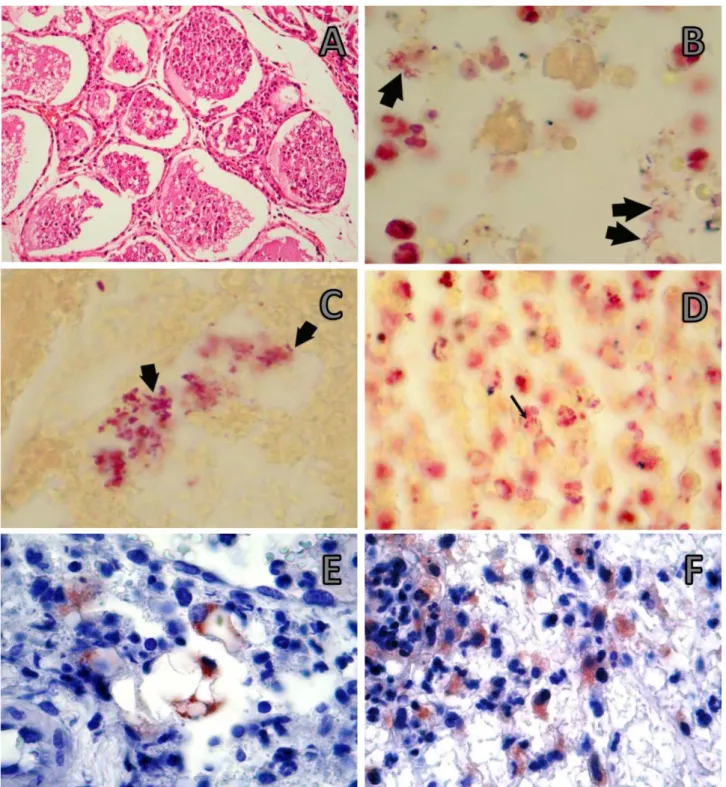
Figure 3. IMG inoculation group A) Intensive cellular debris in alveolary structures. Thoracic mammary gland. x10 inset: x40, HxE. B) Red, spiral shaped bacteria (arrows) of various sizes detectable within the cytoplasm of macrophages in the interalveolar tissue, x 100, Gram stain. C-D) Spiral shaped bacteria (arrows) detectable within inflammatory areas in the mammary gland, x 100, Gram stain. E-F) Immunohistochemical staining (×100 oil), ABC method. E) Arcobacter positive reaction present within macrophages. F) Intracytoplasmatic Arcobacter-positive reaction in macrophages around vessels.
Şekil 3. Meme bezine inokulasyon grubu A) Alveoler yapılarda yoğun hücresel döküntüler.Torasik meme bezi, x10, küçük ekli resim: x40. B) İnteralveoler dokuda makrofaj sitoplazmalarında spiral şekilli bakteriler x 100, Gram boyama. C) Meme bezinde yangısal alanlarda spiral şekilli bakteri, x 100, Gram boyama . D) Makrofaj sitoplazmasında immunopozitif hücreler, x 100, Gram boyama. E-F) Immunohistokimyasal boyama (×100 oil), ABC method. E) Makrofaj sitoplazmasında Arcobacter pozitif reaksiyon. F) Damarlar etrafında makrofajlarda Arcobacter pozitif reaksiyon.
In IP group, the passive hyperemia was observed in hepatocytes of all mice. Fat vacuoles of varying sizes were noted in the cytoplasm of hepatocytes, the majority of these were small droplets. The liver tissue had been replaced with fibrosis and chronic inflammation in a mouse, (Figure 2C). Dense plasma cells and lymphocytes infiltration were present in portal veins, periportal areas and necrotic areas. Similar cells settled as the periglomerular, perivascular and intertubular in nearly all of kidney tissue. Chronic inflammation was evident in the liver of one case. Also, wide pyogranulomatous foci were evident in the skin of the abdominal wall where the liver stuck to and in breast. Diffuse hemorrhage in the spleen was a significant finding. Lymphocytes in white pulp were markedly reduced compared to that of control animals. The endothelium of vascular sinuses was enlarged. The nucleus of lumen was expanded by dark colored lymphoblastic cells and by desquamated endothelial cells. (Figure 2D).
In IMG group, in the infected mammary glands, a
reaction ranging from diffuse pyogranulomatous
inflammation to the necrosis of nearly all of the mammary tissues was characterized as the main finding (Figure 2E). Alveolar luminae were filled with erythrocytes and dense neutrophil leukocytes infiltration (Figure 2F and 3A). In the mammary tissues without necrotic areas, degenerative changes developed in secretoric epithelium but alveolar structures were not lost. In addition, inter-acinar edema was present next to these areas. Cell debris was noticed in nipple ducts, in alveolar structures and in the interalveolar tissue.
Immunohistochemistry and Gram staining results:
The infectious agent was identified in tissues by means of Gram staining methods (Figure 3B-3D) and Avidin-Biotin Complex Peroxidase Stainings (Figure 3E and 3F). Intra-cytoplasmic Arcobacter-positive antigen was
detected in macrophages around vessels in mammary
gland. Arcobacter antigen was present within
macrophages. The immunohistochemistry stain most frequently identified bacteria in the lung and gastrointestinal tract. Occasionally positive staining was also found in crypt epithelium. Strong immunopositive extracellular and intracellular materials were found in the necrotic areas in the propria mucosa. In addition, strong immunopositive reaction was observed extracellularly as well as in the cytoplasm of hepatocytes. Necrosis developed in lymphoid follicles, and the intracellular and extracellular weak immunopositive reactions were observed in the surroundings of necrotic areas.
Bacteriological findings: The agent was isolated
from the lung, liver, spleen, stomach, intestine, kidney,
brain and mammary gland until the 5th day of pi, no
isolation was made thereafter Results of immuno-histochemistry and culture presented semiquantitatively. (Table 1-4).
Discussion and Conclusion
Arcobacter species, which are morphologically
similar to Campylobacter species but differ from
Campylobacter species in terms of their growth
characteristics (16), have been isolated from several diseases in human (22, 25, 29) and in animals (15, 23, 24, 28). However, knowledge on the pathogenicity of
Arcobacter is still very limited (2, 3, 13, 14, 16).
Therefore, in this study, tissue colonization and pathogenicity of A. butzleri in BALB/c mice inoculated by peroral, intraperitoneal and intramammary routes were determined. In some in vivo studies conducted on 1-day-old piglets (27) and on several days of old chickens (28) concerning the invasion and virulence of Arcobacter strains, the results varied with the bacterial strains as well as with the species and breed of the animals. Wesley et
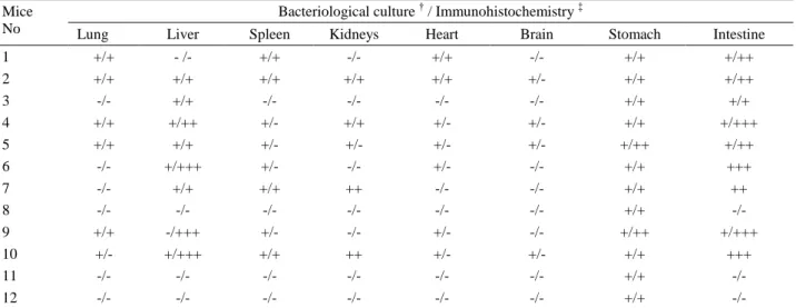
Table 1 Relationship between cultural (†) and immunohistochemical (‡) results obtained from organs of BALB/c mice infected by PO inoculation.
Tablo 1. PO inokulasyonla infekte BALB/ c farelerin organlarına ait immunohistokimyasal ve kültür sonuçları arasındaki ilişki. Mice
No
Bacteriological culture † / Immunohistochemistry ‡
Lung Liver Spleen Kidneys Heart Brain Stomach Intestine
1 +/+ - /- +/+ -/- +/+ -/- +/+ +/++ 2 +/+ +/+ +/+ +/+ +/+ +/- +/+ +/++ 3 -/- +/+ -/- -/- -/- -/- +/+ +/+ 4 +/+ +/++ +/- +/+ +/- +/- +/+ +/+++ 5 +/+ +/+ +/- +/- +/- +/- +/++ +/++ 6 -/- +/+++ +/- -/- +/- -/- +/+ +++ 7 -/- +/+ +/+ ++ -/- -/- +/+ ++ 8 -/- -/- -/- -/- -/- -/- +/+ -/- 9 +/+ -/+++ +/- -/- +/- -/- +/++ +/+++ 10 +/- +/+++ +/+ ++ +/- +/- +/+ +++ 11 -/- -/- -/- -/- -/- -/- +/+ -/- 12 -/- -/- -/- -/- -/- -/- +/+ -/-
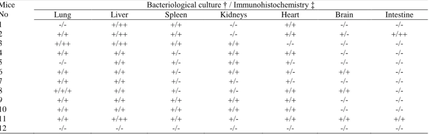
Table 2 Relationship between cultural (†) and immunohistochemical (‡) results obtained from organs of BALB/c mice infected by IP inoculation.
Tablo 2.İP inokulasyonla infekte BALB/ c farelerin organlarına ait immunohistokimyasal ve kültür sonuçları arasındaki ilişki. Mice
No
Bacteriological culture † / Immunohistochemistry ‡
Lung Liver Spleen Kidneys Heart Brain Intestine
1 -/- +/++ +/+ -/- +/+ -/- -/- 2 +/+ +/++ +/+ -/- +/+ +/- +/++ 3 +/++ +/++ +/+ +/+ -/- -/- -/- 4 +/+ +/+ +/- +/+ +/+ -/- -/- 5 -/- +/+ +/- +/+ +/- -/- -/- 6 +/+ +/+ +/- +/+ +/- +/+ -/- 7 +/+ +/+ +/- +/- +/- -/- -/- 8 +/+/+ +/+ +/- +/- +/+ +/+ -/- 9 +/+ +/+ +/+ +/+ +/+ -/- -/- 10 +/+ +/+ +/+ +/+ +/+ -/- -/- 11 +/+ +/++ +/+ +/- +/+ +/+ +/+ 12 -/- -/- -/- -/- -/- -/- -/-
Table 3 Relationship between cultural (†) and immunohistochemical (‡) results obtained from organs and absces of BALB/c mice infected by IMG inoculation.
Tablo 3. IMG inokulasyonla infekte BALB/ c farelerin organlarına ait immunohistokimyasal ve kültür sonuçları arasındaki ilişki. Mice
No
Bacteriological culture † / Immunohistochemistry ‡
Lung Liver Spleen Kidneys Heart Brain Mammary Glands *Abscess
1 +/+ +/+ +/+ -/- -/- -/- +/++ +/+ 2 +/+++ +/+ +/+ -/- -/- -/- +/+++ +/+ 3 +/+++ -/- -/- -/- -/- -/- +/+++ ND** 4 -/- -/- -/- -/- -/- -/- +/+ ND 5 +/+ +/+ +/+ -/- -/- -/- +/++ +/+ 6 +/+ +/++ +/+ -/- -/- -/- +/++ +/+ 7 +/+ +/+ +/- -/- -/- -/- +/+ +/+ 8 +/+ +/+ +/- -/- -/- -/- +/+++ +/- 9 +/+ +/+ +/- -/- -/- -/- +/++ +/+ 10 +/+ +/+ +/- -/- -/- -/- +/++ +/- 11 -/- -/- -/- -/- -/- -/- +/++ ND 12 -/- -/- -/- -/- -/- -/- +/++ ND
*Present in mammary gland and surrounding tissues **ND: Not Detected
†
Culture results: -: culture negative; +: culture positive.
‡ Arcobacter antijen. - : Negatif; low+; ++ : moderate; +++ : severe. *Meme bezi ve çevre dokularda mevcut **Saptanmadı
† Kültür sonuçları: -: kültür negatif; +:kültür pozitif
‡ Arcobacter antigen. -: Negatif +: zayıf; ++: orta şiddette ; +++: şiddetli
Table 4. Differences in the histopathological and immunohistochemical findings of experimental groups. Tablo 4. Deney gruplarınının histopatolojik ve immunohistokimyasal bulgularındaki farklılıklar.
Infection Groups Differences
Histopathological findings Immunohistochemical findings*
PO
Neutrophilic reaction and necrosis of colon Congestion of mesenterial lymph nodes
Propria mucosa and crypt epithelium Surrounding of necrotic areas of mesenterial lymhoid nodes
IP Chronic inflammation of liver and periton Cytoplasm of hepatocyte
IMG Pyogranulamatous inflammation to necrosis of mammary glands
Macrophage cytoplasm in mammary glands Alveoler macrophage
PO, Peroral IP, Intraperitoneal IMG, Intrammary glands
*Immunostaining with Arcobacter positive antigen PO, Peroral
IP, İntraperitoneal IMG, Meme bezi içi
al. (27) induced experimental oral infections, in four one-day-old piglets that were not received colostrum. Two trials were conducted in that study. The authors determined that despite the growth of all strains (A.
butzleri ATCC 49616, A. cryaerophilus 1B ATCC
43159, A. skirrowii CCUG 10374, and three field strains of A. butzleri) in the intestine, only the A. butzleri strains isolated from human faces and pig were colonized in the internal organs. The authors observed the invasion in all of the four piglets infected with A. butzleri strains in the first trail whereas the invasion was determined in only two piglets in the second trial. The occurrence of mortality due to A. butzleri and A. skirrowii in the first trail but not in the second trail was explained with age and individual sensitivity. In this study, inoculation of A.
butzleri by PO, IP and IMG routes to BALB / c mice
resulted in infection. The pathological findings of the induced infection was similar to the findings seen in
Campylobacter infections (13, 27) except higher
mortality rate. The mortality rates that observed as 25 % and 95% in PO, IP and IMG groups respectively was higher than the mortality rates reported in previous studies conducted using Campylobacter species (5, 13). The high mortality in this study might be associated with the sensitivity of laboratory animals to Arcobacter spp. and virulence of the strain used which is a human pathogen. On the other hand, Wesley and Baetz (28) induced oral infections with human strains of A. butzleri in chickens, and infection with turkey field strains in 3 and 5 days old chickens and turkeys. The authors found that the human strain did not colonize and invade White Leghorn chickens and commercial outbred of turkeys whereas, the agent was isolated from the cloacal swabs and the tissues of inbred Beltsville White turkeys. These results have been interpreted as the virulence and invasion capacity of A. butzleri strains are associated with host factors.
In a rat study of Adesiji et al. (2), two strains of A.
butzleri isolated from stool of healthy chickens were
tested on albino rat by giving a single oral challenge of
109CFU/mL to adult male rats, and diarrhea occurred in
all rats from the 5th day post infection, In this study, similar clinical signs were detected in all groups, but they were evident especially in mice infected with intraperitoneal and mammary gland routes which was confirmed with the pathological findings. Diarrhea a significant decrease in body temperature and severe tremors were observed as reported in previous studies (2,
3). The isolation of the agent until the 5th days of pi was
consistent with the results of Adesiji et al. (2). The isolation of A. butzleri in tissue samples from mice with diarrhea, and the determination of colitis with
histopathological examination similar to the
Campylobacter infections (6, 8, 16) might suggest that
the agent colonize and invade to the intestine in PO group as well as to the intestine and the visceral organs in IP group. Severe histopathological lesion such as villous erosion, desquamation, matting and necrosis of the segments of small intestine observed by Adesiji et al. (2) was also observed in the presented study. In some cases in IP and IMG groups remarkable findings were diffuse abscess and cellulitis. Campylobacter fetus subspecies
fetus, similarly to Arcobacter spp. causes systemic
infection in patients with compromised immune systems, in the elderly and in the individuals those with chronic diseases (meningitis, vascular infections) (16, 18). However, in Campylobacter infections abscess formation outside the small intestine associated with bacteremia is rare. In the presented study, abscess and adhesions seen in the liver of mice in IP and IMG group might be the penetration of the agent to bloodstream as indicated by de Vries et al. (12) who speculated that the abscess formed in the liver might be due to transportation of the agent through bloodstream that were penetrated to intestinal mucosa. Some Arcobacter strains play a primary role in abortions and reproductive disorders while others merely are opportunistic pathogens (23, 27). They have also been isolated from raw milk during a mastitis outbreak in cattle (21). It was reported that pregnancy in the systemic Campylobacter infections is a predisposing factor and Campylobacter spp. causes developmental disorders such as disorder of fertilize ovum implantation and resorption of fetus in the laboratory animals in different stages of the pregnancy (16). Diker et al. (13) inoculated the bovine and avian origin Campylobacter coli strains into the mammary gland of mice and examined the induced acute mastitis lesions histopathologically. In present study, diffuse breast abscess formation and conjunctivitis were seen with A. butzleri in addition to acute mastitis reported by these authors. Result of this study revealed that the pregnancy seems to be a predisposing factor in the occurrence of systemic A. butzleri infection. In addition, the mouse provides a conventional model for fundamental studies on A. butzleri mastitis. To the best knowledge,of authors this is the first study reporting the pathology of A. butzleri in BALB / c mice. In conclusion, BALB/c mice can be considered as a model animal in further investigations for the pathogenesis of Arcobacter infections.
Acknowledgements
The authors are grateful to the research scholars Department of Microbiology, Faculty of Veterinary Medicine, University of Erciyes for the providing of strains used in this study and for bacteriological examinations. This study was supported by Erciyes University Research Fund (Project no: SBT-07-49).
References
1. Abay S, Kayman T, Hizlisoy H, et al. (2012): In vitro
antibacterial susceptibility of Arcobacter butzleri isolated from different sources. J Vet Med Sci 74, 613-616.
2. Adesiji YO, Emikpe BO, Olatain JO (2009):
Histopathological changes associated with experimental infection of Arcobacter butzleri in albino rats. SLJB, 1,
4-9.
3. Adesiji YO, Emikpe BO, Opalekunde AB (2011):
Histopathological and functional changes in the testicles of albino rats experimentally infected with Arcobacter butzleri. Acta Med Lituanica, 18, 127-131.
4. Adesiji YO, Seibu E, Emikpe BO, et al. (2012): Serum
biochemistry and heamatological changes associated with graded doses of experimental arcobacter infection in rats.
West Afr J Med, 31, 186-91.
5. Al-banna NA, Junaid TA, Mathew TC, et al. (2008):
Histopathological and ultrastructural studies of a mouse lung model of Campylobacter jejuni infection. J Med
Microbiol, 57, 210-217.
6. Anderson, KF, Kiehlbauch JA, Anderson DC, et al. (1993): Arcobacter (Campylobacter) butzleri associated
diarrheal illness in a nonhuman primate population. Infect
Immun, 61, 2220-2223.
7. Atabay HI, Aydin F, Houf K, et al. (2003): The prevalance of Arcobacter spp. on chicken carcases sold in
retail markets in Turkey, and identification of the isolates using SDS-PAGE. Int J Food Microiol, 81, 21-28.
8. Bucker R, Troeger H, Kleer J, et al. (2009): Arcobacter
butzleri induces barrier dysfunction in intestinal HT-29/B6 cells. J Infect Dis, 200, 756-764.
9. Chandler RL (1970): Experimental bacterial mastitis in
the mouse. J Med Microbiol, 3, 273-282.
10. De Oliveira SJ, Baetz AL, Wesley IV, et al. (1997):
Classification of Arcobacter species isolated from aborted pig fetuses and sows with reproductive problems in Brazil.
Vet. Microbiol, 57, 347-354.
11. De Oliveria SJ, Wesley IV, Baetz AL, et al. (1999):
Arcobacter cryaerophilus and Arcobacter butzleri isolated from preputial fluid of boars and fattening pigs in Brazil. J.
Vet Diagn Invest, 11, 462-464.
12. De Vries JJ, Arents NI, Manson WL (2008):
Campylobacter species isolated from extra-oro-intestinal abscesses: a report of four cases and literature review. Eur
J Appl Microbiol, 27, 1119-1123.
13. Diker KS, Haziroglu R, Diker FS (1992): Experimental
infection of the mouse mammary gland with Campylobacter coli. Res Vet Sci, 52, 123-125.
14. Ellis WA, Neill SD, O’brien JJ, et al. (1977): Isolation of
Spirillum/Vibrio-like organisms from bovine fetuses. Vet
Rec, 100, 451-452
15. Ellis WA, Neill SD, O’brien JJ, et al. (1978): Isolation of
Spirillum-like organisms from pig fetuses. Vet Rec, 102,
106.
16. Ferreira S, Queiroz JA, Oleastro M, et al. (2015): Insights in the pathogenesis and resistance of Arcobacter: A review. Crit Rev Microbiol, 25, 1-20.
17. Kayman T, Abay S, Hizlisoy H, et al. (2012): Emerging
pathogen Arcobacter spp. in acute gastroenteritis: molecular identification, antibiotic susceptibilities and
genotyping of the isolated arcobacters. J Med Microbiol,
61, 1439-1444.
18. Kiehlbauch JA, Brenner DJ, Nicholson MA, et al. (1991): Campylobacter butzleri sp. nov. isolated from
humans and animals with diarrheal illness. J Clin
Microbiol, 29, 376-385.
19. Lau SK, Woo PC, Teng JL, et al. (2002): Identification by 16S ribozomal RNA gene sequencing of Arcobacter butzleri in a patient with acute gangrenous appendicitis. Mol Path, 55, 182-185.
20. Lehner A, Tasara T, Stephan R (2005): Relevant aspects
of Arcobacter spp. as potential foodborn pathogen. Int J
Food Microbiol, 102, 127-135.
21. Logan EF, Neill SD, Mackie DP (1992): Mastitis in dairy
cows associated with an aerotolerant Campylobacter. Vet
Rec, 110, 229-230.
22. Mansfiled LP, Forsythe SJ (2000): Arcobacter butzleri,
A. Skirrowii and A. cryaerophilus—potential emerging human pathogens. Rev Med Microbiol, 11, 161- 170.
23. On SLW, Jensen TK, Bille-Hansen V, et al. (2002):
Prevalence and diversity of Arcobacter spp. isolated from the internal organs of spontaneous porcine abortions in Denmark. Vet Microbiol, 85, 159-167.
24. Vandamme P, Vancanneyt M, Pot B, et al. (1992):
Polyphasic taxonomic study of the emended genus Arcobacter with Arcobacter butzleri comb. nov. and Arcobacter skirrowii sp. nov., an aerotolerant bacterium isolated from veterinary specimens. Int J Syst Bacteriol,
42, 344-356.
25. Vandamme P, Pugina P, Benzi G, et al. (1992b):
Outbreak of recurrent abdominal cramps associated with Arcobacter butzleri in an Italian school. J Clin Microbiol,
30, 2335-2337.
26. Vandenberg O, Dediste A, Houf K, et al. (2004):
Arcobacter species in humans. Emerg Infect Dis, 10,
1863-1867.
27. Wesley IV, Baetz AL, Larson DJ (1996): Infection of
cesarean derived colostrum-deprived 1-day-old piglets with Arcobacter butzleri, Arcobacter cryaerophilus, and Arcobacter skirrowii. Infec Immun, 64, 2295-2299.
28. Wesley IV, Baetz AL (1999): Natural and experimental
infections of Arcobacter in poultry. Poultry Sci, 78,
536-545.
29. Wybo I, Breynaert J, Lauwers S et al. (2004): Isolation
of Arcobacter skirrowi from a patient with chronic diarrhea. J Clin Microbiol, 42, 1851-1852.
30. Yan JJ, Ko WC, Huang AH, et al. (2000): Arcobacter
butzleri bacteremia in a patient with liver cirrhosis. J
Formos Med Assoc, 99, 166-169.
Geliş tarihi: 21.04.2015 / Kabul tarihi: 26.10.2015
Address for correspondence:
Latife Çakır Bayram, DVM, PhD Associate Professor,
University of Erciyes,
Faculty of Veterinary Medicine, Department of Pathology, 38039, Kayseri, Turkey e-mail: [email protected]