Lack of Prognostic Significance of C-erbB-2 in Astrocytomas
Asian Pac J Cancer Prev, 15 (3), 1333-1337
Introduction
Astrocytic tumors (astrocytomas) are the most common primary glial tumors of the central nervous system (CNS), and are classified according to a grading system developed by the World Health Organization (WHO), from low grade astrocytomas (diffuse astrocytoma, pilocytic astrocytoma) to high grade astrocytomas (anaplastic astrocytoma, glioblastoma) (Louis et al., 2007).
Glioblastoma (GBM) is the most common primary malignant tumor of the CNS in adults, representing 50% of all gliomas and 20% of all solid intracranial lesions that undergo surgery. With a mean incidence of 3 per year per 100.000 persons (Mineo et al., 2002; Rainov et al., 2007), it ranks fourth among the causes of death due to cancer in the middle-aged population (Hiesiger et al., 1993). Despite advances in the therapeutic management of GBM, prognosis remains poor and the median overall survival rate is <1 year. Several factors, such as age, performance status, radiotherapy, chemotherapy, quality of surgery, location and nature (de novo or secondary glioma)
Department of Medical Oncology, 1Faculty of Medicine, University of Baskent, Adana, 2Van Training and Research Hospital, Van, 3Mersin State Hospital, Mersin, 4Faculty of Medicine, University of Mersin, Mersin, 5Department of Pathology, Faculty of Medicine, University of Baskent, Adana, Turkey *For correspondence: [email protected]
Abstract
Background: Astrocytic tumors, the most common primary glial tumors of the central nervous system, are classified from low to high grade according to the degree of anaplasia and presence of necrosis. Despite advances in therapeutic management of high grade astrocytic tumors, prognosis remains poor. In the present study, the frequency and prognostic significance of c-erb-B2 in astrocytic tumors was investigated. Materials and Methods: Records of 72 patients with low- and high-grade astrocytic tumors were evaluated. The expression of C-erbB-2 was determined immunohistochemically and intensity was recorded as 0 to 3+. Tumors with weak staining (1+) or no staining (0) were considered Her-2 negative, while tumors with moderate (2+) and strong (3+) staining were considered Her-2 positive. Results: Of the 72 patients, 41 (56.9%) had glioblastoma (GBM), 10 (13.9%) had diffuse astrocytoma, 15 (20.8%) had anaplastic astrocytoma, 6 (8.3%) had pilocytic astrocytoma. C-erbB-2 overexpression was detected in the tumor specimens of 17 patients (23.6%). Six (8.3%) tumors, all GBMs, exhibited strong staining, 2 (2.7%) specimens, both GBMs, exhibited moderate staining, and 9 specimens, 5 of them GBMs (12.5%), exhibited weak staining. No staining was observed in diffuse astrocytoma and pilocytic astrocytoma specimens. Median overall survival of patients with C-erbB-2 negative and C-erbB-2 positive tumors were 30 months (95%CI: 22.5-37.4 months) and 16.9 months (95%CI: 4.3-29.5 months), respectively (p=0.244). Conclusions: Although there was no difference in survival, C-erbB-2 overexpression was observed only in the GBM subtype.
Keywords: Glioblastoma - C-erbB-2 - prognostic factor - astrocytic tumor
RESEARCH ARTICLE
Lack of Prognostic Significance of C-erbB-2 Expression in
Low- and High- grade Astrocytomas
Sadık Muallaoglu
1*, Ali Ayberk Besen
2, Alper Ata
3, Huseyin Mertsoylu
1, Ali
Arıcan
4, Fazilet Kayaselcuk
5, Ozgur Ozyilkan
1of the tumor have been established as prognostic factors (Stummer et al., 2008; Gulati et al., 2011; 2012; Jakola et al., 2012). At the time of tumor recurrence, treatment options are limited and survival is short.
Epidermal growth factor receptors (EGFR), transmembrane receptors located on the surface of epithelial cells, are crucial in the activation of subcellular signal transduction pathways controlling epithelial cell growth differentiation (Haapasala et al., 1996). Overexpression of EGFR has been shown to be a negative prognostic marker for survival in multiple cancer types, including breast, bladder, esophagus, cervix and ovarian carcinomas (Neal et al., 1985; Sainsbury et al., 1987; Ozawa et al., 1989; Pfeiffer et al., 1989; Bauknecht et al., 1989). EGFR targeted therapies have been introduced in carcinomas of the lung and colon (Wan den Eynde et al., 2011). In astrocytic tumors, however, the prognostic and diagnostic role of EGFR has shown divergent results (Pigott et al., 1993; Koka et al., 2003; Potti et al., 2004; Haynik et al., 2007).
of ErbB family, located on chromosome region 17q12, encoding for a 185 kD transmembrane glycoprotein with intracellular tyrosine kinase activity. The c-erb-B2 receptor belongs to the family of EGFRs.
In this study, we investigated the frequency and prognostic significance of c-erb-B2 as well as the effects of tumor characteristics on survival in a small group of patients with low- and high-grade astrocytic tumors.
Materials and Methods
After the study was approved by the Institutional Review Board of Baskent University, the records of 72 patients with low- and high-grade astrocytic tumors diagnosed, followed and treated at the Medical Oncology Department of Adana Hospital of Baskent University, between 2006-2011, were investigated. Gender, age, type of operation, treatment modality, and tumor characteristics as location, grade, histological subtype, Ki-67 levels and C-erbB-2 status were evaluated retrospectively. The vital status of each patient (alive, dead and if dead the death date) was noted or inquired by phone, when required. Tumor specimens were obtained from the pathology laboratory archives and prepared for immunohistochemistry. Four micron thick sections cut from the selected paraffin blocks were immunohistochemically stained by the same pathologist with polyclonal rabbit anti-human c-erbB-2 oncoprotein (Ref: A0485, Lot: 00076694, DAKO, Carpinteria, Ca, USA) in Dako Autostainer Link 48. Brown-red coloration in tumor cytoplasmic membrane by light microscopy was considered c-erbB-2 positive. If there was no staining, it was considered as 0. Pale and partial membranous staining in less than 10% of tumor cells was recorded as weak staining (1+), pale and complete staining in more than 10% of tumor cells was recorded as moderate staining (2+) and strong and complete staining in more than 10% of tumor cells was recorded as strong (3+).
Tumors with weak staining (1+) or no staining (0) were considered to be C-erbB-2 negative, while tumors with moderate (2+) and strong (3+) staining were considered to be C-erbB-2 positive.
Data analysis was performed using Statistical Package for Social Sciences (SPSS) for Windows version 11.5 (SPSS Inc., Chicago, IL, USA) software. Continuous variables were expressed as mean±standard deviation or median (minimum-maximum), while categorical variables were expressed as percentages (%) and numbers. We evaluated the effects of factors such as gender, type of operation, tumor location, histological subtype, C-erbB-2 status, and treatment modality on overall survival by using log rank test with Kaplan Meier survival analysis. Univariate and multivariate analyses were performed using the Cox proportional hazards regression model. A p value<0.05 was considered to be statistically significant.
Results
Between 2006 and 2011, 72 subjects (32 females, 40 males) with a biopsy-proven diagnosis of an astrocytic
tumor were identified. The mean age was 48.6±17.7 years (range, 12-81 years). The median follow-up period was 12 months (range, 1-75 months).
Among 72 patients, 41 (56.9%) were diagnosed as having GBM (grade IV), 15 (20.8%) as anaplastic astrocytoma (grade III), 10 (13.9%) as diffuse astrocytoma (grade II), and 6 (8.3%) as pilocytic astrocytoma (grade I). While 36 of 41 GBM patients had de-novo (primary) GBM, 5 patients had secondary GBM, i.e. transformation of low-grade glioma.
The neoplasm was located in the left cerebral hemisphere in 28 patients (12 in the frontal lobe, 6 in the parietal lobe, 6 in the temporal lobe, 3 in the occipital lobe and 4 in the cerebellar region). Of those located in the right hemisphere (n=44), 18 were in the temporal lobe, 13 were in the frontal lobe, 9 were in the parietal lobe and 1 was in the occipital lobe. Gross total tumor resection was achieved in 12 (16.7%) patients, subtotal resection was achieved in 42 (58.3%) patients, and decompression was achieved in 10 (13.9%) patients. Only biopsies were taken in 8 (11.1%) patients.
While 19 (26.4%) patients had received no treatment, 21 (29.2%) patients had received radiotherapy, 7 (9.7%) patients had received chemoradiotherapy, 17 (23.6%) patients had received chemotherapy (temozolomide) followed by chemoradiotherapy, and 8 (11.1%) patients had received chemotherapy followed by radiotherapy. Forty-eight patients had died by the end of follow-up period. Demographic and clinical characteristics of the patients are shown in Table 1.
Location of the tumor was significantly associated with
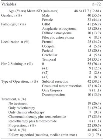
Table 1. Demographic and Clinical Characteristics of Patients
Variables n=72
Age (Years) Mean±SD (min-max) 48.6±17.7 (12-81) Gender, n (%) Male 40 (55.6) Female 32 (44.4) Pathology, n (%) GBM 41 (56.9) Anaplastic astrocytoma 15 (20.8) Diffuse astrocytoma 10 (13.9) Pilocytic astrocytoma 6 (8.3) Localization, n (%) Frontal 25 (34.7) Occipital 4 (5.6) Parietal 15 (20.8) Cerebellar 4 (5.6) Temporal 24 (33.3) Her-2 Staining, n (%) 0 55 (76.4) (+1) 9 (12.5) (+2) 2 (2.8) (+3) 6 (8.3) Type of Operation, n (%) Subtotal resection 42 (58.3) Gross total tumor resection 12 (16.7) Only biopsies 8 (11.1) Decompression 10 (13.9) Treatment, n (%) No treatment 19 (26.4) Only radiotherapy 21 (29.2) Only chemoradiotherapy 7 (9.7) Chemoradiotherapy plus temozolomide 17 (23.6) Radiotherapy plus temozolomide 8 (11.1) Progression, n (%) 22 (30.6) Dead, n (%) 48 (66.7) Follow-up period (months), median (min-max) 12 (1-75) *SD: Standard deviation, GBM: Glioblastoma
Lack of Prognostic Significance of C-erbB-2 in Astrocytomas
survival (p=0.049). The median overall survival for frontal lobe tumors and temporal lobe tumors were 30.2 and 31.9 months, while the median overall survival in occipital lobe tumors and parietal lobe tumors were 15.7 and 15.6 months, respectively. Median overall survival was 17.4 months for GBM patients, 28.1 months for anaplastic astrocytomas, and 55.1 months for diffuse astrocytomas (p<0.001).
For the histological subtype and grade multivariate analysis could not be performed as there was a multiple relation problem. The histological subtype of cases with Grade I tumor was pilocytic astrocytoma, and all the cases were alive, while the histological subtype of cases with Grade IV tumor was GBM, and all the cases were dead. C-erbB-2 overexpression was detected in the tumor specimens of 17 patients (23.6%), 13 patients with GBM and 4 patients with anaplastic astrocytoma. Six tumors (8.3%), all GBMs, showed strong (3+) staining, 2 specimens (2.7%), both GBMs, had moderate staining (2+) and 9 specimens, 5 of them GBMs (12.5%), exhibited weak staining (1+). All the GBM specimens that showed staining were primary GBM. No staining was observed in diffuse astrocytoma and pilocytic astrocytoma specimens. The degree of c-erb-B2 positivity is shown in Table 2. Median overall survival of patients with C-erbB-2
negative and C-erbB-2 positive tumors were 30 months (95%CI: 22.5-37.4 months) and 16.9 months (95%CI: 4.3-29.5 months), respectively (p=0.244). Survival curves are shown in Figure 1.
C-erbB-2 was positive in 8 GBM patients (19.5%) and negative in 33 (80.5%) patients. Survival difference between the 2 groups was not statistically significant (p=0.152).
Discussion
The present study performed in a small sample of low- and high-grade astrocytomas, failed to identify a statistically significant difference in terms of survival between patients with C-erbB-2 positive and C-erbB-2 negative tumors, although the median overall survival in these two groups of patients was 30 and 16.9 months. Astrocytic tumors, particularly anaplastic astrocytomas and GBM, face a poor prognosis despite major efforts to improve radiation therapy, chemotherapy, and surgery. While median survival for patients with anaplastic astrocytoma was reported to be between 3 and 5 years, median survival for patients with GBM was 12 to18 months, with the best available treatment. Although several prognostic factors such as age, performance status, quality of surgery, chemotherapy, radiotherapy and location of the tumor, have been proposed, there is a need for reliable prognostic indicators in patients with brain tumors.
Overexpression of C-erbB-2 has been established as an indicator of poor prognosis in a number of malignancies other than the CNS tumors. In the literature, the rate of C-erbB-2 overexpression is >20% in gastric cancers, 33% in gastroesophageal junction cancers (Albarello et al., 2011) and 31% in non-small cell lung cancers (Hirsch et al., 2012). Similarly, C-erbB-2 is overexpressed in 18-20% of invasive breast cancers (Slamon et al., 1987; Yaziji et al., 2004). However, the rates of C-erbB-2 overexpression in astrocytic tumors vary in the literature. While Reszeć et al. (2011) observed a C-erbB-2 overexpression rate of 47.1% in diffuse astrocytoma, 40% in anaplastic astrocytoma and 34.8% in GBM, Torp et al. (2007) demonstrated C-erbB-2 overexpression in 9 of 21 GBM patients (43%). Haynik et al. (2007), in their study on 49 GBM patients did not show C-erbB-2 overexpression in any of these patients. Haapasalo et al. (1996) screened immunuhistochemically c-erb-B2 protein expression in 94 astrocytic grade 1-4 neoplasm of the brain. Only two
Table 2. Degree of C-erb-B2 Positivity According to Histopathology
GBM Anaplastyic Diffuse Oligo- Pilosytic Astrocytoma Astrocytoma Astrocytoma Astrocytoma c-erb-B2 stain n=72 41 15 6 4 6 Negative 28 11 6 4 6 (1+) positive 5 4 - - (2+) positive 2 - - - (3+) positive 6 - - - -GBM: Glioblastoma
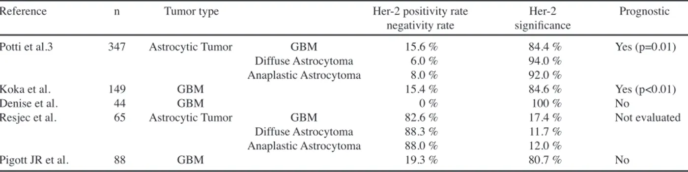
Table 3. Published Studies on Her-2 Overexpression and Prognostic Significance in Astrocytic Tumors
Reference n Tumor type Her-2 positivity rate Her-2 Prognostic negativity rate significance
Potti et al.3 347 Astrocytic Tumor GBM 15.6 % 84.4 % Yes (p=0.01) Diffuse Astrocytoma 6.0 % 94.0 %
Anaplastic Astrocytoma 8.0 % 92.0 %
Koka et al. 149 GBM 15.4 % 84.6 % Yes (p<0.01) Denise et al. 44 GBM 0 % 100 % No
Resjec et al. 65 Astrocytic Tumor GBM 82.6 % 17.4 % Not evaluated Diffuse Astrocytoma 88.3 % 11.7 %
Anaplastic Astrocytoma 88.0 % 12.0 %
Pigott JR et al. 88 GBM 19.3 % 80.7 % No GBM: Glioblastoma
Figure 1. Cumulative Survival Curves for Patients with c-erbB-2 Negative and Positive Tumors
anaplastic (grade 3) astrocytomas and one GBM (grade 4) showed overexpression, interestingly, no amplification of the c-erb-B2 gene was observed by fluorescence in situ hybridization (FISH). In another study, Potti et al. (2004) reported C-erbB-2 overexpression in 36 of 347 patients with primary malignant brain tumors (10.4%). In this study, 23 (64%) of 36 patients with C-erbB-2 overexpression were patients with GBM. In our study, C-erbB-2 positivity (2+ and 3+ staining) was observed in 11% of patients with low and high-grade astrocytomas. It was not surprising to find out that all tumors with Her-2 overexpression were GBMs, 19.5% of patients with GBM showed C-erbB-2 overexpression. C-erbB-2 overexpression was not identified in any of the other astrocytomas.
Similarly, conflicting findings exist regarding the association between prognosis and C-erbB-2 expression. Mineo et al. (2002) showed that survival time was significantly longer when C-erbB-2 expression was low (p=0.04). Koka et al. (2003) in their study, revealed that C-erbB-2 overexpression significantly increased the odds of early mortality, and stated that C-erbB-2 overexpression may be a poor prognostic marker in patients with GBM. In another study, Potti et al. (2004) showed that C-erbB-2 overexpression predicts increased mortality. Published studies on C-erbB-2 overexpression and prognostic significance in astrocytic tumors are shown in Table 3.
In our study, there was no significant difference between the two groups with C-erbB-2 negative (30 months) and C-erbB-2 positive tumors (16.9 months) in terms of median overall survival (p=0.244). Although the difference between the two groups is clinically significant, the small sample size of our study limited our ability to detect a statistically significant difference.
Previous studies have demonstrated a variety of possible clinical and molecular prognostic indicators, but results have not been consistent. Gehan et al. (1997) showed that while young age, radiotherapy, chemotherapy and encapsulated tumor were favorable prognostic factors, parietal location was an unfavorable prognostic factor. In another study, Potti et al. (2004) demonstrated that tumor location (occipital, parietal), tumor histology (GBM), and presenting symptoms (nausea/vomiting) were related to poor prognosis in patients with primary malignant brain tumors. Similar to the literature, our findings suggest that tumor location (occipital and parietal) and tumor histology (GBM) are related to poor prognosis.
This retrospective study has limitations typical of immunohistochemical approaches, including limited technical reproducibility and subjective interpretation. The limited sample size is another important limitation.
Thus far, studies investigating the prognostic value of C-erbB-2 overexpression in human astrocytic tumors have produced mixed findings. In our study, although there was no difference in survival, C-erbB-2 overexpression was observed only in the GBM subtype. Large studies are necessary to fully clarify the prognostic role of C-erbB-2 in human astrocytic tumors.
Acknowledgements
This study was approved by the Baskent University Institutional Review Board (project no: KA11/241), and was supported by the Baskent University Research Fund.
References
Albarello L, Pecciarini L, Doglioni C (2011). HER2 testing in gastric cancer. Adv Anat Pathol, 18, 53-9.
Bauknecht T, Kohler M, Janz I, Pfleiderer A (1989). The occurrence of epidermal growth factor receptors and the characterization of EGF-like factors in human ovarian, endometrial, cervical and breast cancer: EGF receptors and factors in gynecological carcinomas. J Cancer Res Clin
Oncol, 115, 193-9.
Gehan EA, Walker MD (1997). Prognostic factors for patients with brain tumors. Natl Cancer Inst Monogr, 46,189-95. Gulati S, Jakola AS, Johannesen TB, Solheim O (2012). Survival
and treatment patterns of glioblastoma in the elderly: a population-based study. World Neurosurg, 78, 518-26. Gulati S, Jakola AS, Nerland US, Weber C, Solheim O (2011).
The risk of getting worse: surgically acquired deficits, perioperative complications, and functional outcomes after primary resection of glioblastoma. World Neurosurg, 76, 572-79.
Haapasalo H, Hyytinen E, Sallinen P, et al (1996). c-erbB-2 in astrocytomas: infrequent overexpression by immunohistochemistry and absence of gene amplification by fluorescence in situ hybridization. Br J Cancer, 73, 620-3. Hiesiger EM, Hayes RL, Pierz DM, Budzilovich GN (1993).
Prognostic relevance of epidermal growth factor receptor (EGF-R) and c-neu/erbB2 expression in glioblastomas (GBMs). J Neurooncol, 16, 93-104.
Hirsch FR, Franklin WA, Veve R, Varella-Garcia M, Bunn PA Jr (2002). HER2/neu expression in malignant lung tumors.
Semin Oncol, 29, 51-8.
Haynik DM, Roma AA, Prayson RA (2007). HER-2/ neu expression in glioblastoma multiforme. Appl
Immunohistochem Mol Morphol, 15, 56-8.
Jakola AS, Myrmel KS, Kloster R, et al (2012). Comparison of a strategy favoring early surgical resection vs a strategy favoring watchful waiting in low-grade gliomas. JAMA,
308, 1881-8.
Koka V, Potti A, Forseen SE, et al (2003). Role of Her-2/neu overexpression and clinical determinants of early mortality in glioblastoma multiforme. Am J Clin Oncol, 26, 332-5. Lacroix M, Abi-Said D, Fourney DR, et al (2001). A multivariate
analysis of 416 patients with glioblastoma multiforme: prognosis, extent of resection, and survival. J Neurosurg,
95, 190-8.
Louis DN, Ohgaki H, Wiestler OD, et al (2007). The 2007 WHO classification of tumours of the central nervous system. Acta
Neuropathol, 114, 97-109.
Mineo JF, Bordron A, Baroncini M, et al (2007). Low HER2-expressing glioblastomas are more often secondary to anaplastic transformation of low-grade glioma. J
Neurooncol, 85, 281-7.
Mineo JF, Quintin-Roue I, Lucas B, Buburusan V, Besson G (2002). Glioblastomas: clinical study and search for prognostic factors. Neurochirurgie, 48, 500-9.
Neal DE, Marsh C, Bennett MK, et al (1985). Epidermal-growth-factor receptors in human bladder cancer: comparison of invasive and superficial tumours. Lancet, 1, 366-8.
Lack of Prognostic Significance of C-erbB-2 in Astrocytomas
Ozawa S, Ueda M, Ando N, Shimizu N, Abe O (1989). Prognostic significance of epidermal growth factor receptor in esophageal squamous cell carcinomas. Cancer, 63, 2169-73.
Pfeiffer D, Stellwag B, Pfeiffer A, et al (1989). Clinical implications of the epidermal growth factor receptor in the squamous cell carcinoma of the uterine cervix. Gynecol
Oncol, 33, 146-50.
Pigott TJ, Robson DK, Palmer J, Ward LM (1993). Expression of epidermal growth factor receptor in human glioblastoma multiforme. Br J Neurosurg, 7, 261-5.
Potti A, Forseen SE, Koka VK, et al (2004). Determination of HER-2/neu overexpression and clinical predictors of survival in a cohort of 347 patients with primary malignant brain tumors. Cancer Invest, 22, 537-44.
Rainov NG, Dobberstein KU, Bahn H, et al (2007). Prognostic factors in malignant glioma: influence of the overexpression of oncogene and tumor-suppressor gene products on survival. J Neurooncol, 35, 13-28.
Reszeć J, Bernaczyk PS, Milewski R, Chyczewski L, Mariak Z (2011). C-erbB-2 protein expression in astrocytic tumors of the brain. Med Sci Monit, 17, 216-20.
Sainsbury JR, Farndon JR, Needham GK, Malcolm AJ, Harris AL (1987). Epidermal-growth-factor receptor status as predictor of early recurrence of and death from breast cancer.
Lancet, 1, 1398-402.
Slamon DJ, Clark GM, Wong SG, et al (1987). Human breast cancer: correlation of relapse and survival with amplification of the HER-2/neu oncogene. Science, 235, 177-82. Stummer W, Reulen HJ, Meinel T, et al (2008).
ALA-Glioma Study Group: Extent of resection and survival in glioblastoma multiforme: identification of and adjustment for bias. Neurosurgery, 62, 564-76.
Torp SH, Gulati S, Johannessen E, Dalen A (2007). Coexpression of c-erbB 1-4 receptor proteins in human glioblastomas. An immunohistochemical study. J Exp Clin Cancer Res,
26, 353-9.
Van den Eynde M, Baurain JF, Mazzeo F, Machiels JP (2011). Epidermal growth factor receptor targeted therapies for solid tumours. Acta Clin Belg, 66, 10-17.
Yaziji H, Goldstein LC, Barry TS, et al (2004). HER-2 testing in breast cancer using parallel tissue-based methods. JAMA,