78
Research Article
Cytotoxic T lymphocyte antigen-4 (CTLA-4) A49G
polymorphism and autoimmune blood diseases
Sitotoksik T lenfosit antijen-4 (CTLA-4) A49G polimorfizmi ve otoimmün kan
hastalıkları
Faruk Aktürk
1, Veysel Sabri Hançer
2, Reyhan Küçükkaya
31Division of Hematology, Department of Internal Medicine, İstanbul University, İstanbul Faculty of Medicine, İstanbul, Turkey 2Department of Medical Biology and Genetics, İstanbul Bilim University, Faculty of Medicine, İstanbul, Turkey
3Department of Internal Medicine, İstanbul Bilim University, Faculty of Medicine, İstanbul, Turkey
Address for Correspondence: Asst. Prof. Dr. Veysel Sabri Hançer, Istanbul Bilim University, Faculty of Medicine, Department of Medical Biology and Genetics, Esentepe, Istanbul, Turkey Phone: +90 533 634 30 14 E-mail: [email protected]
doi:10.5152/tjh.2010.04
Abstract
Objective: The cytotoxic T lymphocyte associated antigen-4 (CTLA-4) is expressed on T lymphocytes, and inhibits
the T-cell responses. In animal models, it has been shown that complete CTLA-4 deficiency was lethal due to mas-sive infiltration of tissues by polyclonally proliferating lymphocytes. CTLA-4 A49G polymorphism, which has been suggested to reduce the inhibitory function of the CTLA-4 molecule, was found to be associated with various auto-immune diseases in recent studies.
Material and Methods: In this study, we evaluated the frequency of CTLA-4 A49G polymorphism in 46 patients
with autoimmune hemolytic anemia (AIHA), 62 patients with immune thrombocytopenic purpura (ITP), and 150 healthy individuals.
Results: Allele frequencies and genotype distributions were similar in both ITP and AIHA patients compared to
healthy individuals. In subgroup analysis, however, we found that in chronic lymphocytic leukemia (CLL) patients with AIHA (n=4), all patients had CTLA-4 A49G polymorphism (3 had AG, 1 had GG). There was no significant statistical association between G allele and systemic lupus erythematosus (SLE) or AIHA.
Conclusion: These data suggest that CTLA-4 A49G polymorphism does not contribute to the pathogenesis of
lymphoproliferative diseases itself, nor does it increase the risk of autoimmune complications in patients with lymphoproliferative disease. (Turk J Hematol 2010; 27: 78-81)
Key words: Cytotoxic T lymphocyte antigen-4 (CTLA-4), A49G polymorphism, autoimmunity, idiopathic
throm-bocytopenic purpura, autoimmune hemolytic anemia, chronic lymphocytic leukemia
Received: December 26, 2008 Accepted: February 22, 2010
Özet
Amaç: Sitotoksik T lenfosit antijen-4 (CTLA-4) ifadesi T lenfositlerde gerçekleşir ve T hücre cevabını engeller. Hayvan
modellerinde CTLA-4 yokluğunun, dokuların poliklonal çoğalan lenfositler tarafından yoğun infiltrasyonu nedeni ile, ölümcül olduğu gösterilmiştir. Farklı otoimmün hastalıklar ile ilişkilendirilmiş CTLA-4 A49G polimorfizminin, CTLA-4 molekülünün inhibitör fonksiyonunu azalttığı düşünülmektedir.
Yöntem ve Gereçler: Bu çalışmada otoimmun hemolitik anemi (OIHA) hastası 46 kişi, immun trombositopenik
Introduction
Autoimmune disorders are the result of disturbed immune tolerance to self-antigens. After the presentation of an antigen in conjunction with HLA molecules on the surface of an anti-gen-presenting cell to a specific T-cell receptor (TCR), co-stimulatory signals are required for the T-cell responses [1]. A
number of co-stimulatory molecules are present on the T-cell membrane, which may have stimulatory or inhibitory effects on T lymphocytes. The cytotoxic T lymphocyte associated anti-gen-4 (CTLA-4) is expressed on T lymphocytes and inhibits the T-cell responses [2]. Mechanisms of down-regulation of T-cell immunogenic response by CTLA-4 may be due to competitive antagonism with CD28, increment in TCR stimulation thresh-old, and limitation of T-cell division capacity and activation of cell cycle arrest of T-cells after antigenic stimulation [2]. In ani-mal models, it has been shown that complete CTLA-4 defi-ciency was lethal due to severe lymphoproliferative disorders with massive infiltration of tissues by polyclonally proliferating lymphocytes [3]. Blocking the CTLA-4/B7 interaction by mono-clonal antibodies results in an increase in antigen-specific T-cell proliferation, whereas enhancing the inhibitory function of CTLA-4 suppresses T-cell responses [1,2]. These findings
provide strong evidence that CTLA-4 has a crucial role in the negative regulation of T-cell responses.
The CTLA-4 gene is located on chromosome 2 (2q33). Several polymorphisms were identified on the CTLA-4 gene [4]. The A49G polymorphism in exon 1 of the CTLA-4 gene is especially important because it alters the structure of the CTLA-4 protein by causing Thr17Ala amino acid substitution [5]. It has been suggested that this polymorphism reduces the inhibitory function of CTLA-4 [2]. In recent studies, the CTLA-4 A49G polymorphism was found to be associated with autoim-mune diseases such as insulin-dependent diabetes, rheuma-toid arthritis, systemic lupus erythematosus (SLE), multiple sclerosis, primary biliary cirrhosis, Hashimoto’s thyroiditis, and Graves’ disease [1,2,4,6]. In this study, we evaluated the fre-quency of the CTLA-4 A49G polymorphism in patients with autoimmune hemolytic anemia (AIHA) and autoimmune throm-bocytopenic purpura (ITP).
Materials and Methods
Participants: The present study was approved by the
local ethics committee (Istanbul Faculty of Medicine) and all
participants gave informed consent. Patients were selected from cases admitted to the Hematology Division of the Istanbul Faculty of Medicine from May 2003 to June 2006. One hun-dred and fifty healthy Turkish subjects, 46 patients with AIHA and 62 patients with ITP were included in our study. ITP and AIHA patients were diagnosed by standard methods and were being followed by the Hematology Department.
In the AIHA group, median age was 53 (28-66) and male to female ratio was 13/33. Fourteen AIHA patients also had SLE, 4 had chronic lymphocytic leukemia (CLL), 2 had Evans’s syn-drome, and 2 had autoimmune diseases other than SLE (auto-immune thyroiditis and auto(auto-immune hepatitis).
In the ITP group (n=62), median age was 41 (18-56) and male to female ratio was 17/45. Thirty-six patients had primary ITP. Twenty-six patients in the ITP group had antiphospholipid antibodies (aPLA), and 12 of those patients were diagnosed as definite antiphospholipid syndrome (with thrombosis and/or pregnancy morbidity). Fourteen ITP patients had only persis-tent aPLA-positivity.
In the healthy control group (n=150), median age was 38 years (18-55), and male to female ratio was 67/83.
Methods: Genomic DNA was isolated from peripheral
venous blood samples using high pure polymerase chain reac-tion (PCR) template preparareac-tion kit (Roche Diagnostics). For the amplification of exon 1 to show CTLA-4 A49G polymorphism, 5’-GCTCTACTCCTGAAGACCT-3’ and 5’-AGTCTCACTCACTCACTTTGCAG-3’ primers were used. Amplified DNA samples were digested by Bbv I restriction endonuclease enzyme (New England Biolabs, Beverly, MA). Amplified fragment (162 bp) analyzed on 3.5% agarose gel was stained by ethidium bromide. The allele A was not cut by the enzyme and so left 162 bp; on the contrary, G allele was cut into two DNA fragments 88 and 74 bp long [4].
Statistical Analysis: Statistical analysis was performed
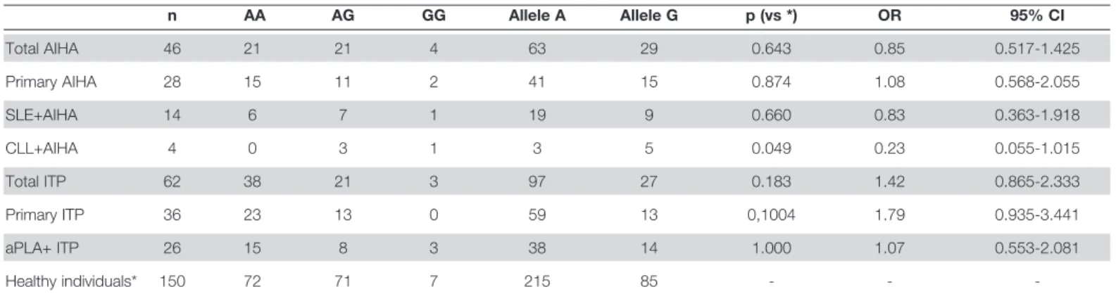
using chi-square and Fisher’s exact tests at ‘Graphpad for Windows Version 3.0’. P values <0.05 were accepted as sta-tistically significant. Results of the statistical analysis of com-parisons of the allele frequencies are shown in Table 1.
Results
Healthy Controls: One hundred and fifty healthy subjects
were investigated in terms of CTLA-4 A49G polymorphism.
Aktürk et al. A49G polymorphism and autoimmune blood diseases
Turk J Hematol 2010; 27: 78-81
79
Bulgular: ITP ve OIHA hastalarından oluşan iki grupta da sağlıklı kontrol bireyleri ile karşılaştırıldığında benzer allel
frekansları ve genotip dağılımları saptanmıştır. Alt grup analizi gerçekleştirildiğinde ise hem OIHA hem de KLL has-talığına sahip 4 bireyin hepsinin polimorfizme sahip olduğu gösterilmiştir (3 AG, 1 GG). Risk alleli olan G OIHA, İTP ve kontrol grubunda incelendiğinde istatistik olarak anlamlı bir farklılık saptanmamıştır.
Sonuç: Bu verilerin ışığında CTLA-4 A49G polimorfizminin lenfoproliferatif hastalıkların patogenezine bir katkısının
olmadığı veya lenfoproliferatif hastalığa sahip bireylerde otoimmün komplikasyonların gelişmesi açısından risk oluştur-madığı sonucuna varılmıştır. (Turk J Hematol 2009; 27: 78-81)
Anahtar kelimeler: Sitotoksik T lenfosit antijen-4 (CTLA-4), A49G polimorfizmi, otoimmünite, idiopatik
trombosito-penik purpura, otoimmün hemolitik anemi, kronik lenfositik lösemi
Genetic variation was distributed according to Hardy-Weinberg equilibrium. Genotype frequencies were 48% for AA, 47.3% for AG and 4.7% for GG. A-allele frequency was 71.6% and G-allele frequency was 28.4% (Table 1). There was no statisti-cal difference between genders and allele frequencies in the healthy control group.
AIHA Group: Twenty-one patients had AA (45.6%), 21 had
AG (45.6%), and 4 had GG (8.8%) genotype in this group. A-allele frequency was 68.4% and G-allele frequency was 31.6%, which was similar to healthy controls. In sub-group analysis, in a small group of patients with CLL and AIHA (n=4), all patients were found to have CTLA-4 A49G polymorphism (3 had AG, 1 had GG) (Table 1). When we statistically com-pared the risk allele G in the AIHA and control groups, there was no statistical difference (p=0.643; odds ratio [OR]: 0.858; 95% confidence interval [CI]: 0.8658-2.33). In sub-group analysis, we found no difference between patients and controls for the risk allele G (Table 1).
ITP Group: Thirty-eight had AA (61.3%), 21 had AG
(33.9%), and 3 had GG (4.8%) genotype in this group. A-allele frequency was 78.4% and G-allele frequency was 21.6%. There was no statistical difference between the ITP group and healthy control group for the risk allele. In sub-group analysis, we similarly found no difference between ITP patients and con-trols for the risk allele G.
Discussion
The role of the CTLA-4 A49G polymorphism in the develop-ment of autoimmune diseases is not well understood. Since CTLA-4 controls the amplitude of immune response and periph-eral tolerance, this polymorphism could have an impact on autoimmunity in some way. The CTLA-4 A49G polymorphism may result in susceptibility to autoimmune disease [1,2,6,7].
The role of the CTLA-4 A49G polymorphism in the develop-ment of autoimmune blood diseases such as ITP and AIHA is not clear. In the only published study, which was done by Pavkovic et al. [5], it was shown that the CTLA-4 A49G polymor-phism was significantly higher in patients with AIHA, but no association was found in patients with ITP. They also showed
that the G-allele frequency was highest among CLL patients who had developed AIHA. In that study, 60% of the AIHA cases were patients with CLL. Our study is the second study investi-gating the role of the CTLA-4 A49G polymorphism in patients with ITP and AIHA. Although we could not demonstrate an association between autoimmune blood diseases and the CTLA-4 A49G polymorphism, we found very interesting results in the CLL+AIHA subgroup, as in Pavkovic’s study. In our study, all CLL patients who had AIHA (n=4) were found to have G allele (3 had AG, 1 had GG). With the knowledge of the development of fatal lymphoproliferative disease in CTLA-4-deficient mice, these data suggest that the CTLA-4 A49G polymorphism may contribute to the pathogenesis of the lymphoproliferative dis-eases itself, or may increase the risk of autoimmune complica-tions in patients with lymphoproliferative diseases. In a meta- analysis study, Lee et al. [6] demonstrated a significant statistical association between the A49G polymorphism and SLE suscep-tibility. Their meta-analysis did not show a significant association of other polymorphisms of CTLA-4 with SLE. Though the esti-mated ORs from the race-specific analysis were very similar, a statistically significant SLE association was found with Asians only (n=522). Similarly, in our study, there was no association between the A49G polymorphism and SLE in the Caucasian population based on this meta-analysis. When we statistically compared groups, there was no association between the AIHA and control group. In contrast to Lee et al. [6], in the present study, our findings demonstrated that there was no significant statistical association between G allele and SLE. Similar findings were seen for ITP (Table 1).
In conclusion, our results showed that there is no associa-tion between the A49G polymorphism of the CTLA-4 gene and AIHA or ITP disease. Further genetic and clinical studies are required to understand the exact role of the CTLA-4 A49G polymorphism in the development of autoimmune and lympho-proliferative diseases.
Conflict of interest
No author of this paper has a conflict of interest, including specific financial interests, relationships, and/or affiliations rel-evant to the subject matter or materials included in this manu-script.
Aktürk et al.
A49G polymorphism and autoimmune blood diseases Turk J Hematol 2010; 27: 78-81
80
Table 1. Patient and control genotype and allele frequencies for the CTLA-4 A49G polymorphism
n AA AG GG Allele A Allele G p (vs *) OR 95% CI Total AIHA 46 21 21 4 63 29 0.643 0.85 0.517-1.425 Primary AIHA 28 15 11 2 41 15 0.874 1.08 0.568-2.055 SLE+AIHA 14 6 7 1 19 9 0.660 0.83 0.363-1.918 CLL+AIHA 4 0 3 1 3 5 0.049 0.23 0.055-1.015 Total ITP 62 38 21 3 97 27 0.183 1.42 0.865-2.333 Primary ITP 36 23 13 0 59 13 0,1004 1.79 0.935-3.441 aPLA+ ITP 26 15 8 3 38 14 1.000 1.07 0.553-2.081 Healthy individuals* 150 72 71 7 215 85 - -
-AIHA: Autoimmune hemolytic anemia; SLE: Systemic lupus erythematosus; CLL: Chronic lymphocytic leukemia; ITP: Immune thrombocytopenic purpura; aPLA: Antiphospholipid antibodies; OR: Odds ratio; CI: Confidence interval
References
1. Chambers CA, Kuhns MS, Egen JG, Allison JP. CTLA-4 mediated inhibition in regulation of T cell responses: mechanisms and manipulation in tumor immunotherapy. Annu Rev Immunol 2001;19:565-94.
2. Teft WA, Kirchhof MG, Madrenas J. A molecular perspective of CTLA-4 function. Annu Rev Immunol 2006;24:65-97.
3. Waterhouse P, Penninger JM, Timms E, Wakeham A, Shahinian A, Lee KP, Thompson CB, Griesser H, Mak TW. Lymphoproliferative disorders with early lethality in mice deficient in CTLA-4. Science 1995;270:985-8.
4. Sahin M, Erdogan MF, Erdogan G. Cytotoxic T lymphocyte-associated molecule-4 polymorphisms in Turkish Graves’ disease patients and association with probability of remission after antithyroid therapy. Eur J Intern Med 2005;16:352-5. 5. Pavkovic M, Georgievski B, Cevreska L, Spiroski M, Efremov
DG. CTLA-4 exon 1 polymorphism in patients with autoimmune blood disorders. Am J Hematol 2003;72:147-9.
6. Lee YH, Harley JB, Nath SK. CTLA-4 polymorphisms and systemic lupus erythematosus (SLE): a meta-analysis. Hum Genet 2005;116:361-7.
7. Dallos T, Kovacs L. CTLA-4 and the genetic predisposition on autoimmunity. Bratisl Lek Listy 2005;106:55-62.
Aktürk et al. A49G polymorphism and autoimmune blood diseases