Full Terms & Conditions of access and use can be found at
https://www.tandfonline.com/action/journalInformation?journalCode=tbcp21
Psychiatry and Clinical Psychopharmacology
ISSN: 2475-0573 (Print) 2475-0581 (Online) Journal homepage: https://www.tandfonline.com/loi/tbcp21
Evaluation of retinal nerve fiber layer, macular,
and choroidal thickness in schizophrenia: spectral
optic coherence tomography findings
Pinar Topcu-Yilmaz, Memduha Aydin & Bilge Cetin Ilhan
To cite this article: Pinar Topcu-Yilmaz, Memduha Aydin & Bilge Cetin Ilhan (2019) Evaluation of retinal nerve fiber layer, macular, and choroidal thickness in schizophrenia: spectral optic coherence tomography findings, Psychiatry and Clinical Psychopharmacology, 29:1, 28-33, DOI: 10.1080/24750573.2018.1426693
To link to this article: https://doi.org/10.1080/24750573.2018.1426693
© 2018 The Author(s). Published by Informa UK Limited, trading as Taylor & Francis Group
Published online: 29 Jan 2018.
Submit your article to this journal Article views: 1110
View related articles View Crossmark data
Evaluation of retinal nerve fiber layer, macular, and choroidal thickness in
schizophrenia: spectral optic coherence tomography findings
Pinar Topcu-Yilmaza, Memduha Aydin band Bilge Cetin Ilhanc
a
Department of Ophthalmology, Ankara Numune Research and Training Hospital, Ankara, Turkey;bDepartment of Psychiatry, Faculty of Medicine, Selcuk University, Konya, Turkey;cDepartment of Psychiatry, Konya Research and Training Hospital, Konya, Turkey
ABSTRACT
OBJECTIVES: The study was performed to evaluate the retinal nerve fiber layer (RNFL), macular, and subfoveal choroidal thickness (SFCT) in patients with schizophrenia and healthy controls using spectral domain optical coherence tomography (SD-OCT).
METHODS: Fifty-nine schizophrenic patients and 36 age- and sex-matched healthy volunteers were enrolled in this cross-sectional clinical study. The RNFL, macular, and SFCT thickness measurements obtained by SD-OCT were compared between the groups.
RESULTS: The mean age of 59 patients included in this study was 34.64 years and the mean age of 36 healthy controls was 32.08 years, mean illness duration was 10.33 years, mean total Positive and Negative Symptom Scale (PANSS) score was 75.18, and mean CGI-S score was 3.88. Macular thickness in the superior inner (350.97 ± 16.51 vs. 341.81 ± 16.35), nasal inner (348.97 ± 17.53 vs. 340.25 ± 17.55), inferior inner (345.5 ± 17.59 vs. 335.47 ± 16.92), temporal inner (333.28 ± 17.96 vs. 321.97 ± 19.96), and temporal outer (289.14 ± 14.10 vs. 281.29 ± 15.31) segments were significantly decreased in schizophrenic patients. However; RNFL thickness and choroidal thickness (CT) measurements between the two groups were similar. No significant correlation was found between illness duration, PANSS score, CGI-S score and RNFL thickness and macular thickness measurements. There was a weak negative correlation between disease duration and CT.
CONCLUSIONS: The findings from this study suggest that macular thickness measurements are reduced in schizophrenic patients but do not indicate any significant change in RNFL or choroids. Further studies are needed to determine the potential application of optical coherence tomography as a tool for the diagnosis and monitoring the progression of this disease.
ARTICLE HISTORY Received 17 November 2017 Accepted 8 January 2018 KEYWORDS
Choroidal thickness; macular thickness; optical coherence tomography; retinal nerve fiber layer thickness; schizophrenia
Introduction
Schizophrenia is a progressive, chronic, and disabling mental disorder characterized by positive, negative, and cognitive symptoms that affect almost all aspects of mental activity, including perception, attention, memory, and emotion [1]. The etiology and funda-mental pathophysiology of schizophrenia remain unclear, but a large body of evidence suggests that many factors, including genetic, environmental, neuro-developmental, and neurodegenerative factors make a contribution to pathophysiological processes [2]. Increasingly advanced neuroimaging technologies and neuropathological methods are used frequently in various types of studies to support the neurodeve-lopmental and neurodegenerative hypothesis of schizo-phrenia [3,4].
The structural brain abnormalities in schizophrenia involve the gray matter volume deficits, white matter abnormalities, and ventricular enlargement observed in these neuroimaging studies support the neurodeve-lopmental hypothesis of schizophrenia [5–8].
Numerous neuropathological and neuroimaging studies of schizophrenia representing the effect of the disease on the brain after it has begun often support neurodegenerative hypothesis of schizophrenia [9]. Decreases in the total brain volume as well as decreases in the numbers and functions of neurons have been demonstrated not only in chronic schizophrenia patients but also in the first episode and prodromal stage patients [10,11]. Findings from neuroimaging studies of patients with schizophrenia also support that neurodegeneration starts at the earliest stages of schizophrenia [5,12–14].
Another hypothesis called the progressive neurode-velopmental model has been proposed to provide a unifying hypothesis that conceptualizes schizophrenia neither entirely neurodevelopmental nor solely neuro-degenerative [9]. It may be said that schizophrenia requires more neurobiological explanations supporting these complementary theories.
The retina is anatomically and developmentally an extension of the central nervous system, and the retina
© 2018 The Author(s). Published by Informa UK Limited, trading as Taylor & Francis Group
This is an Open Access article distributed under the terms of the Creative Commons Attribution License (http://creativecommons.org/licenses/by/4.0/), which permits unrestricted use, distribution, and reproduction in any medium, provided the original work is properly cited.
and the brain are connected by the optic nerve [15]. Thus, the retina and its subcortical projections have become a crucial site of research for schizophrenia in the last decade. Visual evoked potential studies demon-strated a selective dysfunction of the magnocellular pathway [16]. Electroretinogram studies have shown that the cone a-wave amplitude was decreased indicat-ing a photoreceptor dysfunction in the acute phase of psychosis [17,18].
Over the past years, researchers have extensively focused on the role of functional and structural neuroi-maging in schizophrenia in order to understand the underlying neurobiology and develop reliable bio-markers for diagnosis [19]. As one of these, optical coherence tomography (OCT) is a relatively new, con-tactless, non-invasive imaging method that permits in vivo visualization of the optic nerve and can easily pro-vide high resolution, cross-sectional images of the ret-ina and choroid [20]. While OCT is primarily used for the follow-up of ocular pathologies, it has also gained importance for the detection of retinal and choroidal changes that might be valuable as a biomarker of sev-eral neurodegenerative diseases [21].
The diseases classically described as neurodegenera-tive are Alzheimer’s disease, Huntington’s disease, Par-kinson’s disease, and Multiple Sclerosis. Several studies have reported retinal nerve fiber layer (RNFL) thinning in Alzheimer’s disease, mild cognitive impairment, Parkinson’s disease, and Multiple Sclerosis [22–25]. However; very few studies have investigated the RNFL and macular thickness changes in psychiatric disorders as schizophrenia and depression [4,26–30].
It was concluded that peripapillary RNFL thinning is associated with neuronal cell loss and axonal loss [31]. The studies of OCT in patients with schizo-phrenia demonstrated a decrease in the RNFL thick-ness which can be explained by neuronal cell and axonal loss as a process of neurodegeneration. The RNFL is a beneficial structure for detecting the severity and duration of the disorder, and it has been suggested that OCT, measuring the RNFL thickness, may be used as a diagnostic tool in schizophrenia.
Data from clinical, pathological, and neuroimaging studies have also revealed evidence of vascular involve-ment in schizophrenia [32–34]. Recently, Meier et al. [35] have shown that wider retinal venules were a dis-tinguishing feature of schizophrenia. This finding was also supported by a study on twins that found an association with wider retinal venules and psychosis symptoms [36]. Choroid is the other vascular tissue supplying more than 70% of blood flow in the eye and can also be affected by microvascular changes in schizophrenia.
The purpose of this study was to evaluate the RNFL, macular, and subfoveal choroidal thickness (SFCT) in patients with schizophrenia and to compare them with healthy controls. We also aimed to investigate
the associations between OCT measurements and clinical parameters of the schizophrenic patients.
Methods
Participants
A total of 65 patients who had been consecutively admitted to the psychiatry inpatient clinic of Konya Training and Research Hospital with the diagnosis of schizophrenia and fulfilled DSM-IV criteria between December 2014 and May 2015 were included in this cross-sectional, observational study [37]. Six patients were excluded for clinical reasons. Four patients were excluded due to refractive errors with a spherical equiv-alent greater than 2.00 D, one patient was excluded due to cataract that precluded OCT examination and one patient was excluded due to strabismic amblyopia that could affect OCT results. The final sample in the study included 59 patients with schizophrenia. The control group consisted of 37 age- and gender-matched healthy volunteers. The study protocol was approved by the Institutional Review Board of Selcuk University Faculty of Medicine, Konya and adhered to the tenets of the Declaration of Helsinki (18 November 2014; 2014/305). Informed consent was obtained from all subjects prior to their participation in the study. Cri-teria for the exclusion of a subject from the study were as follows: (1) A history of an ophthalmological, neurological, or systemic condition that could affect the RNFL or macula (such as amblyopia, glaucoma, age-related macular degeneration, diabetes mellitus, hypertension, optic neuropathy, ocular trauma, ocular surgery, intraocular pressure (IOP) greater than 21 mmHg, etc.), (2) refractive errors greater than ± 2.0 diopters of spherical equivalence, and (3) media opaci-ties that preclude OCT examination.
Measures
Sociodemographic and Clinical Data Form: Semi-structured sociodemographic and clinical data form was developed by the researchers of the study. Patients were interviewed face-to-face by psychiatrists who were experienced in working with schizophrenic patients on basis of this sociodemographic and clinical data form. Medical records, including patient sociode-mographic and clinical variables, and reports from relatives and/or care-givers are also used as infor-mation sources.
The PANSS: PANSS is a semi-structured interview scale involving 30 items and a seven-point severity rat-ing. Seven parameters are included in the positive syn-drome subscale, 7 parameters are included in the negative syndrome subscale, and the remaining 16 par-ameters are included in the general psychopathology subscale [38]. The reliability and validity of the PSYCHIATRY AND CLINICAL PSYCHOPHARMACOLOGY 29
Turkish-language version of the scale were established by Kostakoglu et al. [39].
Clinical Global Impression Scale: Global severity of psychopathology was determined by using the Clinical Global Impression Scale-Severity (CGI-S) [40]. This scale has ratings from 1 (not ill) to 7 (extremely ill).
Procedure
Patients were interviewed face-to-face by psychiatrists who were experienced in working with schizophrenic patients. Those who are clinically partially stable have been directed to ophthalmologic examination after psychiatric interventions within the second week of hospitalization. Lack of aggression/negativism was the most important item taken into consideration before ophthalmologic evaluation. Subjects included in the study underwent a complete ophthalmologic evalu-ation including retinoscopy, slit lamp biomicroscopy, IOP measurement, fundus examination. Spectral domain optical coherence tomography (SD-OCT) images for the measurement of RNFL, macular, and choroidal thickness (CT) were taken by an experienced technician (Spectralis OCT, Heidelberg Engineering, Heidelberg, Germany). In order to avoid the diurnal variation in CT, all OCT examinations were performed between 1:00 pm and 2:00 pm. RNFL imaging was per-formed using circular scans of 3.4 mm diameter at the center of the optic disc. RNFL thickness at six sectors (nasal (N), superonasal (SN), superotemporal (ST), temporal (T), inferotemporal (IT), inferonasal(IN), and global (G) RNFL thickness) were automatically calculated by the SD-OCT device. The macular thick-ness results were noted for nine subfields as defined by the ETDRS study [Central foveal subfield (CFS), superior inner macula (SIM), superior outer macula (SOM), nasal inner macula (NIM), nasal outer macula (NOM), inferior inner macula (IIM), inferior outer macula (IOM), temporal inner macula (TIM), tem-poral outer macula (TOM)]. Only well-centered, high-quality images with a signal strength >15 dB were included in the study.
The CT was measured manually from the outer edge of the retinal pigment epithelium to the inner scleral surface at the fovea and at positions 500, 1000, and 1500 µ nasal and temporal to the fovea. Measurements were done by the same ophthalmologist (P.T.Y.) who was masked to the diagnosis of study participants. Images with low image quality resulting in an indis-cernible chorioscleral border were excluded from the study.
Statistical analyses
Statistical analysis was performed using SPSS ver. 17.0 (Chicago, IL, USA). Only one eye of each subject was included for the analysis. Once the normality of the
data was determined via Kolmogorov Smirnov test; Mann–Whitney U-test was used to compare the RNFL, macular, and CT values in patients with schizo-phrenia and healthy volunteers. The categorical vari-ables were analyzed by the Pearson χ2 test. Spearman’s rank correlation analysis tests were used for correlative analyses. Effect sizes (r2) reported for significant findings for between-group comparisons. For all evaluations, a p-value of less than .05 was con-sidered as statistically significant.
Results
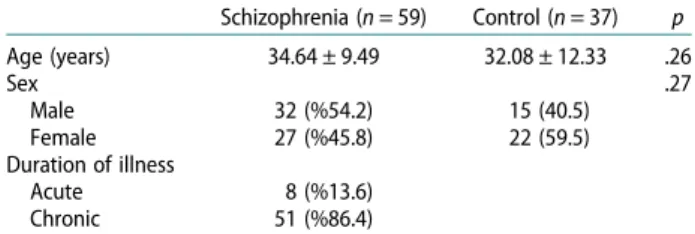
Fifty-nine patients with schizophrenia and 37 healthy volunteers were included in the study. The mean age of schizophrenic patients was 34.64 ± 9.49 and the mean age of healthy controls was 32.08 ± 12.33. There was no statistically significant difference in the mean age (p = .26) and gender (p = .27) of the two groups. Detailed demographic characteristics of the study groups can be found inTable 1.
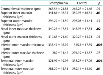
The mean total PANSS score was 75.19 ± 20.14 and mean CGI-S score was 3.88 ± 1.22 in the schizophrenia group. Ten patients were treated with typical chotics, 38 patients were treated with atypical antipsy-chotics and 11 patients were drug naïve on admission. The mean peripapillary RNFL thickness at all sec-tors was similar in schizophrenic patients and healthy controls (Table 2). Comparison of the macular thick-ness between the two groups showed that macular thickness was decreased in schizophrenia patients, and this difference was statistically significant for superior inner (341.81 ± 16.35 vs. 350.97 ± 16.51; r2= 0.063; p = .01), nasal inner (340.25 ± 17.55 vs. 348.97 ± 17.53; r2= 0.09; p = .02), inferior inner (335.47 ± 16.92 vs. 345.5 ± 17.59; r2= 0.140; p = .008), temporal inner (321.97 ± 19.96 vs. 333.28 ± 17.96; r2= 0.145; p = .006), and temporal outer (281.29 ± 15.31 vs.
Table 2.Comparison of RNFL thickness in schizophrenia and healthy controls. Schizophrenia Control p Nasal RNFL thickness 75.37 ± 12.80 74.49 ± 12.55 .74 Superonasal RNFL thickness 104.73 ± 22.33 102.51 ± 17.38 .73 Superotemporal RNFL thickness 135.31 ± 18.95 136.40 ± 16.14 .28 Temporal RNFL thickness 73.64 ± 10.54 74.70 ± 9.63 .62 Inferotemporal RNFL thickness 152.64 ± 16.35 156.22 ± 15.01 .76 Inferonasal RNFL thickness 118.37 ± 23.88 116.73 ± 21.59 .59 Global RNFL thickness 101.32 ± 8.53 101.27 ± 6.98 .97 Table 1.Demographic characteristics of the study population.
Schizophrenia (n = 59) Control (n = 37) p Age (years) 34.64 ± 9.49 32.08 ± 12.33 .26 Sex .27 Male 32 (%54.2) 15 (40.5) Female 27 (%45.8) 22 (59.5) Duration of illness Acute 8 (%13.6) Chronic 51 (%86.4)
289.14 ± 14.10; r2= 0.142; p = .01) macular thickness (Table 3). The CT measurements at all measurement points were similar between the study and control groups (Table 4).
No correlation was noted between the RNFL thick-ness, macular thickness and PANNS score or disease duration. Subfoveal, 500 Nasal, 1500 Nasal, and 500 Temporal CT measurements had a weak negative cor-relation with disease duration (Table 5).
Discussion
In the present study, we used SD-OCT to evaluate the RNFL, macular, and subfoveal CT in 59 patients with schizophrenia and 37 healthy controls. Our findings suggest that macular thickness measurements are reduced in schizophrenic patients, but showed no differences between patients with schizophrenia and
healthy controls in terms of RNFL thickness and CT measurements.
While our findings are in line with an earlier study [27], several other groups have reported significant RNFL thinning in schizophrenia [4,26,28–30]. The ear-liest study investigating the retinal changes in schizo-phrenia was a short report on 10 patients and found that overall RNFL thickness and nasal RNFL thickness were reduced in the patient group [26]. A larger study by Lee at al. [28] have shown that overall peripapillary RNFL thickness, superior, temporal, and inferior RNFL thickness were significantly reduced in schizophrenia. This reduction was correlated with the duration of ill-ness and was much more significant in patients with longer disease duration. The thinning in RNFL was also confirmed by Yilmaz et al. [30] and Celik et al. [4]. Celik et al. [4] also reported that decreases in specific cell layers (i.e. ganglion cell layer, inner plexi-form layer) volumes correlate better with disease sever-ity in schizophrenia patients than RNFL thickness. Recently, Ascaso et al. [29] have evaluated the retinal OCT findings in schizophrenia patients with recent and non-recent illness episode. Interestingly, patients with a recent illness episode (RIE) had similar RNFL thickness with controls and the reduction in RNFL thickness was only significant for patients clinically stable for more than six months. They suggested that the ongoing inflammatory processes during the acute psychotic episodes might also affect the retina, increase its thickness, and mask the RNFL thinning in these patients. As all of our patients were recruited from the inpatient clinic of the Psychiatry Department and were having a recent psychotic episode, the absence of RNFL thinning in our study might be comparable to the lack of significant difference in RNFL thickness of RIE patients and healthy controls in the study by Ascaso et al. [29]. Future longitudinal studies would be helpful to prove the role of inflammation on RNFL changes of schizophrenia patients.
The second parameter investigated in this study was the macular thickness and it was found to be reduced in patients with schizophrenia. This reduction was stat-istically significant for superior inner, nasal inner, inferior inner, temporal inner, and temporal outer seg-ments. Several other studies have also reported decreased macular thickness in schizophrenia [28,29]. Lee et al. [28] have found a negative correlation between disease duration and macular thickness. How-ever; such an association was lacking in the study by Ascaso et al. [29] and our study.
The association between microvascular abnormal-ities and schizophrenia has been an interesting research topic since the 1960s [41]. Demonstration of wider reti-nal venules in individuals with psychotic symptoms or schizophrenia has drawn attention to the possible role of retinal imaging for clarifying the pathophysiology in these patients [35,36,41]. As a highly vascular tissue Table 5. Correlation between CT and disease duration in
schizophrenia. Disease duration r p-Value Subfoveal CT −0.38 .006 500 nasal CT −0.33 .02 1000 nasal CT −0.26 .07 1500 nasal CT −0.27 .05 500 temporal CT −0.27 .05 1000 temporal CT −0.26 .07 1500 temporal CT −0.24 .09 Note: CT: choroidal thickness. The significance of bold value indicates
p <.05.
Table 4. Comparison of CT of schizophrenia patients and healthy controls. Schizophrenia Controls p Subfoveal CT 341.18 ± 63.42 329.89 ± 69.03 .20 500 nasal CT 322.63 ± 65.05 309.97 ± 71.13 .21 1000 nasal CT 305.92 ± 67.37 294.11 ± 72.19 .27 1500 nasal CT 278.02 ± 66.95 272.94 ± 71 .55 500 temporal CT 325.92 ± 60.39 319.83 ± 67.92 .39 1000 temporal CT 316.16 ± 62.31 309.14 ± 69.92 .40 1500 temporal CT 303.78 ± 66.21 297.83 ± 69.72 .54 Note: CT: choroidal thickness.
Table 3.Comparison of macular thickness in schizophrenia and healthy controls.
Schizophrenia Control p Central foveal thickness (µm) 265.56 ± 24.83 265.28 ± 21.66 .95 Superior inner macular
thickness (µm)
341.81 ± 16.35 350.97 ± 16.51 .01 Superior outer macular
thickness (µm)
294.22 ± 13.50 298.03 ± 11.44 .15 Nasal inner macular thickness
(µm)
340.25 ± 17.55 348.97 ± 17.53 .02 Nasal outer macular thickness
(µm)
312.63 ± 21.60 320.22 ± 15.73 .05 Inferior inner macular thickness
(µm)
335.47 ± 16.92 345.5 ± 17.59 .008 Inferior outer macular thickness
(µm)
289 ± 14.42 294.19 ± 12.37 .07 Temporal inner macular
thickness (µm)
321.97 ± 19.96 333.28 ± 17.96 .006 Temporal outer macular
thickness (µm)
281.29 ± 15.31 289.14 ± 14.10 .01 Note: The significance of bold value indicatesp <.05.
supplying oxygen and nutrients to the outer retina, choroid is another candidate for detection of microvas-cular abnormalities. Recently, several investigators have focused on the choroidal changes in neurodegen-erative diseases. Gharbiya et al. [42] and Bayhan et al. [43] have found that CT was significantly reduced in patients with Alzheimer’s disease. Choroidal thinning was also reported in Parkinson’s disease [44]. Our study was the second study to evaluate the choroidal structure in schizophrenia and was unable find a sig-nificant difference in the CT of patients with schizo-phrenia and healthy controls. This was consistent with the findings of Celik et al. [4]. Interestingly, our results showed a weak negative correlation between CT and disease duration, but this result should be con-firmed by further studies before drawing a conclusion. We recognized several limitations to the present study. The lack of confirmation of diagnoses and schizophrenia subtypes with a structured clinical inter-view such as SCID-I is among the major limitations of our study. The control subjects were not evaluated with a clinical interview also. The heterogeneity of patients in terms of disease progression (i.e. acute/ chronic) and in terms of treatments received (i.e. type of anti-psychotics used for treatment, total dose of antipsycho-tics received since disease onset in chlorpromazine equivalents, etc.) is another limitation that make it dif-ficult to generalize the results and to make comparisons between different subgroups. It was speculated that longer periods of the recent depressive episode cause more retinal atrophy by Yıldız et al. [45]. Recent depressive episode of patients is another issue that should be considered while evaluating retinal degener-ation due to schizophrenia. The lack of informdegener-ation about depressive episodes of our patient group is one of the limitations. Last of all, evaluating the PANSS scores as total may cause ignorance of the specific effect of some symptoms. Relative importance of specific symptoms (i.e. visual hallucinations) was pointed in a recent review study by Bernardin et al. [46] Assessment of positive syndrome subscale, negative syndrome sub-scale, and general psychopathology subscale of PANSS separately could help confirm the hypothesis that suggests correlations between symptom severity and retinal degeneration in schizophrenia patients.
In conclusion, this study confirmed that macular thickness was reduced in patients with schizophrenia. Though most of the previous reports found thinner RNFL thickness measurements in schizophrenia, we were unable to find such a change in our study popu-lation. Longitudinal studies with a long-term follow-up may be useful to establish the role of OCT in the fol-low-up of these patients.
Disclosure statement
No potential conflict of interest was reported by the authors.
Funding
The study protocol was approved by the Institutional Review Board of Selcuk University Faculty of Medicine, Konya, TURKEY.
ORCID
Memduha Aydin http://orcid.org/0000-0001-7679-6568
References
[1] Schultz SK, Andreasen NC. Schizophrenia. Lancet.
1999;353:1425–1430.
[2] Siever LJ, Davis KL. The pathophysiology of schizo-phrenia disorders: perspectives from the spectrum. Am J Psychiatry.2004;161(3):398–413.
[3] Wong AH, Van Tol HH. Schizophrenia: from phe-nomenology to neurobiology. Neurosci Biobehav Rev.2003;27(3):269–306.
[4] Celik M, Kalenderoglu A, Sevgi Karadag A, et al. Decreases in ganglion cell layer and inner plexiform layer volumes correlate better with disease severity in schizophrenia patients than retinal nerve fiber layer thickness: findings from spectral optic coherence tom-ography. Eur Psychiatry.2016;32:9–15.
[5] Shenton ME, Dickey CC, Frumin M, et al. A review of MRI findings in schizophrenia. Schizophr Res.
2001;49:1–52.
[6] Brans RG, van Haren NE, van Baal GC, et al. Longitudinal MRI study in schizophrenia patients and their healthy siblings. Br J Psychiatry.2008;193:422–423. [7] Bracht T, Horn H, Strik W, et al. White matter path-way organization of the reward system is related to positive and negative symptoms in schizophrenia. Schizophr Res.2014;153:136–142.
[8] Gupta CN, Calhoun VD, Rachakonda S, et al. Patterns of gray matter abnormalities in schizophrenia based on an international mega-analysis. Schizophr Bull.
2015;41:1133–1142.
[9] Gupta S, Kulhara P. What is schizophrenia: A neurode-velopmental or neurodegenerative disorder or a com-bination of both? A critical analysis. Indian J Psychiatry.2009;51:21–27.
[10] Perkins DO, Gu H, Boteva K, et al. Relationship between duration of untreated psychosis and outcome in first-episode schizophrenia: a critical review and meta-analysis. Am J Psychiatry.2005;162:1785–1804.
[11] Kochunov P, Hong LE. Neurodevelopmental and neu-rodegenerative models of schizophrenia: white matter at the center stage. Schizophr Bull. 2014;40(4):721– 728.
[12] Cahn W, Pol HE, Lems EB, et al. Brain volume changes in first-episode schizophrenia: a 1-year follow-up study. Arch Gen Psychiatry.2002;59:1002–1010. [13] Ho BC, Andreasen NC, Nopoulos P, et al. Progressive
structural brain abnormalities and their relationship to clinical outcome: a longitudinal magnetic resonance imaging study early in schizophrenia. Arch Gen Psychiatry.2003;60:585–594.
[14] Palaniyappan L. Progressive cortical reorganisation: A framework for investigating structural changes in schizophrenia. Neurosci Biobehav Rev.2017;79:1–13. [15] Hoon M, Okawa H, DellaSantina L, et al. Functional
architecture of the retina: development and disease. Prog. Retin Eye Res.2014;42:44–84.
[16] Butler PD, Javitt DC. Early-stage visual processing def-icits in schizophrenia. Curr Opin Psychiatry.
2005;18:151–157.
[17] Warner R, Laugharne J, Peet M, et al. Retinal function as a marker for cell membrane omega-3 fatty acid depletion in schizophrenia: a pilot study. Biol Psychiatry.1999;45:1138–1142.
[18] Balogh Z, Benedek G, Kéri S. Retinal dysfunctions in schizophrenia. Prog Neuropsychopharmacol Biol Psychiatry.2008;32:297–300.
[19] Zarogianni E, Moorhead TW, Lawrie SM. Towards the identification of imaging biomarkers in schizophrenia, using multivariate pattern classification at a single-sub-ject level. Neuroimage Clin.2013;3:279–289.
[20] Schuman JS, Pedut-Kloizman T, Hertzmark E, et al. Reproducibility of nerve fiber layer thickness measure-ments using optical coherence tomography. Ophthalmology.1996;103:1889–1898.
[21] Gupta S, Zivadinov R, Ramanathan M, et al. Optical coherence tomography and neurodegeneration: are eyes the windows to the brain? Expert Review of Neurotherapeutics.2016;16(7):765–775.
[22] Moreno-Ramos T, Benito-León J, Villarejo A, et al. Retinal nerve fiber layer thinning in dementia associ-ated with Parkinson’s disease, dementia with Lewy bodies, and Alzheimer’s disease. J Alzheimers Dis.
2013;34:659–664.
[23] Kirbas S, Turkyilmaz K, Tufekci A, et al. Retinal nerve fiber layer thickness in Parkinson disease. J Neuroophthalmol.2013;33:62–65.
[24] Shi Z, Zhu Y, Wang M, et al. The utilization of retinal nerve fiber layer thickness to predict cognitive deterio-ration. J Alzheimers Dis.2015;49:399–405.
[25] Thomson KL, Yeo JM, Waddell B, et al. A systematic review and meta-analysis of retinal nerve fiber layer change in dementia, using optical coherence tomogra-phy. Alzheimers Dement (Amst).2015;1:136–143.
[26] Ascaso F, Cabezon L, Quintanilla MA, et al. Retinal nerve fiber layer thickness measured by optical coher-ence tomography in patients with schizophrenia: a short report. Eur J Psychiatry.2010;24:227–235.
[27] Chu EM, Kolappan M, Barnes TR, et al. A window into the brain: an in vivo study of the retina in schizo-phrenia using optical coherence tomography. Psychiatry Res.2012;203:89–94.
[28] Lee WW, Tajunisah I, Sharmilla K, et al. Retinal nerve fiber layer structure abnormalities in schizophrenia and its relationship to disease state: evidence from opti-cal coherence tomography. Invest Ophthalmol Vis Sci.
2013;54:7785–7792.
[29] Ascaso FJ, Rodriguez-Jimenez R, Cabezón L, et al. Retinal nerve fiber layer and macular thickness in patients with schizophrenia: influence of recent illness episodes. Psychiatry Res.2015;229:230–236.
[30] Yılmaz U, Küçük E, Ülgen A, et al. Retinal nerve fiber layer and macular thickness measurement in patients with schizophrenia. Eur J Ophthalmol.2016;26:375–378.
[31] Burkholder BM, Osborne B, Loguidice MJ, et al. Macular volume determined by optical coherence tom-ography as a measure of neuronal loss in multiple sclerosis. Arch Neurol.2009;66(11):1366–1372.
[32] Curtis CE, Iacono WG, Beiser M. Relationships between nailfold plexus visibility and clinical, neurop-sychological, and brain structural measures in schizo-phrenia. Biol Psychiatry.1999;46:102–109.
[33] Andreasen NC, Calarge CA, O’Leary DS. Theory of mind and schizophrenia: a positron emission tomogra-phy study of medication-free patients. Schizophr Bull.
2008;34:708–719.
[34] Uranova NA, Zimina IS, Vikhreva OV, et al. Ultrastructural damage of capillaries in the neocortex in schizophrenia. World J Biol Psychiatry.
2010;11:567–578.
[35] Meier MH, Shalev I, Moffitt TE, et al. Microvascular abnormality in schizophrenia as shown by retinal ima-ging. Am J Psychiatry.2013;170:1451–1459.
[36] Meier MH, Shalev I, Moffitt TE, et al. Retinal microves-sels reflect familial vulnerability to psychotic symp-toms: A comparison of twins discordant for psychotic symptoms and controls. Schizophr Res.
2015;164:47–52.
[37] American Psychiatric Association. Diagnostic and stat-istical manual of mental disorders. 4th ed. Washington (DC): American Psychiatric Association;2004. [38] Kay SR, Fiszbein A, Opler LA. The positive and
nega-tive syndrome scale (PANSS) for schizophrenia. Schizophr Bull.1987;13:261–276.
[39] Kostakoglu AE, Batur S, Tiryaki A. Positive and nega-tive syndrome scale (PANSS) the validity and reliability of the Turkish version. Türk Psikoloji Dergisi.1999;14(44):23–32. Turkish.
[40] Guy W. Clinical global impression scale. The ECDEU Assessment Manual for Psychopharmacology-Revised. 1976: Volume DHEW Publ No ADM 76,338, 218–222.
[41] Malaspina D. Looking schizophrenia in the eye. Am J Psychiatry.2013;170:1382–1384.
[42] Gharbiya M, Trebbastoni A, Parisi F, et al. Choroidal thinning as a new finding in Alzheimer’s disease: evi-dence from enhanced depth imaging spectral domain optical coherence tomography. J Alzheimers Dis.
2014;40:907–917.
[43] Bayhan HA, Aslan Bayhan S, Celikbilek A, et al. Evaluation of the chorioretinal thickness changes in Alzheimer’s disease using spectral-domain optical coherence tomography. Clin Exp Ophthalmol.
2015;43:145–151.
[44] Eraslan M, Cerman E, Yildiz Balci S, et al. The choroid and lamina cribrosa is affected in patients with Parkinson’s disease: enhanced depth imaging optical coherence tomography study. Acta Ophthalmol.
2016;94:e68–e75.
[45] Yıldız M, Alim S, Batmaz S, et al. Duration of the depressive episode is correlated with ganglion cell inner plexifrom layer and nasal retinal fiber layer thicknesses: optical coherence tomography find-ings in major depression. Psychiatry Res.
2016;251:60–66.
[46] Bernardin F, Schwan R, Lalanne L, et al. The role of the retina in visual hallucinations: A review of the literature and implications for psychosis. Neuropsychologia.
2017;99:128–138.