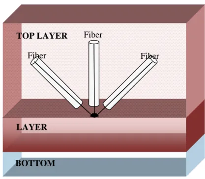
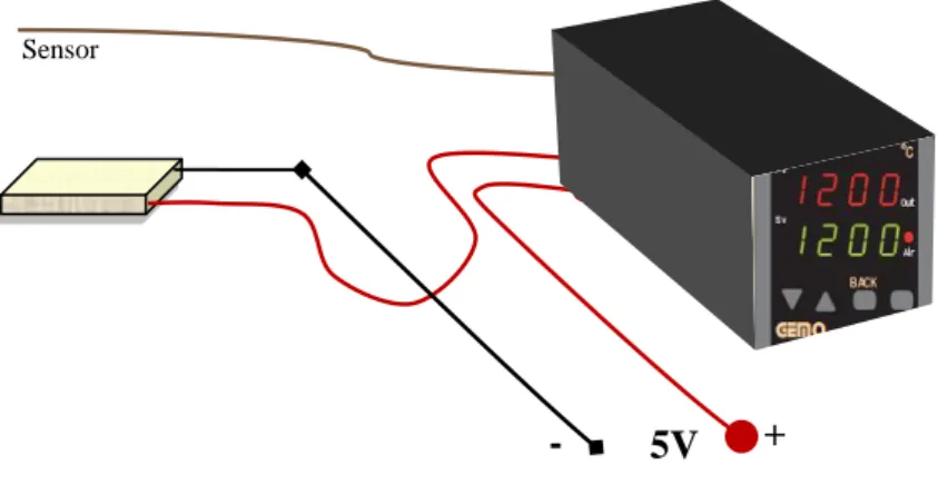
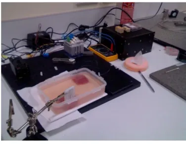
Imitation of radiofrequency ablation with fiber delivered laser system for magnetic resonance guided treatment of atrial fibrillation
Tam metin
Şekil


Benzer Belgeler
- 294 -.. According to Adnan Omran: 'Remember f that] nuclear war will eliminate Israel from the map but it will not eliminate the Arab nation f r om the map.' This is
Our detection algorithm consists of three main steps: i) green colored moving region detection in video, ii) analysis of the motion trajectories in the wavelet domain, and iii)
1948 ‹stanbul Üniversitesi Orman Fakültesi 2018 ‹stanbul Üniversitesi-Cerrahpafla Orman Fakültesi 95 1963 Karadeniz Teknik Üniversitesi Orman Fakültesi 85 1992 Abant ‹zzet
This chapter conclude that the performance of marine fisheries in India, Tamilnadu and Ramanathapuram include the market wise exports, port wise exports and
In addition to providing drivers and passengers with a wide range of information, this method of communication allows safety applications to enhance road safety
Scavenging Activities 中文摘要 我們利用 DNA 鬆懈實驗、DPPH
Sinyal değeri: Sinyal değeri için p değeri 0,025 < 0,05 olduğu için en kısa mesafeli turu bulma üzerinde sinyal değerinin etkili olduğunu söyleyebiliriz. İterasyon
ij IJ ߖپӵҏऋߞຨϛี౪Οඁএᝒ१Ֆޟ ޱȂࠉپ൷ຨкौޟϚ݈ᓞཷȃᓞฮȃڳ֜
