Generation of Singlet Oxygen by Persistent Luminescent
Nanoparticle–Photosensitizer Conjugates: A Proof of
Principle for Photodynamic Therapy without Light
Tugba Ozdemir
+,
[a]Yu-Chen Lu
+,
[b]Safacan Kolemen,
[a]Esra Tanriverdi-Ecik,
[c]and
Engin U. Akkaya*
[a, d]The broader application of photodynamic therapy as a treat-ment procedure for cancer is hampered by the limited pene-tration of light through mammalian tissues. Since the photo-sensitized generation of cytotoxic singlet oxygen requires ef-fective excitation of the tumor-localized photosensitizer, pho-todynamic action can only be guaranteed for the first few milli-meters of the irradiated tissues. In this work, we demonstrated that the phenomenon of persistent luminescence, that is, de-layed emission from certain metal-ion excited states (with crys-tal defects acting as energy traps), can provide an alternative excitation possibility. Thus, persistent luminescent nanoparti-cles functionalized by FRET-matching Bodipy sensitizers (FRET= Fçrster resonance energy transfer) were excited in situ before administration into a cell culture or an organism. It was found that this system continues to produce singlet oxygen re-gardless of their location and without any need for continuous photonic excitation.
Photodynamic therapy (PDT) of cancer, which is based on the photosensitized generation of singlet oxygen in tumor tissues, has attracting renewed interest in recent years.[1–4] Unlike the
previous decades, most of this recent work is focused on the selective activation of photosensitizers and the precise delivery of the cytotoxic agent, singlet oxygen, rather than the synthe-sis of newer photosensitizers.[5–13]However, there are a few
lin-gering problems associated with PDT which hinder its develop-ment into a broadly applicable therapeutic procedure.[14–16]
One of these problems is the issue of light penetration through tissues.[17] Although the term “therapeutic window”
implies that light with a wavelength in the l=650–800 nm range is more penetrating inside mammalian tissues, a closer inspection reveals that even at these optimal wavelengths, ef-fective penetration is limited to a few millimeters.[18] This
would limit the applicability to superficial lesions.
We surmised that materials with a long afterglow may offer a solution, eliminating altogether the need for external excita-tion. Naturally, most appropriate materials for this purpose would be persistent luminescent nanoparticles (PLNP). There has been considerable progress in the design of such materi-als.[19–24] Some of these systems have even been proposed as
in vivo imaging agents.[25–34]Persistence luminescence is an
in-teresting phenomenon resulting from the embedding of metal ions in certain inorganic matrices with particular energy-trap states, which can relax by thermal equilibration or photonic emission with emissive states of other ions present in the same matrix.[34,35] Compared to bulk persistent luminescent
materials, nanoparticles are less emissive as a result of the rela-tive increase in the defect structures.[26] Nevertheless, efficient
energy transfer to dyes on PLNPs have been amply document-ed.[32,34]
Our proposed design concept is shown schematically in Figure 1. Thus, excitation of the photosensitizer-functionalized PLNPs can be done outside of the biological medium (in vivo or in vitro), and then transferred to the location of the tumor. As result of the long afterglow, or in this case, delayed energy transfer, the energy of the excited ionic species in the PLNPs will be transferred to the photosensitizer (PS) in the dark, and most likely, even minutes after the UV irradiation has stopped. Generation of singlet oxygen inside the tumor would lead to apoptosis and cell death, as it takes place in regular PDT.[36–39]
The great advantage here would be the fact that once the (1)-PLNP conjugate is activated by irradiation, it will be capable of producing singlet oxygen regardless of the depth of the tumor.
The zinc-germanogallate-based PLNP sample used in this study was previously characterized and reported,[40] with the
preparation procedure involving a citrate sol–gel method fol-lowed by calcination. The intensity and the persistence of the PLNP emission were improved by co-doping with Pr3+/Cr3+.
The PLNPs had a composition of Zn2.78Ga1.68Ge1.00O8:Cr0.01,Pr0.02
based on X-ray fluorescence spectrometry. The average size of [a] Dr. T. Ozdemir,+Dr. S. Kolemen, Prof. E. U. Akkaya
UNAM-Institute of Material Science and Nanotechnology Bilkent University
Ankara 06800 (Turkey) E-mail: [email protected] [b] Y.-C. Lu+
Research Center for Analytical Science College of Chemistry and Nano Science Nankai University
94 Weijin Road, Tianjin 300071 (P.R. China) [c] Dr. E. Tanriverdi-Ecik
Department of Chemistry Gebze Technical University Kocaeli 41400 (Turkey) [d] Prof. E. U. Akkaya
Department of Chemistry Bilkent University Ankara 06800 (Turkey)
[++] These authors contributed equally to this work.
Supporting Information and the ORCID identification number(s) for the author(s) of this article can be found under:
the NPs were approximately 50 nm based on TEM, with hydro-dynamic diameter of 70 nm calculated on the basis of hydro-dynamic light scattering (DLS) data. The luminescence band maximum of the nanoparticles occurred at l=695 nm. The PLNPs were then coated by the reaction of 3-aminopropyltriethoxysilane (APTES) with the surface hydroxy groups on the PLNPs.
In order to experimentally investigate the premise of this work, we targeted compound 1 as the photosensitizer (Figure 2). Here, to ensure an efficient energy-transfer from
PLNP to compound 1, a long-wavelength-absorbing Bodipy dye (distyryl-Bodipy)[41] that has significant spectral overlap
with the PLNP emission was selected. Heavy atoms (iodines) were placed on the Bodipy chromophore to facilitate intersys-tem crossing to the triplet manifold for an efficient singlet oxygen generation.[42,43]A meso-substituent was chosen for
po-tential further derivatization. The 3-position of the Bodipy core carries an aliphatic carboxylic acid function, placed for
straight-forward conjugation to amine-modified PLNPs. Compound 1 was synthesized in nine steps from commercially available materials (detailed procedures and the synthetic scheme are available in the Supporting Information). The coupling reaction of the carboxylic acid functionalized photosensitizer 1 to the amino-ligated PLNP was carried out in DMF using BOP as a re-agent (BOP =benzotriazol-1-yloxy)tris-(dimethylamino)-phos-phonium hexafluorophosphate). The (1)-PLNP conjugated nanoparticles were purified by centrifugation and washing.
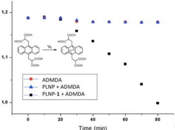
The singlet-oxygen generation capacity of the (1)-PLNP con-jugates was first investigated by looking for singlet-oxygen phosphorescence at l=1270 nm following UV excitation of the nanoparticles. The data unequivocally shows that bare PLNPs do not generate singlet oxygen, whereas the (1)-PLNP conjugate show the characteristic phosphorescence band (see the Supporting Information). Further experiments with the se-lective singlet-oxygen trap compound 2’-(anthracene-9,10-diyl-bis(methylene))dimalonic (ADMDA) in aqueous solutions (Figure 3) also corroborate the activity of the (1)-PLNP conju-gate for singlet-oxygen generation.
In order to confirm that singlet-oxygen generation continues in darkness, we carried out the following experiment: Instead of following absorbance changes, the1O
2generation capability
of (1)-PLNP was examined by recording the decrease in fluo-rescence emission of the singlet-oxygen probe 1,3-diphenyl-isobenzofuran (DPBF; Figure 4). In a typical experiment, the solvent acetonitrile was bubbled with air for 30 min to ensure the availability of dissolved oxygen during1O
2detection. Then,
the DPBF (200 mL, 20 mm in acetonitrile) was added to the dis-persion of pre-excited (254 nm, 6W) (1)-PLNP (2 mL, 1 mgmL@1) in the dark. The fluorescence of DPBF at l=
480 nm was recorded at two minute intervals (lex= 410 nm).
The results (Figure 4) clearly indicate that singlet oxygen pro-Figure 1. Ex situ “charging” of the PLNP by UV irradiation, followed by
energy-transfer excitation of the tethered photosensitizers (PS), leads to sin-glet oxygen generation in situ long after the excitation source is turned off and PLNPs have been transferred to a different locale (cell culture or a tumor model).
Figure 2. The structure and synthesis of the (1)-PLNP conjugate.
Figure 3. Relative singlet-oxygen efficiency of just the trap (red circles), PLNP alone (blue triangles) and the (1)-PLNP conjugate (black squares) in aqueous solutions. The efficiency was detected by the absorbance intensity decrease of ADMDA at l=376 nm with time. During the first 20 min, the samples were kept in the dark and for the following 60 min, the samples were irradi-ated with 254 nm light using a UV lamp.
duction continues in darkness only for the PLNP-Bodipy com-pound 1 conjugate.
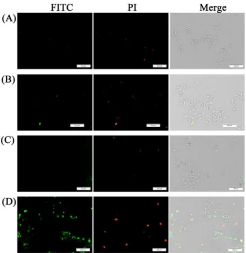
Cell culture studies were done using HepG2 cells. The cells were incubated with PLNP (Bare PLNPs) or (1)-PLNP and sub-jected to UV irradiation at 254 nm for 5 min (Figure 5). Without irradiation, the cells showed no staining with fluorescein-la-beled Annexin V (FITC-Annexin V). This indicates that there is no apoptotic cell death under those conditions, as FITC-Annex-in V is a specific marker of phosphatidylserFITC-Annex-ine flipped to the outer leaf of the cellular membrane during apoptosis.[44] The
situation is not much different with PNLP and UV light expo-sure. Only when (1)-PLNP derivative was used, significant An-nexin V labeling and propidium iodide (PI) staining was ob-served. PI enters the cells only when membrane integrity is se-verely compromised, another indication of cell death.
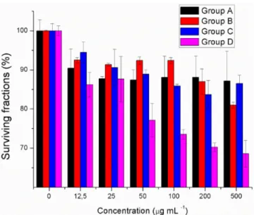
In order to quantify the effectiveness of the applied method-ology, standard MTT assays were carried out. In Figure 6, the surviving fraction of the HepG2 cells was shown as a function of the concentration of the agent used. As expected, UV irradi-ation and PLNPs alone result in some cell death under the ex-perimental conditions. However, the cytotoxic effect is signifi-cantly enhanced when (1)-PLNP conjugates were used. Figure 4. Persistent-luminescence-sensitized generation of1O
2. The time course of the relative fluorescence emission intensity change of DPBF moni-toring emission at l=480 nm caused by the generation of1O
2in the dark was monitored. The PLNP-compound 1 or the bare-PLNPs were pre-excited at 254 nm for 5 min before addition of DPBF in acetonitrile. F0is the DPBF emission intensity at t=0.
Figure 5. (1)-PLNP induced cell apoptosis as determined by fluorescence imaging using Annexin V-FITC/PI staining on HepG2 cells. A) HepG2 cells with Bare-PLNP (200 mgmL@1) treatment and without irradiation. B) HepG2 cells incubated with Bare-PLNP (200 mgmL@1) and exposed to UV light for 5 min. C) HepG2 cells treated with (1)-PLNP (200 mgmL@1) without irradiation. D) HepG2 cells incubated with (1)-PLNP (200 mgmL@1), and exposed to UV light for 5 min. Scale bar =100 mm.
The PDT efficacy of the (1)-PLNP conjugate was investigated using tumor-bearing mice models (adult athymic BALB/c mice). Tumor-bearing mice were established by subcutaneous inocu-lation of a HepG2 cell suspension (5x 10⁶cells per mouse) into
the flank region of 3–4-week-old male nude mice. The experi-ment included two groups for comparison: Bare PNLP-NH2
with irradiation as the control, and Bodipy-conjugated (1)-PLNP conjugate as the experiment group. The nanoparticles solutions (40 mL,15 mg mL@1in PBS at pH 7.4) were irradiated
with a UV lamp for 2 min at 254 nm before they were injected intratumorally to mice. The nanoparticles were injected into the tumor site every 24 h for 10 days after pre-excitation under UV light irradiation.
To evaluate the therapeutic efficacy, tumor growth was monitored, measuring the tumor size with a digital caliper. On day 11, a modest but statistically significant reduction in tumor volume (15%) was recorded.
Considering the fact that the PLNP afterglow was short, it is not surprising that the in vivo PDT effect is modest. However, persistence luminescence is a vibrant field, thus new and longer afterglow nanoparticles are expected. On the other hand, singlet-oxygen trap experiments, cell culture and MTT experiments all validated our proposal, and as a proof of prin-ciple, the (1)-PLNP conjugate was shown to function in accord-ance with the design parameters. Needless to say, we expect further work along this line to bring more persistent genera-tors of singlet oxygen in the dark. It is also possible to block the singlet-oxygen generation by a disulfide-tethered quench-er, which would be cleaved under the reductive conditions of the tumors. Thus, this proof of principle may offer a unique po-tential for addressing a persistent problem of PDT. Our work in that direction is in progress.
Note on Animal Experiments Described
herein:
Animal treatment and maintenance were performed in accordance with the Principle of Laboratory Animal Care (NIH Publication number 85-23, revised 1985). All animals were treated in accord-ance with the guidelines of the Committee on Animals of Nankai University (Tianjin, Peoples’ Republic of China). All animal proce-dures were approved by the Nankai University Experimental Animal Ethics Committee.
Acknowledgements
E.U.A. gratefully acknowledges support from Bilkent University. E.T.-E. is grateful for a postdoctoral scholarship from TUBITAK.
Conflict of interest
The authors declare no conflict of interest.
Keywords: nanoparticles · persistent luminescence · photodynamic therapy · photosensitizers · singlet oxygen
[1] S. Ozlem, E. U. Akkaya, J. Am. Chem. Soc. 2009, 131, 48– 49.
[2] I. Simsek Turan, D. Yildiz, A. Turksoy, G. Gunaydin, E. U. Akkaya, Angew. Chem. Int. Ed. 2016, 55, 2875 –2878; Angew. Chem. 2016, 128, 2925 – 2928.
[3] S. Kolemen, T. Ozdemir, D. Lee, G. M. Kim, T. Karatas, J. Yoon, E. U. Akkaya, Angew. Chem. Int. Ed. 2016, 55, 3606 –3610; Angew. Chem. 2016, 128, 3670 – 3674.
[4] Y. Cakmak, S. Kolemen, S. Duman, Y. Dede, Y. Dolen, B. Kilic, Z. Kostereli, L. T. Yildirim, A. L. Dogan, D. Guc, E. U. Akkaya, Angew. Chem. Int. Ed. 2011, 50, 11937–11941; Angew. Chem. 2011, 123, 12143 –12147. [5] M. Bio, P. Rajaputra, G. Nkepang, Y. You, J. Med. Chem. 2014, 57, 3401 –
3409.
[6] A. Shao, Y. Xie, S. Zhu, Z. Guo, S. Zhu, J. Guo, P. Shi, T. D. James, H. Tian, W. H. Zhu, Angew. Chem. Int. Ed. 2015, 54, 7275 –7280; Angew. Chem. 2015, 127, 7383 – 7388.
[7] S. Erbas-Cakmak, E. U. Akkaya, Angew. Chem. Int. Ed. 2013, 52, 11364 – 11368; Angew. Chem. 2013, 125, 11574 – 11578.
[8] I. Simsek-Turan, F. Pir-Cakmak, D. C. Yildirim, R. Cetin-Atalay, E. U. Akkaya, Chem. Eur. J. 2014, 20, 16088 – 16092.
[9] S. Kolemen, M. Isik, G. M. Kim, D. Kim, H. Geng, M. Buyuktemiz, T. Kara-tas, X.-F. Zhang, Y. Dede, J. Yoon, E. U. Akkaya, Angew. Chem. Int. Ed. 2015, 54, 5340 –5344; Angew. Chem. 2015, 127, 5430 –5434.
[10] B. Wang, H. Yuan, Z. Liu, C. Nie, L. Liu, F. Lv, Y. Wang, S. Wang, Adv. Mater. 2014, 26, 5986 – 5990.
[11] E. J. Kim, S. Bhuniya, H. Lee, H. M. Kim, C. Cheong, S. Maiti, K. S. Hong, J. S. Kim, J. Am. Chem. Soc. 2014, 136, 13888 –13894.
[12] G. Kong, G. Anyarambhatla, W. P. Petros, R. D. Braun, O. M. Colvin, D. Needham, M. W. Dewhirst, Cancer Res. 2000, 60, 6950 –6957.
[13] N. Huebsch, C. J. Kearney, X. Zhao, J. Kim, C. A. Cezar, Z. Suo, D. J. Mooney, Proc. Natl. Acad. Sci. USA 2014, 111, 9762 –9767.
[14] R. Sullivan, C. H. Graham, Cancer Metastasis Rev. 2007, 26, 319 –331. [15] J. A. Bertout, S. A. Patel, M. C. Simon, Nat. Rev. Cancer 2008, 8, 967 –975. [16] T. M. Busch, S. M. Hahn, S. M. Evans, C. J. Koch, Cancer Res. 2000, 60,
2636 –2642.
[17] S. Stolik, J. A. Delgado, A. Perez, L. Anasagasti, J. Photochem. Photobiol. B 2000, 57, 90– 93.
[18] W. J. Cheong, S. A. Prahl, A. J. Welch, IEEE J. Quantum Electron. 1990, 26, 2166 –2185.
[19] J. Hçls-, Electrochem. Soc. Interf. 2009, 18, 42– 45.
[20] W. M. Yen, S. Shionoya, H. Yamamoto, Phosphor Handbook, CRC Press, Boca Raton, FL, 2007.
Figure 6. MTT assays (performed after 24 h): Surviving fractions of HepG2 cells incubated with Bare-PLNP for 8 h, followed by UV light irradiation (5 W, 254 nm). A) HepG2 cells with Bare-PLNP treatment, without irradiation. B) HepG2 cells incubated with Bare-PLNP, exposed to UV light for 5 min. C) HepG2 cells treated with (1)-PLNP, without irradiation. D) HepG2 cells in-cubated with (1)-PLNP, exposed to UV light for 5 min. Data correspond to mean : S.D. values.
[21] T. Aitasaloa, P. Deren´c, J. Hçls-a, H. Jungnerd, J.-C. Krupae, M. Lastusaa-ria, J. Legendziewiczf, J. Niittykoskia, W. Stre˛kc, J. Solid State Chem. 2003, 171, 114– 122.
[22] T. Matsuzawa, Y. Aoki, N. Takeuchi, Y. Murayama, J. Electrochem. Soc. 1996, 143, 2670 – 2673.
[23] W. Chen, J. Zhang, J. Nanosci. Nanotechnol. 2006, 6, 1159– 1166. [24] K. Van den Eeckhout, P. F. Smet, D. Poelman, Materials 2010, 3, 2536 –
2566.
[25] Q. le Masne de Chermont, C. Chaneac, J. Seguin, F. Pelle, S. Maitrejean, J. P. Jolivet, D. Gourier, M. Bessodes, D. Scherman, Proc. Natl. Acad. Sci. USA 2007, 104, 9266 –9271.
[26] T. Maldiney, C. Richard, J. Seguin, N. Wattier, M. Bessodes, D. Scherman, ACS Nano 2011, 5, 854– 862.
[27] T. Maldiney, A. Lecointre, B. Viana, A. Bessiere, M. Bessodes, D. Gourier, C. Richard, D. Scherman, J. Am. Chem. Soc. 2011, 133, 11810 –11815. [28] T. Maldiney, M. U. Kaikkonen, J. Seguin, Q. le Masne de Chermont, M.
Bessodes, K. J. Airenne, S. Yla-Herttuala, D. Scherman, C. Richard, Opt. Mater. Express 2012, 2, 261–268.
[29] F. Liu, W. Yan, Y.-J. Chuang, Z. Zhen, J. Xie, Z. Pan, Sci. Rep. 2013, 3, 1554.
[30] T. Maldiney, M. U. Kaikkonen, J. Seguin, Q. le Masne de Chermont, M. Bessodes, K. J. Airenne, S. Yl--Herttuala, D. Scherman, C. Richard, Bio-conjugate Chem. 2012, 23, 472 –478.
[31] T. Maldiney, G. Byk, N. Wattier, J. Seguin, R. Khandadash, M. Bessodes, C. Richard, D. Scherman, Int. J. Pharm. 2012, 423, 102–107.
[32] B.-Y. Wu, H.-F. Wang, J.-T. Chen, X.-P. Yan, J. Am. Chem. Soc. 2011, 133, 686– 688.
[33] Z.-J. Li, H.-W. Zhang, M. Sun, J.-S. Shen, H.-X. Fu, J. Mater. Chem. 2012, 22, 24713 – 24720.
[34] T. Maldiney, A. BessiHre, J. Seguin, E. Teston, S. K. Sharma, B. Viana, A. J. J. Bos, P. Dorenbos, M. Bessodes, D. Gourier, D. Scherman, C. Ri-chard, Nat. Mater. 2014, 13, 418 –426.
[35] Z. Pan, Y.-Y. Lu, F. Liu, Nat. Mater. 2012, 11, 58– 63.
[36] P. Mroz, A. Yaroslavsky, G. B. Kharkwal, M. R. Hamblin, Cancers 2011, 3, 2516 –2539.
[37] D. E. Dolmans, D. Fukumura, R. K. Jain, Nat. Rev. Cancer 2003, 3, 380 – 387.
[38] T. J. Dougherty, J. E. Kaufman, A. Goldfarb, K. R. Weishaupt, D. Boyle, A. Mittleman, Cancer Res. 1978, 38, 2628 – 2635.
[39] J. Moan, Q. Peng, Anticancer Res. 2003, 23, 3591 –3600.
[40] A. Abdukayum, J.-T. Chen, Q. Zhao, X.-P. Yan, J. Am. Chem. Soc. 2013, 135, 14125 –14133.
[41] A. Loudet, K. Burgess, Chem. Rev. 2007, 107, 4891 –4932.
[42] A. Gorman, J. Killoran, C. O’Shea, T. Kenna, W. M. Gallagher, D. F. O’Shea, J. Am. Chem. Soc. 2004, 126, 10619– 10631.
[43] T. Yogo, Y. Urano, Y. Ishitsuka, F. Maniwa, T. Nagano, J. Am. Chem. Soc. 2005, 127, 12162–12163.
[44] I. Vermes, C. Haanen, H. Steffens-Nakken, C. P. Reutelingsperger. J. Immu-nol. Methods 1995, 184, 39– 51.
Manuscript received: December 24, 2016 Accepted Article published: February 16, 2017 Final Article published: March 29, 2017