East J Med 25(1): 68-72, 2020 DOI: 10.5505/ejm.2020.72692
ORIGINAL ARTICLE
An overview of SMAS; Anatomic and Radiologic
Approach
Selma Çalişkan1*, Emre Can Çelebioğlu2, Sinem Akkaşoğlu1, İbrahim Tanzer Sancak3
1Department of Anatomy, Faculty of Medicine, Ankara Yıldırım Beyazıt University, Ankara, Turkey 2Department of Radiology, Faculty of Medicine, Ankara University, Ankara, Turkey
3Department of Radiology, Faculty of Medicine, TOBB University, Ankara, Turkey
Introduction
SMAS (superficial musculo aponeurotic system) is a network of collagen fibers, elastic fibers and fat interconnecting facial muscles and dermis (1). Besides its rich neurovascular content it facilitates the movements of the skin (2). Different definitions of SMAS are found in the literature because of the region specific histologic morphology of this structure (1, 2). Subcutaneous morphology of face is more organized than subcutaneous layer of any part of body. SMAS has distinctive features in forehead, parotid region, zygomatic region, temporal region, cheek, infraorbital region, nasolabial fold, and lower lip (1, 3). Medial to nasolabial fold SMAS is in form of a net including dense collagen and muscle fibers. SMAS lateral to nasolabial fold consists of fibrous septa enclosing lobules of fat (1). Detailed analysis of SMAS enlightens disputed points in rhytidectomy and face lifting procedures.
Because SMAS is a key feature for either percutaneous and surgical aesthetic interventions neurovascular structure embedded in this tissue must be well known. Besides the histologic differences,
anatomical relationship between facial nerve and SMAS should be well known by medical aesthetic professionals. After leaving cranial cavity from stylomastoid foramen facial nerve splits into its peripheral branches in parotid gland. These branches penetrate the parotid fascia and become close related to SMAS (3,4). This relationship is extremely important when performing a SMAS-platysma rhytidectomy (4). Anterior to parotid gland they are located more superficially. From ear to mouth nerve fibers increase in number. They lie in different depths in different regions. Despite Gray’s, Mitz et al suggested that facial nerve fibers lie deeper than SMAS in the cheek and indicated that sensory nerves of cervical plexus go through SMAS (3, 5). Motor facial nerve branches reach superficial layer of facial muscles through their deeper aspect (3). SMAS encloses voluntary muscles in its deep portion and extends into the fibers of risorius, frontalis, platysma and peripheral part of orbicularis oculi muscle. (2, 3, 5) Facial artery and vein lie deep to SMAS. Vessels supplying skin branch in the SMAS plane before reaching the subdermal level. Perforating branches of
ABSTRACT
Superficial Musculo-Aponeurotic System (SMAS) is a network of collagen fibers, elastic fibers and fat interconnecting facial muscles and dermis. Subcutaneous morphology of face is more organized than subcutaneous layer of any part of body. SMAS has distinctive features in forehead, parotid region, zygomatic region, temporal region, cheek, infraorbital region, nasolabial fold, and lower lip. Because SMAS is a key feature for either percutaneous and surgical aesthetic interventions, neurovascular structure embedded in this tissue must be well known.
Radiologic views were enrolled from the archive system of radiology department. Images of 50 patients were randomly selected. (29 ale, 21 female). Age width of the participants was in between 18 and 78 years (mean age 40.34± 15.32).
Thickness of SMAS for each region is measured. Continuity of the tissue was followed between the junctions of the regions in a proper sequence with MR images. Thickness of SMAS in the zygomatic region was measured in 50 patients and mean value was 0.12 mm. Left and right side measures were compared between genders and no statistical difference between gender groups was found. Correlation between measures and age was analyzed statistically and no correlation was found.
A radiologically and clinically neglected tissue: SMAS deserves more attention because of its continuous course interconnecting distinct regions of face and acting as a functional unit for the expressions. Age and gender related changes in SMAS morphology studied in healthy individuals by means of radiology provides an important contribution to the literature
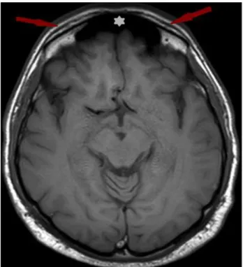
Fig 1. SMAS at forehead region on axial T1 weighted
images. Note: White star indicates the frontal sinus
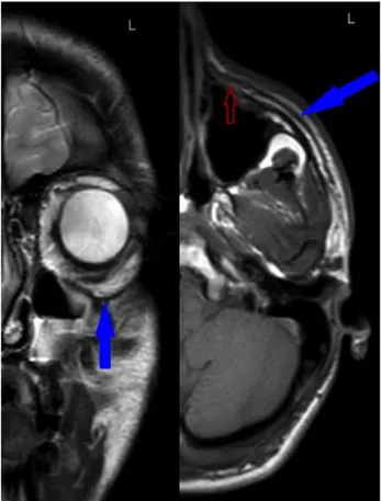
Fig 2. On the axial T1 weighted images obtained at a 63
years old female patient green star indicates SMAS that merges with parotid fascia
these vessels go through SMAS to form subdermal vascular network (3).
Temporal region has distinctive characteristics. SMAS in this region is extremely thin. Superficial temporal artery and vein and temporal and zygomatic branches of facial nerve are within the SMAS in temporal region (4).
In this study distinctive characteristics and thickness of SMAS accommodating neurovascular network of face is radiologically studied in different parts of face.
Materials and Methods
Radiologic views were enrolled from the archive system of radiology department.
Fig 3. Yellow arrows indicates the SMAS at zygomatic
region on axial T1 weighted images
Fig 4. On T2 weighted coronal images red arrow shows
SMAS at the zygomatic region which merges the temporoparietal fascia at a 63 years old female patient
Patients with BMI>25 were not included in the present study. Radiologic views of patients with no history of thyroid gland diseases, renal, cardiovascular or rheumatologic diseases were included in the study. Patients with all types of dermatologic conditions (cutaneous lymphoma, sarcoidosis, dermatomyositis) were not involved in the study.
MR images were performed with 1.5 T Siemens Magnetom Symphony device. All images were acquired with a GE 1.5 T MRI unit. Imaging
Fig 5. SMAS at infraorbital region at a 19 years old male
blue arrows at coronal T2 weighted images and axial T1 weighted images clearly depicts SMAS. Red arrow indicates SMAS near nasolabial fold
parameters are TR: 3000-4500 msec/ TE: 80-150 msec for T2 weighted sequences and TR:600-650 msec/ TE: 15-25 msec for T1 weighted sequences with a 3 mm slice thickness and %20 interslice gap. Image analyses were made with Clear Canvas Dicom Viewer. All SMAS thickness measurements were made 3 x magnification level.
Statistical Analysis: Descriptive statistics were given
as mean (std.deviation) values were used for continuous ones. To compare measures between gender categories, Student’s t test was used because measures are normal distributed within each group and variances of two groups are equal (Table 1). Pearson-rho correlation coefficient was used to calculate the correlation between age and measures (Table 2). Type-I error rate was taken as 0.05 to test statistical hypotheses. SPSS 20.0 was used to run statistical analyses (IBM Corp. Released 2011. IBM SPSS Statistics for Windows, Version 20.0. Armonk, NY: IBM Corp.)
Results
Thickness of SMAS for each region was measured bilaterally in forehead, parotid region, zygomatic region, infraorbital area and nasolabial region.
Continuity of the tissue was followed between the junctions of the regions in a proper sequence with MR images.
50 patients who admitted to hospital from 2012 to 2015 were subsequently enrolled to the study. Of the 50 patients; 29 were male and 21 were female. Age width of the participants was in between 18 and 78 years (mean age 40.34± 15.32).
SMAS thickness in forehead was measured at a point one centimeter superior and medial to lateral end of corrugator supercilii muscle (Fig 1). It was measured at the midpoint of anterior margin of parotid gland in parotid region (Fig 2). In the zygomatic region thickness was measured on attachment site of zygomaticus major muscle on the zygomatic bone (Fig 3, Fig 4). We indicated the point one centimeter inferior and one centimeter lateral to superior margin of lower eyelid on the mid pupillary line to measure SMAS thickness in the infraorbital area. Midpoint of the nasolabial fold was the measurement point of SMAS in the nasolabial region (Fig 5).
Thickness of SMAS in the zygomatic region was measured in 50 patients and mean value was 0.12 mm. SMAS ın the parotid region was evaluated in 46 patients and mean thickness was found 0.7mm. There was no statistically significant difference between regions according to thickness measures. Left and right side measures were compared between genders and no statistical difference between gender groups was found (Table 2). Correlation between measures and age was analyzed statistically and no correlation was found (Table 1).
Discussion
Clear understanding of age related changes in SMAS tissue is basis for application of appropriate procedure even in surgical procedures or in medical aesthetic and reconstructive interventions.
There are studies in the literature based on cadaver dissections and histological specimens from cadavers (1, 4). In the present study we aimed to visualize the living tissue in terms of radiology.
Different comments on regional continuity of SMAS are clearly visible in the literature. Mitz et al stated that there is superficial muscular and aponeurotic system in the parotid and cheek areas while Wassef et al found a continuous fibromuscular layer deep to skin (3, 6). Thaller et al concluded that SMAS is a distinct structure covering face which superiorly attaches to frontalis muscle and inferiorly to platysma. According to Thaller et al SMAS attaches posteriorly to pericondrium of tragal cartilage and sternocleidomastoid muscle on mastoid bone,
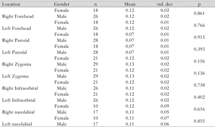
Table 1. Comparison of left and right measures between gender categories
Location Gender n Mean std. dev p
Right Forehead Female 18 0.12 0.02 0.861 Male 26 0.12 0.02 Left Forehead Female 18 0.12 0.01 0.766 Male 26 0.12 0.02 Right Parotid Female 18 0.07 0.01 0.915 Male 28 0.07 0.01 Left Parotid Female 18 0.07 0.01 0.393 Male 28 0.07 0.01 Right Zygoma Female 21 0.12 0.02 0.156 Male 29 0.13 0.02 Left Zygoma Female 21 0.12 0.02 0.136 Male 29 0.13 0.02 Right Infraorbital Female 21 0.12 0.02 0.738 Male 26 0.11 0.02 Left Infraorbital Female 21 0.12 0.02 0.402 Male 26 0.12 0.02 Right nasolabial Female 10 0.12 0.09 0.616 Male 17 0.11 0.05 Left nasolabial Female 10 0.11 0.07 0.855 Male 17 0.11 0.06
Table 2. Correlation matrix between age and measures
forehe
ad R Forehead L Parotid R Parotid L zygoma R zygoma L infraorbital R infraorbital L nasolabial R nasolabial L Age rho
* -0.12 -0.13 0.03 0.03 -0.14 -0.07 -0.16 -0.08 -0.28 -0.25 p 0.45 0.39 0.85 0.84 0.33 0.65 0.27 0.58 0.15 0.21 n 44 44 46 46 50 50 47 47 27 27 *Pearson correlation coefficient
There is no correlation between age and measures (p>0.05) (Table 2). R: right L: left
anteriorly it is contiguous with platysma muscle (4). Owsley postulated that SMAS ends at nasolabial fold (7). Aforementioned cadaveric studies found in the literature have some limitations. Because of the thin and curvilinear structure of the SMAS mingling with some of the expression muscles and deep fascia of the face in certain areas it is hard to follow non interrupted course of this tissue during dissection. Sequenced radiologic sections enable us to follow the continuity of tissue. In the present study we presented the results from MR images and evaluated the tissue in each region. This is an additional value of the present study. Despite Owsley we defined no interruption in the nasolabial region. Besides, our study is not in consistent with the study of Mitz et al who concluded that SMAS is visible in parotid and cheek areas. We presented radiologic images of SMAS in all regions of face.
Facial expression muscles and SMAS are neglected in radiology reports. It is hard to identify expression
muscles on serial radiologic images because of the oblique courses and overlapping parts. Location and morphology of a muscle changes on serial images (8). SMAS encloses voluntary muscles in its deep portion and extends into the fibers of risorius, frontalis, platysma and peripheral part of orbicularis oculi muscle (2, 3, 5). Although SMAS and expression muscles are anatomically defined individual structures they act as single functional unit. SMAS magnifies the action of the expression muscles. Som et al were the first to identify SMAS on imaging (8). Different from the study of Som et al we presented measures of thickness and analyzed age related and gender related changes of SMAS.
Owsley et al reported that SMAS-platysma face lift procedure provides a natural appearing result which is not excessively tight or pulled in appearance. In comparison with the standard face lift procedure SMAS-platysma face lift has advantage for lifting submental area with acceptably low incidence of
complications (7). In recent years incision-needing cosmetic surgery such as facelift procedures are substituted by medical aesthetic procedures. Most common one of these procedures is the dermal filler injections. Perioral area, periocular region, nasolabial folds, malar fat pad, marionette lines, glabella, and lips are the most commonly dermal filler injected sites on the face. Filler-related complications like abscess, cellulitis, non-inflammatory nodules and foreign body granulomas are common complications of dermal fillers. Radiologists may be asked to evaluate these complications and make differential diagnosis (9). From this point of view previously neglected structures in routine radiologic reports such as SMAS and expression muscles become important to indicate the landmarks and evaluate the pathology. Our study will enlighten this point of view for radiologists. To the best of our knowledge there are studies in the literature evaluating mid facial fat pads on CT images but no attribute to SMAS layer was detected in these studies (10, 11). MRI has a high capacity in evaluation of soft tissue and in differential diagnosis of inflammation, abscess, and also foreign material in the soft tissues (9). Dermis, superficial fat compartment, deep fat compartment and expression muscles with respect to SMAS may be well defined on MRI sections (9). In the present study SMAS was evaluated on MRI images of patients who have no diagnosis of collagen tissue disease or edema.
We measured thickness of the tissue in each region. Age related changes and differences between genders in terms of thickness were evaluated on radiologic images. Our study has some limitations. We found that thickness of SMAS in the zygomatic region was greatest but future studies with greater sample size are required to support this idea statistically. Current popular injectable antiaging therapies focus on enhancement of skin and SMAS. In the present study we found no link between age and SMAS thickness. Future studies with sufficient sample size will enlighten this point. We think present study will give idea to researches for detailed future studies.
A radiologically and clinically neglected tissue: SMAS deserves more attention because of its non-interrupted course interconnecting distinct regions of face and acting as a functional unit for the
expressions. Age and gender related changes in SMAS morphology studied in healthy individuals by means of radiology provides an important contribution to the literature. Knowledge of detailed anatomical morphology of SMAS will enlighten upcoming studies and new surgical approaches.
References
1. Ghassemi A, Prescher A, Riediger D, Axer H. Anatomy of the SMAS revisited. Aesthetic Plast Surg 2003; 27: 258-264.
2. Hwang K, Choi JH. Superficial Fascia in the Cheek and the Superficial Musculoaponeurotic System. J Craniofac Surg 2018; 29: 1378-1382. 3. Mitz V, Peyronie M. The superficial
musculo-aponeurotic system (SMAS) in the parotid and cheek area. Plast Reconstr Surg 1976; 58: 80-88. 4. Thaller SR, Kim S, Patterson H, Wildman M,
Daniller A. The submuscular aponeurotic system (SMAS): a histologic and comparative anatomy evaluation.Plast Reconstr Surg 1990; 86: 690-696. 5. Standring S, Gray’s Anatomy: The Anatomical
Basis of Clinical Practice, 40th Edition, Churchill
Livingstone 2009: 468.
6. Wassef M. Superficial and muscular layers in the face and neck:a histological study. Aesthetic Plast Surg 1987; 11: 171-176.
7. Owsley JQ Jr. Plast Reconstr Surg. SMAS-platysma face lift 1983; 71: 573-576.
8. Som PM, Su ANG, Stuchen C, Tang C Y. , Lawson W, Laitman JT. The MR Imaging Identification of the Facial Muscles and the Subcutaneous Musculoaponeurotic System. Neurographics 02:35-43 March 2012.
9. Mundada P, Kohler R, Boudabbous S, et al. Injectable facial fillers: imaging features, complications, and diagnostic pitfalls at MRI and PET CT. Insights Imaging DOI 10.1007/s13244-017-0575-0.
10. Gierloff M, Stöhring C, Buder T, Gassling V, Açil Y, Wiltfang J. Aging changes of the midfacial fat compartments: a computed tomographic study. Plast Reconstr Surg 2012; 129: 263-273.
11. Tart RP, Kotzur IM, Mancuso AA, Glantz MS, Mukherji SK. CT and MR imaging of the buccal space and buccal space masses. Radiographics 1995; 15: 531-550.