545
Journal of Natural and Applied Sciences Volume 22, Issue 2, 545-551, 2018 Fen Bilimleri Enstitüsü Dergisi
Cilt 22, Sayı 2, 545-551, 2018
DOI: 10.19113/sdufbed.38076
Enhancement Physical Performance of Nanostructured CuO Films via Surfactant
TX-100
Bünyamin ŞAHİN1, Raşit AYDIN*2
1Mustafa Kemal Üniversitesi, Fen-Edebiyat Fakültesi, Fizik Bölümü, 31034, Hatay 2Selçuk Üniversitesi, Fen Fakültesi, Fizik Bölümü, 42075, Konya
(Alınış / Received: 26.03.2018, Kabul / Accepted: 16.05.2018, Online Yayınlanma / Published Online: 25.05.2018)
Keywords
Copper Oxide (CuO), Triton X-100, XRD, Band Gap
Abstract: In this study, we informed a systematic approach to obtain CuO films
with and without TX-100 surfactant by the SILAR procedure. Morphological, structural and optical features of the CuO films were researched by metallurgical microscope, scanning electron microscopy, X-ray diffraction analysis and ultraviolet–visible spectrophotometry respectively with respect to concentrations of TX-100 agent. Metallurgical and scanning electron microscope photographs displayed that the morphology of the film surface was impressed by surfactant TX-100. X-ray diffraction patterns verified that all CuO films have monoclinic crystal lattice structure with preferential orientations of (1 11) and (111) planes. Ultraviolet–visible spectra demonstrated that the optical bandgap and transmittance values of the films were altered with TX-100 content.
Nanoyapılı CuO Filmlerin Fiziksel Performansının Surfaktan TX-100 Yoluyla
Geliştirilmesi
Anahtar Kelimeler
Bakır Oksit (CuO), Triton X-100, XRD, Bant Aralığı
Özet: Bu çalışmada TX-100 surfaktan içeren ve içermeyen CuO filmler sistematik
bir yaklaşım gözeterek SILAR yöntemi yardımıyla elde edilmişlerdir. Elde edilen CuO filmlerinin morfolojik, yapısal ve optik özellikleri, TX-100 konsantrasyonuna bağlı olarak sırasıyla metal mikroskobu, taramalı elektron mikroskobu, X-ışını difraksiyon analizi ve ultraviyole-görünür spektrometresi ile incelendi. Metal ve taramalı elektron mikroskobu fotoğrafları, film yüzey morfolojisinin yüzey aktif madde TX-100 tarafından etkilendiğini ortaya koydu. X-ışını kırınım desenleri, tüm CuO filmlerinin (1 11) ve (111) düzlemlerin tercihli yönelimleriyle monoklinik kristal kafes yapısına sahip olduğunu doğruladı. Ultraviyole – görünür spektrum, filmlerin optik bant boşluğu ve geçirgenlik değerlerinin TX-100 içeriği ile değiştiğini gösterdi.
1. Introduction
Recently, nanostructured transparent metal oxide semiconductor materials have been attracting a great deal of attention not only for different practical applications in electronic and optoelectronic technology due to important chemical and physical features but also for basic scientific research [1-3]. Among the different these materials, copper oxide (CuO) is hopeful p-type semiconductor metal oxide material with band gap energy of about 1.4 eV and monoclinic crystalline form [4, 5]. Due to its significant properties, such as abundance, economic, nontoxic, well electrical and optical characteristic and perfect thermal stabilities, CuO films are widely used in many different applications, such as gas sensors,
solar cells, photocatalysis, lithium batteries, biosensors and transistors [6-10].
Various physical and chemical growth procedures have been improved for the preparation of nanostructured CuO films including sol–gel, sputtering, thermal oxidization, electrochemical, evaporation, hydrothermal, co-precipitation and successive ionic layer adsorption and reaction (SILAR) [11-15]. Among these solution-based synthesis techniques SILAR is based on the submersion of the substrate into individually embed precursor solutions. It is also simple, inexpensive and low-temperature method. In addition, it does not need vacuum and complicated setup system [16-18].
546
Surfactants like sodium dodecyl sulfate, ethylene glycol, cetyl trimethyl ammonium bromide and triethanolamine can work a significant duty in the growth of the film in different attractive properties. The optoelectronic, electrical, structural and morphological properties of the nanostructured films can be enhanced by using surfactants in the aqueous solutions. The supplement of surfactant in synthesis solution decreases the surface strain of the solution and checks the formation of new crystal structures [19-22]. Among these surfactant diversities Triton-X 100 (TX-100) with the chemical equation (C14H22O(C2H4O)n) is one of the non-ionic surfactants. TX-100 is extensively used surfactant agents in industrial applications such as wetting agents, emulsifiers, detergents and dispersants [23, 24]. Triton X-100 has been used by many researchers as a surfactant to examine the properties of oxide films [25-27].In our previous review, we notified the influence of Triton X-100 as a surfactant on the growth of SILAR deposited CdO films [28]. When we examined the literature, there is no report of obtain of nanostructured CuO films with TX-100 as non-ionic surfactants grown SILAR method. Therefore, in this work, for the first time CuO films with and without TX-100 were synthesized on glass substrates by using a SILAR procedure. Then, the concentration (0.5 and 1.0 M%) effects of TX-100 surfactant on the structural, morphological and optical properties of CuO nanoparticles were examined. The exhaustive investigations of the CuO films are presented in the following parts.
2. Material and Method
All chemical materials used in the synthesis experiment of films are analytical reagent quality and were procured Merck Company. Copper (II) chloride dehydrate (CuCl2·2H2O), sulfuric acid solution (H2SO4), acetone (CH3COCH3), double distilled water (18.2 MΩ cm), ammonium, magnetic fish and soda lime glass substrates were used all the syntheses. TX-100 was prefered as surfactant for the deposition. Three series of CuO films with and without TX-100 were prepared on glass slides by the SILAR technique. To acquire the Cu2+ ion solution, 1.7 g CuCl2·2H2O was stirred with 100 ml double distilled water and was prepared 0.1 M copper chloride solution. The pH of the solution bath was arranged to ≈10.0 by adding ammonium and the aqueous solution was warmed about 85 oC. The substrates were immersed the solution for 20s, then it was dipped into deionized water for another 20 s. This period was iterated for 6 times. TX-100 was included to the solution baths in certain quantities (0.5 and 1 M%) to investigate the impression of TX-100 as a surfactant agent.
The surface morphological properties of the CuO films were analyzed by scanning electron microscope (SEM: Zeiss evo Ls 10) and metallurgical microscope (Nikon Eclipse MA100), respectively. The crystal qualities and structural features of the CuO films were studied by X-ray diffractometer (Bruker D8 Advance with Cu Kα radiation (λ = 0.15406 nm)). UV– visible absorption spectra of the deposited CuO films were measured with a UV-Vis. spectrometer (Thermo Scientific Genesys 10S UV-Vis.).
3. Results
3.1. Metallurgical microscope (MM) studies
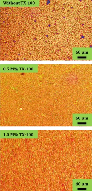
Figure 1. Metallurgical microscope photos of CuO films at
varied TX-100 contents.
The physical performance of nanostructured CuO films exceedingly depends on the morphological surface properties. Variance in surface structures of CuO films with and without TX-100 were examined
547
nanoparticles with increasing TX-100 accumulations of 0, 0.5 and 1.0 M %, respectively. When the concentration of TX-100 is 0 M% (without TX-100), it is seen from the Figure 1 that there are some dark regions on the surface of CuO film. The surface morphologies of CuO films are denser, smoother at higher TX-100 concentrations such as 0.5 and 1.0 M %. In other words, the surface uniformity of films was improved with increasing TX-100 addition in the solution bath. This advance in surface morphology is clearly seen in Figure 1. For this reason, we can say that the surface properties of the films highly depends on the TX-100 content. As is seen from photos, TX-100 as a non-ionic agent has a favourable impact on morphologic characteristics of the CuO nanoparticles.3.2. Scanning electron microscope(SEM) studies
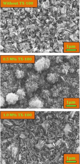
Figure 2. SEM photographs of the nanostructured CuO
films deposited without and with TX-100.
The examination of the morphological properties of CuO films is extremely important because it also affects their optical properties. SEM was exploited to investigate the surface characterisitcs of the CuO particles grown with and without TX-100. Figure 2 demonstrates the SEM surface photographs of these films with TX-100 concentration of 0, 0.5 and 1.0 M%. As seen from the SEM photographs, the synthesized films are homogenous, riftless and dense surface structures. Also film surfaces have occurred from plate-like structures as in [29, 30]. The SEM pictures illustrate that the surface morphologies of the films are affected by the content of TX-100. On addition of TX-100 to the solution bath leads to the formation of CuO film with varied morphological surface with more regular growth of films (indicated in Figure 2). These variances in the surface morphological structures of CuO films may be due to the entity of steric effect by TX-100 [31, 32].
3.3. XRD studies
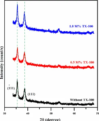
The effects of non-ionic TX-100 surfactant concentration on the structural properties such as crystallite size and preferred orientation of nanostructured CuO films were investigated by XRD. Figure 3 indicates the XRD diffractograms of CuO nanoparticles as a function of TX-100 concentration in the synthesis solutions. All these prepared CuO films with and without TX-100 are polycrystalline monoclinic crystal structures (JCPDS Card No. 05-0640). XRD patterns in Figure 3 demonstrated dominant preferential orientation in , planes. Adding of TX-100 to the growth solution (0.5 M %,) caused a decrement in the (1 11) and (111) peak densities. But then, increment in TX-100 concentration (1.0 M %,) caused a rise in these peak intensities. Parallel results reported by the different researchers [24, 26]. In order to investigate the TX-100 effects on the crystallite size (D) values of the CuO films are calculated from their XRD patterns using the following Scherer equations [33].
𝐷 = 0.94𝜆
𝛽𝑐𝑜𝑠𝜃 (1)
where λ, β and θ are wavelength of the X-ray, full width at half-maximum and Bragg diffraction angle in radian, respectively. The average crystallite size values are listed in Table 1. When the concentration of Triton X in the aqueous solution was 0.5 M%, the crystallite size value decreased from 17.11 nm to 16.57 nm. An increase in TX-100 concentration (1.0 M%) resulted in rise of crystallite size value (19.58 nm). This change in the crystallite sizes might be due to lattice expansion of CuO films when surfactant is added in aqueous solution [22, 34]. This result can also be verified by SEM analysis.
548
Figure 3. X-ray diffractograms of CuO films at different
TX-100 concentrations.
The values of peak intensity of the CuO nanoparticles deposited at various concentrations of TX-100 are shown given in Table 1. As seen in Table 1, the peak intensities of these films change with an increment in TX-100 content.
Tablo 1.Crystallite size, recorded peak intensity and band gap values of the CuO films as a function of TX-100 concentration in the growth solutions of the SILAR process
TX-100 concentration (M %) Crystallite Size (nm) Recorded Peak
Intensity Band gap
(eV) (𝟏 𝟏𝟏) (111) 0 17.11 1210 861 1.45 0.5 16.57 1055 890 1.52 1.0 19.58 1430 845 1.49 3.4. Optical studies
The band gap energy values of the CuO particles were specified by using the below equation [33]:
𝛼ℎ𝜐 = 𝐶(ℎ𝜐 − 𝐸𝑔)𝑚 (2)
where Eg is the band gap energy, C is an energy independent constant, hυ is the photon energy, α is the optical absorption coefficient and m is an index (m is ½ for direct permitted transitions). To analyze the influence of TX-100 as surfactant on the optical band gap graphs of (αhυ)2 versus (hυ) were plotted Figure 4 as a function of TX-100 concentration. By using this graph the Eg values were determined to be 1.45, 1.52 and 1.49 eV for without, 0.5 and 1.0 M% TX-100, respectively.
Figure 4. The optical band gap energy values of CuO films
with different TX-100 concentrations.
M% TX-100 amounts, respectively. Similar results were noticed by Afzal et al. [35]. These values are also displayed in Table1. From the Figure 4 it is seen that the optical band gap energy of the CuO films was increased with a TX-100 adding (0.5 M%) and then the energy value diminished with increasing TX-100 amount (1.0 M%). This alteration in band gap energy may be because of change in crystal lattice of copper by adding TX-100 [36,37]. The results demonstrated that the TX-100 as a surfactant may be used as an adjusting of the energy of band gap of a nanostructured semiconductor CuO films. Also these results have good adaptation with XRD data and SEM results.
Figure 5. Comparison of optical transmittance spectra of
the CuO films prepared with different molar concentrations of TX-100.
The spectrum of optical transmittance of the CuO films with and without TX-100 in the wavelength range of 600–1100 nm is displayed in Figure 5. As seen from the figure, the CuO film without TX-100 as
549
a surfactant has the lowest optical transmittance (6%). The optical transmittance increased quickly with augmenting TX-100 concentration then the transmittance value decreased with increasing TX-100 accumulation. The change in optical transmittance can be imputed to altered crystallinity features and crystallite size.
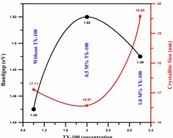
Figure 6. Variation of bandgap and crystallite size of CuO
films synthesized without and with TX-100
The influence of TX-100 surfactant on the band gap and crystallite sizes is illustrated in Figure 6. From Figure 6, it can be seen that the band gap energy increased but the crystallite size decreased with increases TX-100 concentrations. The value of optical band gap of nanostructured films depends heavily on the crystal property. Therefore, the change in the band gap of the metaloxide materials can be clarified by the crystallite size alteration in accordance with the quantum size effects [38, 39]. The results displayed that the TX-100 has a critical significance for the improvement of optical characteristics of CuO films.
4. Discussion and Conclusion
In summary, CuO films with and without TX-100 as non-ionic surfactants were successfully grown through the SILAR method. We have concentrated on the impression of TX-100 concentration on the physical characteristics of CuO nanoparticles. Changes in the surface morphology of the films, structural parameter like crystallite size and the bandgap and transmittance values were examined. As a consequence of all analyses, it has been deduced that TX-100 supplementation has a major effect on the properties of nanostructured CuO films. Therefore, the nanostructured CuO films would be promising for different device technology like electronic, photovoltaic and optoelectronic.
References
[1] Iqbal, T., Aziz, A., Khan, M.A., Andleeb, S., Mahmood, H., Khan, A. A., Khan, R., Shafique M.
2018. Surfactant assisted synthesis of ZnO nanostructures using atmospheric pressure microplasma electrochemical process with antibacterial applications. Materials Science & Engineering B, 228 (2018), 153–159.
[2] Balmuri, S. R., Selvaraj, U., Kumar, V. V., Anthony, S. P., Tsatsakis, A. M., Golokhvast, K. S., Raman T. 2017. Effect of surfactant in mitigating cadmium oxide nanoparticle toxicity: Implications for mitigating cadmium toxicity in environment. Environmental Research, 152 (2017), 141–149. [3] Hu, J., Li, H., Muhammad, S., Wu, Q., Zhao, Y., Jiao
Q. 2017. Surfactant-assisted hydrothermal synthesis of TiO2/reduced graphene oxide nanocomposites and their photocatalytic performances. Journal of Solid State Chemistry, 253 (2017), 113–120.
[4] Zhang, Q., Zhang, K., Xu, D., Yang, G., Huang, H., Nie, F., Liu, C., Yang, S. 2014. CuO nanostructures: Synthesis, characterization, growth mechanisms, fundamental properties, and applications. Progress in Materials Science, 60 (2014), 208–337.
[5] Yathisha, R. O., Nayaka, Y. A. 2018. Structural, optical and electrical properties of zinc incorporated copper oxide nanoparticles: doping effect of Zn. J Mater Sci, 53(2018), 678– 691.
[6] Gopalakrishnan, N., Balakrishnan, L., Arunkumar, B., Gowrishankar S. 2014. Optimization of CuO Ultra Thin Film for Gas Sensor Application by RF Magnetron Sputtering. J. Nanoelectron. Optoelectron., 9:4 (2014), 1-6. [7] Sharma, J. K., Akhtar, M. S., Ameen, S., Srivastava,
P., Singh, G. 2015. Green synthesis of CuO nanoparticles with leaf extract of Calotropis gigantea and its dye-sensitized solar cells applications. Journal of Alloys and Compounds, 632 (2015), 321–325.
[8] Huang, J., Fu, G., Shi, C., Wang, X., Zhai, M. 2014. Novel porous CuO microrods: synthesis, characterization, and their photocatalysis property. Journal of Physics and Chemistry of Solids, 75 (2014), 1011–1016.
[9] Hameed, M. U., Khan, Y., Ali, S., Wu, Z., Dar, S. U., Song, H., Ahmad, A., Chen, Y. 2017. Tween-80 guided CuO nanostructures: Morphology-dependent performance for lithium ion batteries. Ceramics International, 43 (2017), 741–748.
[10] Sahin, B., Alomari, M., Kaya,T., Hydration Detection through use of artificial sweat in doped- and partially-doped nanostructured CuO films. Ceramics International 41 (2015) 8002– 8007 .
550
[11] Wu, J., Hui, K. S., Hui, K. N., Li, L., Chun, H. H., Cho,Y. R. 2016. Characterization of Sn-doped CuO thin films prepared by a sol–gel method. J Mater Sci: Mater Electron, 27(2016), 1719–1724. [12] Wang, Y., Jiang, T., Meng, D., Wang, D., Yu, M.
2015. Synthesis and enhanced photocatalytic property of feather-like Cd-doped CuO nanostructures by hydrothermal method. Applied Surface Science, 355 (2015), 191– 196.
[13] Jan, T., Iqbal, J., Farooq, U., Gul, A., Abbasi, R., Ahmad, I., Malik, M. 2015. Structural, Raman and optical characteristics of Sn doped CuO nanostructures: A novel anticancer agent. Ceramics International, 41 (2015), 13074– 13079.
[14] Lai, M., Mubeen, S., Chartuprayoon, N., Mulchandani, A., Deshusses, M. A., Myung, N. V. 2010. Synthesis of Sn doped CuO nanotubes from core–shell Cu/SnO2 nanowires by the Kirkendall effect. Nanotechnology, 21 (2010), 295601, 1-5.
[15] Wanjala, K. S., Njoroge, W. K., Makori, N. E., Ngaruiya, J. M. 2016. Optical and Electrical Characterization of CuO Thin Films as Absorber Material for Solar Cell Applications. American Journal of Condensed Matter Physics, 6(1) 2016, 1-6.
[16] Mitzi, D. B. 2009. Solution processing of inorganic materials. 1st Edition. John Wiley & Sons, Inc., Publication, 501p.
[17] Shei, S. C., Lee, P. Y., Chang, S. J. 2012. Effect of temperature on the deposition of ZnO thin films by successive ionic layer adsorption and reaction. Applied Surface Science, 258 (2012), 8109– 8116.
[18] Sahin, B., Physical Properties of Nanostructured CdO Films from Alkaline Baths Containing Saccharin as Additive, The Scientific World Journal (2013) 1-5.
[19] Singh, I., Kaur, G., Bedi, R. K. 2011. CTAB assisted growth and characterization of nanocrystalline CuO films by ultrasonic spray pyrolysis technique. Applied Surface Science, 257 (2011), 9546– 9554.
[20] Siddiqui, H., Qureshi, M. S., Haque, F. Z. 2016. Surfactant assisted wet chemical synthesis of copper oxide (CuO) nanostructures and their spectroscopic analysis. Optik, 127 (2016), 2740–2747.
[21] Hosseini, S. R., Ghasemi, S., Ghasemi, S. A. 2016. Effect of surfactants on electrocatalytic performance of copper nanoparticles for hydrogen evolution reaction. Journal of Molecular Liquids, 222 (2016), 1068–1075.
[22] Muiva, C. M., Juma, A. O., Lepodise, L. M., Maabong, K., Letsholathebe, D. 2017. Surfactant assisted chemical bath deposition based synthesis of 1-D nanostructured CuO thin films from alkaline baths. Materials Science in Semiconductor Processing, 67 (2017), 69–74. [23] Khalili, E., Tabrizi, S. A. H. 2017. ZnO–CdO
nanocomposite: microemulsion synthesis and dye removal ability. J Sol-Gel Sci Technol, 81(2017), 475–482.
[24] Andronic, l. 2013. Investigation of the effect of surfactant on dip-coating TiO2 photocatalyst. Bulletin of the Transilvania University of Braşov Series I: Engineering Sciences, 6:55 No.1(2013), 39-44.
[25] Selvakumar, D., Dharmaraj, N., Kadirvelu, K., Kumar, N. S., Padaki, V. C. 2014. Effect of sintering temperature on structural and optical properties of indium(III) oxide nanoparticles prepared with Triton X-100 by hydrothermal method. Spectrochimica Acta Part A: Molecular and Biomolecular Spectroscopy, 133 (2014), 335–339.
[26] Sanguanruang, S., Leotphayakkarat, R., Fangern, N., Koonsaeng, N., Chawengkijwanich, C. 2011. Preparation and Characterization of Thin Films TiO2 Prepared by Various Amount of Triton X-100 Surfactant for Photodegradation of a Dye Pollutant. Advanced Materials Research Vols. 233-235 (2011), 2863-2870.
[27] Hajra, P., Shyamal, S., Bera, A., Mandal, H., Sariket, D., Kundu, M., Pande, S., Bhattacharya, C. 2015. Optimization of Triton-X 100 surfactant in the development of Bismuth Oxide thin film semiconductor for improved photoelectrochemical water oxidation behavior. Electrochimica Acta, 185 (2015), 229–235.
[28] Aydin, R., Şahin, B. 2017. The role of Triton X-100 as a surfactant on the CdO nanostructures grown by the SILAR method. Journal of Alloys and Compounds, 705 (2017), 9-13.
[29] Novikova, A. A., Moiseeva, D. Y., Karyukov, E. V., Kalinichenko, A. A. 2016. Facile prepation photocatalytically active CuO plate-like nanoparticles from brochantite. Materials Letters, 167 (2016), 165-169.
[30] Zhang, Q., Zhang, K., Xu, D., Yang, G., Huang, H., Nie, F., Liu, C., Yang, S. 2014. CuO nanostructures: Synthesis, characterization, growth mechanisms, fundamental properties and applications. Progress in Materials Science, 60 (2014), 208-337.
[31] Saien, J., Asadabadi, S. 2011. Synergistic adsorption Triton X-100 and CTAB surfactants
551
at the toluene + water interface. Fluid Phase Equilibria, 307(2011), 16-23[32] Barry, F. J., Cunnane V. J. 2002. Synergistic effects of organic additives on the discharge, nucleation and growth mechanisms of tin at polycrystalline copper electrodes. Journal of Electroanalytical Chemistry, 537 (2002), 151-163.
[33] Gürbüz, E., Aydin, R., Şahin, B. 2018. A study of influences of transition metal (Mn, Ni) co-doping on the morphological, structural and optical properties of nanostructured CdO films. J Mater Sci: Mater Electron, 29(2018), 1823-1831. [34] Ganesan K. P., Anadhan, N., Dharuman, V. , Sami,
P., Pannerselvam, R., Marimuthu, T. 2017. Electrochemically modified crystal orientation, surface morphology and optical properties using CTAB on Cu2O thin films. Results in Physics, 7(2017), 82-86.
[35] Afzal, M., Naik, P. S., Nadaf, L. I., Shaikh, I. N. 2016. SnO2-Surfactant Composite Films for Superior Gas Sensitivity. SSRG International
Journal of Applied Physics (SSRG-IJAP), 3:5 (2016), 1-5.
[36] Farahmandjou, M. 2010. Effect of LABS and Triton X-100 surfactants on the particle size of nanocrystalline ITO powder. Optoelectronics And Advanced Materials – Rapid Communications, 4:7(2010), 986-988
[37] Suwanchawalit, C., Buddee, S., Wongnawa, S. 2017. Triton X-100 induced cuboid-like BiVO4 microsphere with high photocatalytic performance. Journal Of Environmental Sciences, 55 (2017) 257 – 265
[38] Gupta, R. K., Serbetci, Z., Yakuphanoglu, F. 2012. Bandgap variation in size controlled nanostructured Li–Ni co-doped CdO thin films. Journal of Alloys and Compounds, 515 (2012), 96–100.
[39] Marotti, R.E., Giorgi, P., Machado, G., Dalchiele, E.A. 2006. Crystallite size dependence of band gap energy for electrodeposited ZnO grown at different temperatures, Solar Energy Materials & Solar Cells, 90 (2006), 2356–2361.