Hakan SABUNCUO⁄LU Selçuk ÖZDO⁄AN Erdener T‹MURKAYNAK
Ufuk University School of Medicine, Departmert of Neurosurgery, Ankara, Turkey
Received : 04.10.2008 Accepted : 01.11.2008
Correspondence address: Hakan SABUNCUOĞLU Koza Sokak No:72/36 İkizler Sitesi 06700 Gaziosmanpaşa/Ankara-Turkey Phone : 0.312.2044181
E-mail : [email protected]
Spontaneous Regression of
Extruded Lumbar Disc
Herniation: Report of Two
Illustrative Case and Review
of the Literature
Spontan Regrese Olan Ekstrude
Lomber Disk Hernisi: 2 Olgu
Sunumu ve Literatürün Gözden
Geçirilmesi
ABSTRACT
Although the first reported surgery for lumbar disc herniation was published many years ago, there still remains little agreement for the most effective treatment protocol for symptomatic cases. Many patients with extruded lumbar disc herniation require surgical intervention due to radiculopathy of lower extremities but some neurological symptoms of intervertebral disc herniation may frequently improve with conservative treatment. In this paper, two cases of spontaneous regression of extruded lumbar herniated discs are presented. The disc regressions of two patients were correlated with clinical improvement and documented with follow up MRI studies. Additionally the clinical course of lumbar disc herniation was discussed with mechanisms, features of MRI, immunohistological pathology and treatment options of past clinical studies.
KEY WORDS:Spontaneous, Intervertebral, Disc, Herniation, Regression
ÖZ
Lomber disk hernisi cerrahisine ait ilk yayın yıllar önce yayınlanmış olmasına rağmen, semptomatik olguların en etkili tedavisi konusu hala tartışmalıdır. Ekstrude lomber disk hernili bir çok hasta alt ekstremitelere ait radikülopati nedeniyle cerrahiye gereksinim duymaktadır ancak intervertebral disk herniasyonunun birçok nörolojik semptomu da konservatif tedavi ile düzelmektedir. Bu makalede, spontan regresyon gösteren iki ekstrude lomber disk hernisi hastası sunulmaktadır. İki hastaya ait disk regresyonları klinik iyileşme ile koreledir ve MRG çalışmaları ile dökümante edilmiştir. Ek olarak lomber disk herniasyonu kliniği; geçmiş klinik çalışmaların mekanizmaları, MRG özellikleri, immmünohistolojik patoloji ve tedavi seçenekleri ile tartışılmıştır.
ANAHTAR SÖZCÜKLER: Spontan, Intervertebral, Disk, Herniasyon, Regresyon
INTRODUCTION
Although the first reported surgery for lumbar disc herniation was done more than 60 years ago, the exact treatment method still remains doubtful. Some neurological symptoms due to intervertebral disc herniation may frequently improve without surgical intervention (2,3,4,8,13,15,17,19,21) and over a period of time, the size of the disc can be reduced by itself or via nonoperative methods. Many reports have described spontaneous regression of intervertebral disc herniation and conservative treatment is not a rare option for this problem (10,14,20,23).
In this paper, two patients who experienced large extruded lumbar disc herniations are presented. Each of the herniations regressed spontaneously with nonoperative treatment, as demonstrated by magnetic resonance imaging (MRI), with corresponding remission symptoms. The clinical course of lumbar disc disease and radiographic studies of the patients were discussed with mechanisms, features of MRI, immunohistological pathology and treatment options of past clinical studies.
CASE REPORT 1
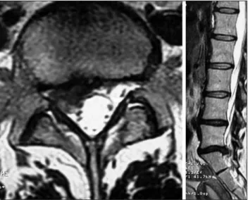
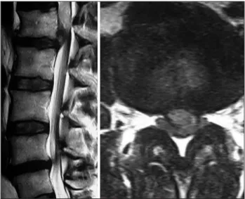
A 33-year-old woman with a 7-months history of low back and right leg pain was admitted to our department on October 2007. The patient's history was notable for intermittent low back pain over the previous 3 years. Straight leg raising test was positive on the right side and the neurological exam was normal except for a hypoactive right achilles reflex. The patient was able to ambulate normally but reqired over the counter analgesics for pain control. MRI of the lumbar spine was obtained after her admission to our department. The large extruded disc fragment was found on the right side of spinal canal at the L5-S1 level on T2-weighted MRI and the dural sac was seen to be compressed by the fragment (Figure 1). After being offered operative treatment, the patient preferred to have conservative treatment that included physical and medical therapy with bed rest. Over the next several weeks, the patient’s complaints were resolved and normal achilles reflex was found in next neurological exam. After visiting our outpatient clinic for follow up, MRI of the lumbar spine was performed on June 2008 and total regression of the extruded disc fragment was found without
compression of the dural sac and S1 root on T2-weighted MRI (Figure 2). The height of L5-S1 disc space was not reduced significantly compared to other levels and was unchanged from the previous lumbar spine MRI examination.
CASE REPORT 2
A 44-year-old man with a 6-month history of untreated left thigh and low back pain was admitted to our hospital in April 2007. He also complained of bilateral inguinal pain which spreaded to the scrotum. The neurological exam was normal except decreased sensation of touch and pain in the left ilioinguinal area. T2-weighted MRI of the lumbar spine was performed and a large extruded and Figure 1: Axial (left) and sagittal (right) T2-weighted MRI of the lumbar spine images show a large extruded disc fragment on the right side at the L5-S1 level (October 2007).
Figure 2: Lumbar disc herniation is seen to be resorbed on follow up axial (left) and sagittal (right) T2 weighted MRI of the lumbar spine at the L5-S1 level (June 2008).
upmigrated disc fragment was found at the L1-2 level. The dural sac was compressed and displaced by the fragment (Figure 3). Surgical intervention was offered to the patient but he refused. Then he was referred to the physical therapy and rehabilitation department for conservative treatment. The patient came to our outpatient clinic for folow-up neurological examination on June 2008 without any complaint. On follow up MRI of the lumbar spine, the totally regressed extruded disc fragment was found at the L1-2 level and the dural sac was free of compression (Figure 4).
DISCUSSION
The reported 2 patients’ case histories demonstrate 2 sequential, independent and spontaneously resolving lumbar disc herniations. As per the patients’ choice, each event had nonoperative treatment and as shown on MRI, each demonstrated full remission in both the abatement of radicular symptoms and the radiologic resolution of the intervertebral disc herniations. It has been shown that extruded disc herniations have great potential for regression (2,3,4).
Mechanisms of Spontaneous Disc Regression
The literature contains several reports of the spontaneous regression or disappearance of herniated intervertebral discs without surgical management. Regression of herniated discs has been described at different levels and with various clinical presentations, including lumbar radiculopathy (7,8,13,21). The exact mechanism of spontaneous disc regression remains unknown. Three popular mechanisms are present in the literature. The first hypothesis states that the herniated disc retracts back into the intervertebral space and protrudes through the annulus fibrosus but is not separated from it. The second hypothesis states that the disc regression is due to gradual dehydration and shrinkage. The last hypothesis takes into account enzymatic degradation and phagocytosis of cartilaginous tissue due to inflammatory reaction and neovascularization of disc herniation (9,13,21). It seems that vascular supply plays a major role in resorption of the disc material (2,3). The vascular mechanism of the resorption is the local reaction around the disc fragments, proliferation of the blood vessels and migration of the phagocytosis of the disc material. On the other hand, production of matrix proteinases and increased cytokine levels play a role in the spontaneous regression process (10, 20).
Features of Magnetic Resonance Imaging
Magnetic resonance imaging (MRI) is a current technique to document the regression of herniated intervertebral disc fragments. Several studies with lumbar spine MRI have demonstrated that the largest lumbar disc herniations are most likely to show the greatest regression in size over time (2,16,17). Rim enhancement on MR images has been reported to be related to the accumulation of contrast material within the vascularized granulation tissue surrounding the avascular sequestrated disc Figure 3: Axial (left) and sagittal (right) T2-weighted MRI of
the lumbar spine show a large extruded and up migrated disc fragment at the L1-2 level. The dural sac is compressed and displaced by the fragment (April 2007).
Figure 4: On follow-up T2-weighted MRI of the lumbar spine, the totally regressed extruded disc fragment is seen at the L1-2 level and the dural sac is free from compression (June 2008).
(14,20,23). According to the current studies of extradural lumbar disc herniation, a herniated mass that shows rim enhancement on Gd-enhanced MR images disappears or markedly decreases in 75-100% of cases (14,20,23). Furthermore, when rim enhancement is evident on Gd-enhanced MR images, there is possibility of spontaneous regression even though the herniated mass may be within the intradural space (20).
Immunohistological Pathology
Intervertebral disc herniations act as foreign bodies in the epidural space. Several immunohistological studies have demonstrated that the immune system attempts to remove the invading disc tissue (6,11,12,17). Hirabayashi et al reported that vessels from the epidural fat infiltrate the disc material. Accompanying the vessels are granulation tissue and the eventual transformation into scar tissue (11,17). Doita et al demonstrated that significant levels of endothelial growth factor in the extruded discs, as compared with smaller protruded discs, presumably enhance the proliferation of endothelial cells (6,17). In an animal study by Haro et al, knockout mice were used to demonstrate that macrophage induction of a chondrocyte enzyme (matrix metalloproteinase-3) plays a key role in disc resorption though several mechanisms (17). These studies have showed that macrophages play a vital role in both resorption and cytokine signaling to promote endothelial cell proliferation and neovascularization.
Clinical Studies
Among the patients choosing nonoperative treatment, some may eventually require operative intervention for unremitting or progressive symptoms. The Weber study estimated this number to be as high as 25% after 1 year (17,22). In the larger Maine Lumbar Spine Study, it was shown that 15% of patients initially treated nonoperatively crossed over and underwent surgical intervention for the treatment for their intervertebral disc herniations by 3 months (1,17). As in prior studies, Buttermann found that larger presenting disc herniations (especially sequestered and extruded types) demonstrated a greater percentage of decrease in size (5). In his study, he found that patients who were successfully treated nonoperatively and whose pain decreased significantly within the first 6 weeks had a larger number of resorbed extruded and sequestered disc herniations on follow up MRI.
Patients who improved during the first 6 weeks may experience more rapid regression of their disc herniation with concomitant decrease in nerve root inflammation and pain (5).
In a retrospective cohort study, Saal and Saal demonstrated that lumbar disc herniation with radiculopathy can be successfully treated with nonoperative procedures resulting in “good to excellent” outcomes for approximately 90% of patients (17,18). In another study, Takada and Takahashi reported that the time taken for spontaneous regression of the herniated mass by >50 % varied from 3 to 12 months (3).
CONCLUSION
In our two cases, the regression of herniated discs may be due to the high water content of larger fragments and their dehydration/shrinkage and inflammation assisted resorption. Although the clinical improvement of our patients was demonstrated with radiological regression, it sometime does not correlate with morphological or radiological changes in different patients. This discrepancy may be explained by the progressive reduction of pressure of the herniated fragments on neighboring neural structures and the increasing of inflammatory response that accompanies the herniation.
Medical management may reduce the clinical symptoms, particulary pain, and radiological intervention may provide morphological regression of extruded disc fragments. These case reports confirm earlier findings demonstrating that a large lumbar disc herniations can be managed successfully by nonoperative methods. Although the successful management of these patients by nonoperative treatment has been demonstrated, many aspects of the disease process remain unknown. Finally, conservative treatment may be the best option when radiculopathy is acceptable and cauda equina syndrome is absent.
REFERENCES
1. Atlas SJ, Keller RB, Chang Y, Deyo RA, Singer DE: Surgical and nonsurgical management of sciatica secondary to a lumbar disc herniation: five-year outcomes from the Maine Lumbar Spine Study. Spine 26(10):1179-1187, 2001
2. Bozzao A, Gallucci M, Masciocchi C, Aprile I, Barile A, Passariello R: Lumbar disk herniation: MR imaging assessment of natural history in patients treated without surgery. Radiology 185(1):135-141, 1992
3. Borota L, Jonasson P, Agolli A: Spontaneous resorption of intradural lumbar disc fragments. Spine J (2):397-403, 2008
4. Bush K, Cowan N, Katz DE, Gishen P: The natural history of sciatica associated with disc pathology. A prospective study with clinical and independent radiologic follow-up. Spine 17(10):1205-1212, 1992
5. Buttermann GR: Lumbar disc herniation regression after successful epidural steroid injection. J Spinal Disord Tech 15(6):469-476, 2002
6. Doita M, Kanatani T, Harada T, Mizuno K: Immunohistologic study of the ruptured intervertebral disc of the lumbar spine. Spine 21(2):235-241, 1996
7. Ellenberg M, Reina N, Ross M, Chodoroff G, Honet JC, Gross N: Regression of herniated nucleus pulposus: two patients with lumbar radiculopathy. Arch Phys Med Rehabil 70(12):842-844, 1989
8. Fager CA: Observations on spontaneous recovery from intervertebral disc herniation. Surg Neurol 42(4):282-286, 1994 (1-9)
9. Guinto FC Jr, Hashim H, Stumer M: CT demonstration of disk regression after conservative therapy. AJNR Am J Neuroradiol 5(5):632-633, 1984
10. Haro H, Shinomiya K, Komori H, Okawa A, Saito I, Miyasaka N, Furuya K: Upregulated expression of chemokines in herniated nucleus pulposus resorption. Spine (21)14:1647-1652, 1996
11. Hirabayashi S, Kumano K, Tsuiki T, Eguchi M, Ikeda S: A dorsally displaced free fragment of lumbar disc herniation and its interesting histologic findings. A case report. Spine 15(11):1231-1233, 1990
12. Ito T, Yamada M, Ikuta F, Fukuda T, Hoshi SI, Kawaji Y, Uchiyama S, Homma T, Takahashi HE: Histologic evidence of absorption of sequestration-type herniated disc. Spine 21(2):230-234, 1996
13. Komori H, Shinomiya K, Nakai O, Yamaura I, Takeda S, Furuya K: The natural history of herniated nucleus pulposus with radiculopathy. Spine 15;21(2):225-229, 1996
14. Komori H, Okawa A, Haro H, Muneta T, Yamamoto H, Shinomiya K: Contrast-enhanced magnetic resonance imaging in conservative management of lumbar disc herniation. Spine 23(1):67-73, 1998
15. Maigne JY, Deligne L: Computed tomographic follow-up study of 21 cases of nonoperatively treated cervical intervertebral soft disc herniation. Spine 19(2):189-191, 1994 16. Maigne JY, Rime B, Deligne B: Computed tomographic
follow-up study of forty-eight cases of nonoperatively treated lumbar intervertebral disc herniation. Spine 17(9):1071-1074, 1992 17. Reyentovich A, Abdu WA: Multiple independent, sequential,
and spontaneously resolving lumbar intervertebral disc herniations: A case report. Spine 27(5):549-553, 2002
18. Saal JA, Saal JS: Nonoperative treatment of herniated lumbar intervertebral disc with radiculopathy. An outcome study. Spine 14(4):431-417, 1989
19. Saal JA, Saal JS, Herzog RJ: The natural history of lumbar intervertebral disc extrusions treated nonoperatively. Spine 15(7):668-683, 1990
20. Sakai T, Tsuji T, Asazuma T, Yato Y, Matsubara O, Nemoto K: Spontaneous resorption in recurrent intradural lumbar disc herniation: Case report. J Neurosurg Spine 6(6):574-578, 2007 21. Slavin KV, Raja A, Thornton J, Wagner FC Jr: Spontaneous regression of a large lumbar disc herniation: Report of an illustrative case.Surg Neurol 56(5):333-336, 2001
22. Weber H: Lumbar disc herniation. A controlled, prospective study with ten years of observation. Spine 8(2):131-140, 1983 23. Yamashita K, Hiroshima K, Kurata A:
Gadolinium-DTPA--enhanced magnetic resonance imaging of a sequestered lumbar intervertebral disc and its correlation with pathologic findings. Spine 15;19(4):479-482, 1994