http://journals.tubitak.gov.tr/medical/ © TÜBİTAK
doi:10.3906/sag-1407-67
A comparison of hair and serum trace elements in patients with Alzheimer disease and
healthy participants
Emine Rabia KOÇ1,*, Atilla İLHAN2, Zübeyde AYTÜRK3, Burcu ACAR3, Mukaddes GÜRLER4, Aynur ALTUNTAŞ5, Mustafa KARAPİRLİ5, Abdurrahman Said BODUR6
1Department of Neurology, Faculty of Medicine, Balıkesir University, Balıkesir, Turkey 2Department of Neurology, Faculty of Medicine, Gazi University, Ankara, Turkey 3Department of Neurology, Faculty of Medicine, Turgut Özal University, Ankara, Turkey 4Department of Medical Biochemistry, Faculty of Medicine, Hacettepe University, Ankara, Turkey
5Department of Chemistry, Ankara Branch of the Council of Forensic Medicine, Ankara, Turkey 6Department of Public Health and Medical Statistics, Faculty of Medicine, Balıkesir University, Balıkesir, Turkey
1. Introduction
Trace elements play an important role in numerous metabolic processes, and they are known to be essential for the activity of some enzymes. Their homeostasis is managed tightly by cells, and any disturbance leads to severe consequences. Alzheimer disease (AD) is a multifactorial disease in which several genetic and environmental factors have been implicated. Amyloid-β (Aβ) is a major constituent of senile plaques and one of the principal hallmarks of AD. Increased production and aggregation of the Aβ peptide is associated with disease pathology. Recent studies have emphasized not only that the etiology of AD involves Aβ aggregation, but also that metal ions contribute to this process by activating or inhibiting enzymatic reactions, by increasing reactive oxygen species (ROS), by competing with other elements, and by affecting the permeability of cell membranes (1,2).
An imbalance of trace elements in the human body might be a possible indicative factor for AD. Therefore, it is important to detect metal dyshomeostasis that occurs with AD, as it could offer possibilities for effective and novel therapeutic approaches. Although many authors have studied the relationship between some trace elements and AD, there are conflicting reports in the literature on this issue. Some researchers have suggested that low trace element levels contribute to the pathogenesis of AD, while others have proposed that elevated element levels are responsible for this process (2–5). However, all of these studies emphasized that patients with AD might show some abnormalities in serum and brain trace element concentrations (1,6–8). It has been proposed that scalp hair should be used to determine systemic levels of elements, rather than other types of samples. Trace element levels tend to be higher in hair than in sera, and
Background/aim: To determine whether there was a difference between serum and hair trace elements’ concentrations in patients with
Alzheimer disease (AD) and healthy participants.
Materials and methods: Hair and serum copper, selenium, zinc, magnesium, manganese, and iron levels were measured by inductively
coupled plasma-mass spectrometry in patients with AD and healthy participants, and the obtained results were statistically compared.
Results: The mean hair selenium and zinc levels of patients with AD were significantly lower than the levels found for control
participants (P < 0.05). Patients with AD had significantly higher mean hair copper and manganese levels than the controls. There were no significant differences between AD patients and controls with respect to the hair iron and magnesium levels (P > 0.05). Hair and serum trace element (copper, selenium, zinc, magnesium, manganese, and iron) levels in patients with AD showed no significant difference according to mini mental test scores or sex (P > 0.05).
Conclusion: Some trace element levels may change in patients with AD. Due to the more permanent status, the analysis of these element
levels in hair might be superior to blood analysis.
Key words: Alzheimer disease, trace element, hair, serum
Received: 15.07.2014 Accepted/Published Online: 26.01.2015 Printed: 30.10.2015 Research Article
serum element levels can vary widely within a given day, depending on food intake. Stable tissues (e.g., hair or nail) should be used to obtain permanent results due to their slow metabolic turnover rates. Based on these facts, we studied trace element levels in hair of AD patients and compared them with those of controls.
2. Materials and methods
The present study was designed as a hospital-based case-control study. It was approved by the Ethics Committee of the Faculty of Medicine of Turgut Özal University.
2.1. Participants
A total of 45 patients diagnosed with AD (22 women and 23 men; average age of 77.66 ± 9.28 years) and 33 healthy participants (17 women and 16 men; average age of 73.18 ± 10.61 years) were examined. The AD patients met the diagnostic criteria of probable AD, according to the established criteria (National Institute of Neurological and Communicative Disorders and Stroke and the Alzheimer’s Disease and Related Disorders Association (NINDS-ADRDA) and Diagnostic and Statistical Manual of Mental Disorders, 4th edition) (9,10). The Turkish version of the Mini Mental State Examination (MMSE) was used to screen for cognitive impairment (11). Since geographic and environmental conditions may affect the hair and blood levels of trace elements, patients had to have lived in Ankara for at least 1 year. Hair samples were obtained from people who did not have colored or treated hair. Patients who had diabetes, kidney failure, inflammatory disease, or malignancy were excluded. Additionally, those who were receiving polytherapy were excluded. The exclusion criteria for the patients were also applied to the controls. Informed consent was obtained from all patients and healthy participants. For those patients who were incapable of providing informed consent, it was obtained from relatives.
2.2. Blood samples
Blood samples were taken in the morning (0930 hours) from the patients and controls after approximately 12 h of fasting (before breakfast).Venous blood (10 mL) was taken from the patients and controls without using anticoagulant, and samples were centrifuged at 3000 × g for 15 min at room temperature. Serum samples were stored at –80 °C for the measurement of trace elements.
2.3. Hair samples
Hair specimens were taken on the same day as blood sampling. Eighty-eight hair samples, weighing about 2 g, were taken from the nape of the head as near as possible to the scalp with Teflon scissors and stored in polyethylene bags. The scissors were cleaned after each sampling.
2.4. Elemental analysis by ICP/MS
The hair samples were prepared according to the modified method developed by the International Atomic Energy Agency (12). The hair specimen was cut and washed in deionized water. The samples were then rinsed with acetone and allowed to dry in an oven at 40–70 °C. After drying, the hair samples were digested, weighing each sample (25–200 mg) in a microwave (Speedwave, Berghof) in digestion Teflon bottles with 5 mL of nitric acid. Serum samples (0.5–1.5 mL) were also digested with 5 mL of nitric acid in the same way. The microwave parameters are given in Table 1. After digestion, the samples were diluted with deionized water containing 1% nitric acid. The final volume was 10–50 mL and 10–30 mL for hair and serum samples, respectively. The resulting product was a clear liquid with a yellow tint. A PerkinElmer Elan 9000 ICP-MS (Waltham, MA, USA ) was used to analyze the samples in a private laboratory.
The performance of the instrument was checked daily with Nexion Setup Solution 1% nitric acid (PerkinElmer Pure Plus). The tuning and response factor criteria were >20,000 cps for Mg24, >50,000 cps for In115, >40,000 cps for U238, background 220 < 1 cps, CeO/Ce 156/140 < 2.5%, and Ce++/Ce+ 70/140 < 3%. All these parameters were obtained before analyzing the samples.
High purity grade reagents were used. For the preparation of calibrators and samples, 18 M cm–1
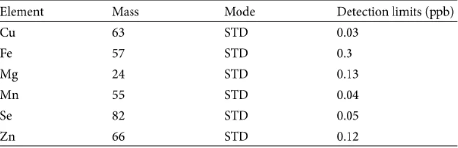
Milli-QTM DI water (Water Purification System, Human Power I) and nitric acid (Suprapur 65%, Merck) were used. Multielement aqueous certified reference material solutions were used for the calibration curve with the standards of 1, 5, 20, 50, 80, and 100 ng/mL for zinc (Zn), copper (Cu), iron (Fe), selenium (Se), and manganese (Mn) and 50, 200, 500, and 800 ng/mL for magnesium (Mg). The correlation coefficient from the initial calibration was ≥0.9988 for each element. A method blank, reagent water acidified with the same acid concentrations present in the standards and samples, was used to determine the concentrations of elements in the reagents used to prepare and analyze the samples as well as the contribution of contamination from the digestion process. The detection limits were calculated from blank standard deviations (Table 1). Indium, germanium, and scandium were used as internal standards. Results are reported as µg/g or µg/ mL for elements in hair and serum samples.
2.5. Statistical analysis
Data were analyzed using SPSS for Windows. The data were first tested using the Shapiro–Wilk test for their distribution. Normally distributed data were analyzed using the Student t-test and data were expressed as mean ± SD. The Mann–Whitney U test was used for data found not to be normally distributed. The results were given as median (min–max) values. A P-value of less than 0.05 was regarded as statistically significant.
3. Results
The results are shown in Table 2. Between groups, there were no differences with respect to the serum levels of Zn, Se, Cu, Fe, and Mg (P > 0.05), while Mn was significantly different (P < 0.05).
The mean hair Se and Zn levels of patients with AD were significantly lower than the levels found for control participants (P < 0.05). Patients with AD had mean hair Cu and Mn levels significantly higher than those found in the controls. There were no significant differences between
Table 1. Detection limits of elements.
Element Mass Mode Detection limits (ppb)
Cu 63 STD 0.03 Fe 57 STD 0.3 Mg 24 STD 0.13 Mn 55 STD 0.04 Se 82 STD 0.05 Zn 66 STD 0.12
Table 2. Hair and serum trace element levels in patients with AD and controls.
Groups N Mean ± SD P
Age (years) Patients 41 77.69 ± 9.29 0.055*
Control 33 73.18 ± 10.61 Cu-hair (µg/g) Patients 41 12.8 ± 4.8 0.013* Control 31 10.3 ± 2.4 Cu-serum (µg/mL) Patients 44 0.9 (0.4–1.3) 0.1** Control 32 1.01 (0.5–1.5) Se-hair (µg/g) Patients 37 0.5 ± 0.1 0.005* Control 31 0.6 ± 0.1 Se-serum (µg/mL) Patients 40 0.75 ± 0.2 0.3* Control 33 0.83 ± 0.4 Zn-hair (µg/g) Patients 41 75 ± 29 0.02* Control 33 98 ± 54 Zn-serum(µg/mL) Patients 44 0.47 ± 0.1 0.4* Control 33 0.52 ± 0.3 Mn-hair (µg/g) Patients 41 0.6 ± 0.3 0.009* Control 33 0.4 ± 0.2 Mn-serum (µg/mL) Patients 44 0.009 (0–0.03) 0.002** Control 33 0.01 (0.01–0.03) Fe-hair(µg/g) Patients 41 24.6 (8.27–121.5) 0.8** Control 33 24.3 (11.4–250) Fe-serum (µg/mL) Patients 44 1.43 (0.5–7) 0.2** Control 33 1.5 (0.6–8.3) Mg-hair (µg/g) Patients 41 39 (12–238) 0.5** Control 33 49 (10–640) Mg-serum (µg/mL) Patients 44 17.3 (6.3–23) 0.6** Control 33 16.01 (11–56)
*: P-values that resulted from independent sample t-test (mean ± SD)
AD patients and control participants with respect to the hair Fe and Mg levels (P > 0.05). Hair and serum trace element (Cu, Se, Zn, Mg, Mn, and Fe) levels in patients with AD showed no significant difference according to MMSE scores or sex (P > 0.05).
4. Discussion
In this study, we compared hair and serum Cu, Se, Zn, Mg, Mn, and Fe levels of patients with AD and healthy participants in order to investigate the relationships between trace elements and AD. Hair and nail tissues, which have slow metabolic turnover rates, are an important source of knowledge regarding the accumulation of elements over a long period, and they are particularly necessary for retrospective research (13). Serum is not suitable for determining the levels of some trace elements, as serum element levels vary widely within a given day, depending on food intake (14). Moreover, hair is easily sampled and it can be preserved indefinitely under appropriate conditions without deteriorating. Therefore, compared to other types of clinical specimens, hair samples are more valuable than blood, serum, urine, or other samples. In the current study, there were no differences between the two groups in terms of serum trace element levels, with the exception of Mn. However, hair trace element levels differed somewhat between the two groups.
AD is a multifactorial disorder; metal dyshomeostasis and oxidative stress contribute to the pathogenesis of neurodegenerative process (15,16). Abnormal aggregations of different metals elicit oxidative stress and result in neuronal damage (17,18). Se, an essential mineral, plays a major role in cellular redox status. Selenoproteins constitute one of the most important classes of antioxidant enzymes, the glutathione peroxidases (GPx). GPx requires Se for its catalytic activity (19,20). Previous reports on serum or cerebrospinal fluid (CSF) Se concentrations in AD patients have produced controversial results. Loef et al. found negative correlations between disease progression and Se levels (21), while Meseguer et al. found no differences between AD patient and control groups in terms of CSF and serum Se levels (22). In our study, hair Se levels were found to be significantly lower in patients with AD. The reduced Se concentrations might cause decreased GPx activity and support the possible role of oxidative stress, resulting in neurodegeneration, in the pathogenesis of AD.
Zinc plays an important role in cellular metabolism, such as proliferation, differentiation, and apoptosis. It is also necessary for axonal transmission and synaptic signaling pathways. Zinc is an antioxidant element and protects tissues against oxidative stress (23,24). Conflicting results were also found in studies evaluating serum Zn levels in individuals with and without AD. For example,
Haines et al. reported no significant differences in Zn levels between patients with AD and controls (25), while previous brain tissue studies showed significantly lower Zn levels in patients with AD compared with controls (26). Similarly, postmortem studies of hippocampus tissue have shown decreased levels of Zn and Se in participants with AD compared with controls (27). On the other hand, animal studies have shown that oral intake of Zn did not accelerate AD pathology in transgenic and nontransgenic mice (28,29). In the present study, we found no significant difference between the serum Zn levels of the two groups, but the AD group displayed lower mean hair serum Zn levels. Decreased hair Zn levels in AD patients might be responsible for decreased antioxidant capacity and cognitive decline.
Cu is a component of several enzymes and proteins, including superoxide dismutase (SOD), cytochrome c oxidase, and ceruloplasmin. It is also a cofactor of Cu/ Zn-SOD, which plays an important role in the antioxidant defense system. Cu deficiency decreases this enzyme’s activity (2). In our study, mean hair Cu levels were found to be significantly higher in patients with AD compared with healthy controls. Increased Cu concentration might also be responsible for the AD etiology. Amyloid plaques, a hallmark of AD, consist of Aβ, which is derived from the amyloid precursor protein, which has binding Cu (8). While Aβ aggregation in the presence of Cu can trigger ROS and consume neuronal glutathione levels, the adult nervous system can also be damaged by Cu deficiency. Several clinical manifestations of Cu deficiency have been described, such as myelopathy, spastic gait, and sensory ataxia, and Cu therapy sometimes improves neurological deficits (2). Cu deficiency may reflect decreased enzyme activity, which can lead to free radical-mediated oxidative damage. Thus, both increased and decreased Cu levels might be responsible for ROS-mediated neural damage in AD patients.
To our knowledge, no prior report on hair Fe levels in patients with AD exists in the literature. Fe is an essential element for maintaining the normal structure and functions of the central nervous system. Neurons and glial cells require Fe for electron transport, for myelination of axons, and as a cofactor for enzymes involved in the synthesis of neurotransmitters (30). Previous studies on serum, plasma, and brain concentrations of Fe in AD patients have reported contradictory results (31–34). The effects of decrease in the bioavailability of Fe in the brain have been shown to affect neurotransmitter production, memory, and cognitive function (35). However, excess Fe accumulation generates cellular damage (36). In our study, there were no differences in serum or hair Fe levels between the groups. This result might be due to insufficient food intake or intestinal malabsorption associated with aging.
Human cells possess enzymatic antioxidant defenses to cope with oxygen free radicals. One example is mitochondrial manganese superoxide dismutase (Mn-SOD), which catalyzes the dismutation of superoxide anions to hydrogen peroxide (37). Mn-SOD was localized by immunocytochemistry in the cerebral cortex and hippocampus of patients with AD. In one study, the Mn-SOD scavenger system was found to be associated with the formation of senile plaques (38). Markesbery et al. measured Mn levels in various brain regions in AD patients and aging individuals using instrumental neutron activation analysis. They found that the differences were not statistically significant for all regions, but the highest Mn levels were found in the basal ganglia in both the controls and the AD patients (39). In a previous study, CSF concentrations of V, Mn, Rb, Sb, Cs, and Pb were significantly lower in patients with AD (N = 264; P ≤ 0.007) than in healthy controls (40). In the current study, hair Mn levels were found to be significantly higher in patients with AD than in the control group. Increased Mn levels may be related to the increased Mn-SOD activity and antioxidant demand of AD patients.
Altered concentrations of hair trace elements might be related to the maintenance of the oxidant/antioxidant
balance of the affected tissue. We did not measure antioxidant status (e.g., SOD, catalase, GPx) in the present study. It would be very interesting to evaluate both element concentrations and antioxidant activity in patients with AD in future studies.
In conclusion, our results demonstrate that some trace elements, especially hair levels of Cu, Se, Mn, and Zn, can change in AD patients. These alterations might suggest a potential role of trace elements in the etiopathogenesis of AD. Previous study results have shown variations in serum trace element levels in AD patients, and we found some differences in hair and serum trace element concentrations between patients with AD and healthy participants. These results suggest that hair trace element levels might supply more accurate trace element level information in AD patients. Normalization of these elements may be critical to the development of preventive and therapeutic strategies for AD. Future evaluation will be useful in understanding the exact roles of trace elements in the etiopathogenesis of AD.
Acknowledgment
This work was supported by the Scientific Research Fund of Turgut Özal University under project number 10494.
References
1. Grasso G, Giuffrida ML, Rizzarelli E. Metallostasis and amyloid beta-degrading enzymes. Metallomics 2012; 4: 937–949. 2. Klevay LM. Alzheimer’s disease as copper deficiency. Med
Hypotheses 2008; 70: 802–807.
3. Brewer GJ. Copper toxicity in Alzheimer’s disease: cognitive loss from ingestion of inorganic copper. J Trace Elem Med Biol 2012; 26: 89–92.
4. Mutter J, Naumann J, Schneider R, Walach H. Mercury and Alzheimer’s disease. Fortschr Neurol Psychiatr 2007; 75: 528– 538.
5. Hung LW, Barnham KJ. Modulating metals as a therapeutic strategy for Alzheimer’s disease. Future Med Chem 2012; 4: 955–969.
6. Bush AI. The metal theory of Alzheimer’s disease. J Alzheimers Dis 2013; 33: 277–281.
7. Craddock TJ, Tuszynski JA, Chopra D, Casey N, Goldstein LE, Hameroff SR, Tanzi RE. The zinc dyshomeostasis hypothesis of Alzheimer’s disease. PLoS One 2012; 7: e33552.
8. Faller P. Copper in Alzheimer disease: too much, too little, or misplaced? Free Radic Biol Med 2012; 52: 747–748.
9. McKhann G, Drachman D, Folstein M, Katzman R, Price D, Stadlan EM. Clinical diagnosis of Alzheimer’s disease: report of the NINCDS-ADRDA Work Group under the auspices of Department of Health and Human Services Task Force on Alzheimer’s Disease. Neurology 1984; 34: 939–944.
10. Trull TJ, Verges A, Wood PK, Jahng S, Sher KJ. The structure of Diagnostic and Statistical Manual of Mental Disorders (4th edition, text revision) personality disorder symptoms in a large national sample. Personal Disord 2012; 3: 355–369.
11. Güngen C, Ertan T, Eker E, Yaşar R, Engin F. Reliability and validity of the standardized Mini Mental State Examination in the diagnosis of mild dementia in Turkish population. Turk Psikiyatri Derg 2002; 13: 273–281 (in Turkish with English abstract).
12. Puchyr RF, Bass DA, Gajewski R, Calvin M, Marquardt W, Urek K, Druyan ME, Quig D. Preparation of hair for measurement of elements by inductively coupled plasma-mass spectrometry (ICP-MS). Biol Trace Elem Res 1998; 62: 167–182.
13. Akyol O, Ersan F, Akcay F, Altuntas I, Senol M, Sasmaz S. Hair, nail, serum, and urine copper levels in users of copper intrauterine devices and interactions between copper and some other trace elements. Trace Elem Electroly 1997; 14: 124–12 9. 14. Hammer DI, Finklea JF, Hendricks RH, Shy CM, Horton RJ.
Hair trace metal levels and environmental exposure. Am J Epidemiol 1971; 93: 84–92.
15. Bolognin S, Messori L, Zatta P. Metal ion physiopathology in neurodegenerative disorders. Neuromolecular Med 2009; 11: 223–238.
16. Johnson WM, Wilson-Delfosse AL, Mieyal JJ. Dysregulation of glutathione homeostasis in neurodegenerative diseases. Nutrients 2012; 4: 1399–1440.
17. Greenough MA, Camakaris J, Bush AI. Metal dyshomeostasis and oxidative stress in Alzheimer’s disease. Neurochem Int 2013; 62: 540–555.
18. Sayre LM, Perry G, Smith MA. Oxidative stress and neurotoxicity. Chem Res Toxicol 2008; 21: 172–188.
19. Weeks BS, Hanna MS, Cooperstein D. Dietary selenium and selenoprotein function. Med Sci Monit 2012; 18: RA127–132. 20. Akman H, Somuncu S, Dikmen G, Ayva Ş, Soyer T, Doğan P,
Çakmak M. Protective effect of selenium on intussusception-induced ischemia/reperfusion intestinal oxidative injury in rats. Turk J Med Sci 2010; 40: 391–397.
21. Loef M, Schrauzer GN, Walach H. Selenium and Alzheimer’s disease: a systematic review. J Alzheimers Dis 2011; 26: 81–104. 22. Meseguer I, Molina JA, Jimenez-Jimenez FJ, Aguilar MV,
Mateos-Vega CJ, Gonzalez-Munoz MJ, de Bustos F, Orti-Pareja M, Zurdo M, Berbel A et al. Cerebrospinal fluid levels of selenium in patients with Alzheimer’s disease. J Neural Transm 1999; 106: 309–315.
23. Lyubartseva G, Lovell MA. A potential role for zinc alterations in the pathogenesis of Alzheimer’s disease. Biofactors 2012; 38: 98–106.
24. Hancock SM, Finkelstein DI, Adlard PA. Glia and zinc in ageing and Alzheimer’s disease: a mechanism for cognitive decline? Front Aging Neurosci 2014; 6: 137.
25. Haines A, Iliffe S, Morgan P, Dormandy T, Wood B. Serum aluminium and zinc and other variables in patients with and without cognitive impairment in the community. Clin Chim Acta 1991; 198: 261–266.
26. Panayi AE, Spyrou NM, Iversen BS, White MA, Part P. Determination of cadmium and zinc in Alzheimer’s brain tissue using inductively coupled plasma mass spectrometry. J Neurol Sci 2002; 195: 1–10.
27. Corrigan FM, Reynolds GP, Ward NI. Hippocampal tin, aluminum and zinc in Alzheimer’s disease. Biometals 1993; 6: 149–154.
28. Akiyama H, Hosokawa M, Kametani F, Kondo H, Chiba M, Fukushima M, Tabira T. Long-term oral intake of aluminium or zinc does not accelerate Alzheimer pathology in AbetaPP and AbetaPP/tau transgenic mice. Neuropathology 2012; 32: 390–397.
29. Becaria A, Lahiri DK, Bondy SC, Chen D, Hamadeh A, Li H, Taylor R, Campbell A. Aluminum and copper in drinking water enhance inflammatory or oxidative events specifically in the brain. J Neuroimmunol 2006; 176: 16–23.
30. Magaki S, Raghavan R, Mueller C, Oberg KC, Vinters HV, Kirsch WM. Iron, copper, and iron regulatory protein 2 in Alzheimer’s disease and related dementias. Neurosci Lett 2007; 418: 72–76.
31. Jomova K, Vondrakova D, Lawson M, Valko M. Metals, oxidative stress and neurodegenerative disorders. Mol Cell Biochem 2010; 345: 91–104.
32. Castellani RJ, Moreira PI, Perry G, Zhu X. The role of iron as a mediator of oxidative stress in Alzheimer disease. Biofactors 2012; 38: 133–138.
33. Rouault TA, Cooperman S. Brain iron metabolism. Semin Pediatr Neurol 2006; 13: 142–148.
34. Greenough MA, Camakaris J, Bush AI. Metal dyshomeostasis and oxidative stress in Alzheimer’s disease. Neurochem Int 2013; 62: 540–555.
35. Youdim MB, Yehuda S. The neurochemical basis of cognitive deficits induced by brain iron deficiency: involvement of dopamine-opiate system. Cell Mol Biol (Noisy-le-grand) 2000; 46: 491–500.
36. González-Domínguez R, García-Barrera T, Gómez-Ariza JL. Homeostasis of metals in the progression of Alzheimer’s disease. Biometals 2014; 27: 539–549.
37. De Leo ME, Borrello S, Passantino M, Palazzotti B, Mordente A, Daniele A, Filippini V, Galeotti T, Masullo C. Oxidative stress and overexpression of manganese superoxide dismutase in patients with Alzheimer’s disease. Neurosci Lett 1998; 250: 173–176.
38. Maeda M, Takagi H, Hattori H, Matsuzaki T. Localization of manganese superoxide dismutase in the cerebral cortex and hippocampus of Alzheimer-type senile dementia. Osaka City Med J 1997; 43: 1–5.
39. Markesbery WR, Ehmann WD, Hossain TI, Alauddin M. Brain manganese concentrations in human aging and Alzheimer’s disease. Neurotoxicology 1984; 5: 49–57.
40. Gerhardsson L, Lundh T, Minthon L, Londos E. Metal concentrations in plasma and cerebrospinal fluid in patients with Alzheimer’s disease. Dement Geriatr Cogn Disord 2008; 25: 508–515.