Background: The increases of free radicals have been pro-posed to be involved in the pathogenesis of sepsis, which leads to multiple-organ dysfunction syndromes. The uses of antioxidants as a complementary tool in the medical care of oxidative stress-related diseases have attracted attention of researchers. Resveratrol (RV) has suggested being anti-oxidant, anti-proliferative, and anti-inflammatory effects in various experimental models and clinical settings.
Aims: This study was undertaken to evaluate the protective effects of RV on oxidative DNA damage induced by sepsis in the liver and kidney tissues of Wistar albino rats.
Study Design: Animal experimentation.
Methods: Four experimental groups consisting of eight ani-mals for each was created using a total of thirty-two male Wistar albino rats. Sham group was given 0.5 mL of sa-line intra-peritoneal (ip) only following laparatomy. Sepsis group was given 0.5 mL saline ip only following the induc-tion of sepsis. RV-treated group was given a dose of 100 mg/kg ip RV in 0.5 mL saline following laparatomy. RV-treated sepsis group was given 100 mg/kg ip RV in 0.5 mL saline following the induction of sepsis. A model of sepsis
was created by cecal ligation and puncture technique. In the liver and kidney tissues, oxidative stress parameters (malo-ndialdehyde (MDA), reduced glutathione (GSH), superox-ide dismutase (SOD), glutathione peroxidase (GPX)) and a proinflammatory cytokine (tumor necrosis factor alpha (TNF-alpha)), were evaluated spectrophotometrically and DNA damage was determined by the alkaline single cell gel electrophoresis (comet assay) technique using formamido-pyrimidine DNA glycosylase protein.
Results: In the RV-treated sepsis group, the levels of MDA and TNF-alpha were lower and GSH levels, SOD and GPX activities were higher than in the septic rats (p<0.05). RV treatment significantly reduced the sepsis-induced oxidative DNA damage in the liver and kidney cells (p<0.05). Conclusion: It is suggested that RV treatment might reduce the sepsis-induced oxidative DNA damages in sepsis-relat-ed diseases; however, there is a nesepsis-relat-ed for more studies to clear up the protective mechanisms of RV against sepsis. Keywords: Resveratrol, sepsis, tumor necrosis factor alpha, oxidative stress, DNA damage, alkaline single cell gel elec-trophoresis
Resveratrol Protects Sepsis-Induced Oxidative DNA Damage
in Liver and Kidney of Rats
1Department of Pharmaceutical Toxicology, Hacettepe University School of Pharmacy, Ankara, Turkey 2Department of Surgery, Kastamonu University School of Medicine, Kastamonu, Turkey
3Department of Bioengineering, Bursa Technical University School of Natural Sciences, Architecture and Engineering, Bursa, Turkey 4Department of Pharmacognosy, Hacettepe University School of Pharmacy, Ankara, Turkey
5Department of Infectious Diseases and Clinical Microbiology, Başkent University School of Medicine, İstanbul Hospital, İstanbul, Turkey
Sevtap Aydın
1, Tevfik Tolga Şahin
2, Merve Bacanlı
1, Gökçe Taner
3, Arif Ahmet Başaran
4, Mehtap Aydın
5,
Nurşen Başaran
1Address for Correspondence: Dr. Sevtap Aydın, Department of Pharmaceutical Toxicology, Faculty of Pharmacy, Hacettepe University, Ankara, Turkey Phone: +90 538 543 76 57 e-mail: [email protected]
Received: 1 June 2015 Accepted: 31 March 2016 • DOI: 10.5152/balkanmedj.2016.15516 Available at www.balkanmedicaljournal.org
Cite this article as:
Sepsis, a serious medical condition, causes mortality and morbidity in intensive care units. It is characterized by the production of proinflammatory cytokines and reactive oxy-gen species (ROS) oxy-generated by macrophages and neutrophils after infection (1). Oxidative stress occurs due to the exces-sive formation of ROS and inadequate antioxidant defense system in sepsis. ROS is a crucial issue in the progression of
sepsis-induced multi-organ damages, which leads to the in-flammatory effects, lipid peroxidation, and DNA damage (2). Antioxidants maintain the redox balance in sepsis. In many studies, it was shown that antioxidant status was reduced in septic patients and antioxidants such as vitamin C, vitamin E, and other natural antioxidants ameliorated the oxidative stress markers changed in sepsis. Antioxidants can also interfere This study was presented at the 13th International Congress of Toxicology, 30 June - 4 July 2013, Seul, Korea.
with inflammatory responses by regulating proinflammatory and inflammatory cytokines and endotoxins, thus modulating cellular activation (1,2).
Resveratrol (3,4′,5-trihydroxystilbene) (RV) is a radical-scav-enger. It ameliorates antioxidant status, reduces the expression of inflammatory biomarkers and prevents or cures cardiovascu-lar diseases and improves microcirculatory disorders (3,4). RV, a natural antioxidant, has been suggested to reduce DNA dam-age and oxidative organ injury (5). Several studies demonstrat-ed the hepatoprotective effects of RV in vivo (6,7). RV (10 mg/ kg body weight (bw)) pretreatment was found to suppress the lipid peroxidation in the kidneys of ferric nitrilotriacetate-treat-ed rats (5). The enzyme Fpg is the major enzyme for removal of oxidized purines in bacteria and has been recommended to determine the oxidative DNA damage, in particular 8-hydroxy-guanine in conjunction with the comet assay (8).
This present study was undertaken to investigate the ame-liorative properties of RV on the oxidative DNA damage in-duced by sepsis in the liver and kidney tissues of Wistar albino rats. Our hypothesis is that RV reduces or prevents the sepsis-induced oxidative stress and DNA damage in the main target tissues. This study may clarify the possible mechanisms of RV on the sepsis-induced oxidative DNA damage in vivo.
MATERIALS AND METHODS Animals
In this study, a total of 32 male Wistar albino rats weigh-ing 200-300 g (3 months old) were housed in plastic cages with stainless-steel grid tops. Before the experiments, they were kept under a controlled environment regarding humidity (50%), temperature (23±2°C), and light (12-hour light-dark cycle). All rats were fed with standard laboratory animal feed and allowed to access drinking water and ad libitum feed be-fore and after operation. All surgical procedures were carried out under anesthesia with intraperitoneal (ip) injection of 90 mg/kg ketamine hydrochloride (Ketalar, Eczacıbaşı, Warner-Lambert, İstanbul, Turkey) (9). The local Animal Ethical Committee (Hacettepe University Animal Ethical Committee) of this study was approved.
Experimental design
A model of sepsis created by cecal ligation and puncture (CLP) technique was applied to rats as described previously (10,11). Four experimental groups consisted of eight animals for each was carried out. Sham group was given 0.5 mL of saline intraperitoneal (ip) only following laparatomy. In the sham group applied laparotomy; the cecum without ligated or perforated was manipulated. Sepsis group, where only CLP
was performed, was given 0.5 mL saline ip following the in-duction of sepsis. RV-treated group was immediately given a dose of 100 mg/kg bw ip RV in 0.5 mL saline following laparatomy. RV-treated sepsis group was immediately given 0.5 mL of a solution containing 100 mg/kg bw ip RV follow-ing the induction of sepsis. The treatment dose of RV was se-lected from the study of Della-Morte et al. (12). At the studied dose, 100% of the animals remained alive. In our previous studies with the phenolic compounds pycnogenol®, ferulic acid, and rosmarinic acid, the dosages of 100 mg/kg bw ip were also used in the experimental sepsis model (13-15). In these studies evaluated independently form the same local ani-mal ethic approval to reduce aniani-mal numbers according to the principles of 3Rs, sham and sepsis groups were common; thus the data from sham and sepsis groups were the same in these studies carried out at the same time (13-16).
All animals were decapitated under the anesthesia, 24 hours following the treatment. Blood samples obtained from cardiac puncture were taken in preservative-free heparin tubes. The tissues of liver and kidney were gently removed as a whole. The liver and kidney tissues were cut into several pieces to perform the structural analysis of the tissues and to determine of antioxidant parameters and DNA damage. The organs were examined for the changes in size, color, and texture, in which there were no changes in the histological structures of the liver and kidney tissues.
Determination of oxidative stress parameters The liver and kidney tissues were extracted following a ho-mogenization and sonication procedure (17). The tissue sam-ples were rinsed with cold phosphate buffered saline (PBS) solution, pH 7.4 to remove any red blood cells and clots. The tissues were homogenized in 5 volumes of fractionation buffer (10 mM HEPES, 10 mM KCl, 1 mM EDTA, 1.5 mM MgCl2, 0.5 mM dithiothreitol, cOmplete Protease Inhibitor Cocktail (Roche Diagnostics; Indianapolis, IN)] using a Potter-Elve-hjem homogenizer. A 10% tissue homogenate was centrifuged at 10000xg for 15 min at 4°C. The homogenized tissue sam-ples were stored at -80°C until the analysis. The supernatant was deproteinized for GSH analysis according to the manu-facturer’s instructions before storing.
The reduced glutathione (GSH) levels, superoxide dis-mutase (SOD) and glutathione peroxide (GPX) enzyme ac-tivities in these tissues were evaluated spectrophotometrically using GSH , SOD, and GPX assay kits (Cayman Cemicals Co.; Ann Arbor, MI, USA) at 440, 340, and 405 nm, respec-tively, according to the manufacturer’s instructions. Results were given as mmol/min/mg tissue.
The level of malondialdehyde (MDA), an indicator of lipid peroxidation, was analyzed by thiobarbituric acid-reactive substances (TBARS) assay (18). The results were given as nmol/g tissue.
Determination of plasma tumor necrosis factor alpha (TNF-alpha) levels
The plasma from whole blood sample was kept at -80°C until the time of analysis. The level of TNF-alpha was measured at 30 min in duplicate at 450 nm. The protocol from the manufac-turer’s instructions using highly sensitive enzyme-linked immu-nosorbent assay (ELISA) kits containing Invitrogen KRC0011 (USA) was performed. The results were given as pg/mL.
Determination of DNA damage
Standard and formamidopyrimidine-DNA glycosylase (Fpg) modified comet assay described by Singh et al. (19), as further described by Anderson et al. (20)and Collins et al. (8) was per-formed. The single-cell suspension from the liver and kidney tissue samples was prepared according to a standard proce-dure (21). Each organ (about 0.2 g) was gently placed in 1 mL cold HBSS containing 20 mM EDTA-Na2 and 10% dimethyl sulfoxide (DMSO) purchased from Sigma-Aldrich Chemicals (Sigma-Aldrich Chemicals; St. Louis, USA) in a petri plate and sliced into small parts using a steel scissor. The pieces were let to settle and the single-cell suspension in the supernatant contain-ing was used. The number of cells was adjusted to 2x10⁶ cells/ mL. The cells were embedded on agarose gel, lysed, and frag-mented DNA strands revealed by the alkaline electrophoresis to create a comet. The details of the alkaline comet assay with and without Fpg protocol was published previously (9). After electrophoresis, the slides were neutralized and then dried. In the dried microscopic slides, DNA damage was investigated at 40x magnification. The microscope was connected to a charge-coupled device camera and a personal computer-based analysis system (Comet Analysis Software, version 3.0, Kinetic Imag-ing Ltd.; Liverpool, UK) to determine DNA damage. Results were presented as DNA tail length, DNA tail intensity (% tail DNA), and DNA tail moment. One hundred cells from each of 2 replicate slides were counted. The difference of DNA damage between the Fpg-modified comet assay and the standard comet assay indicated the oxidative DNA damage in a single cell.
Statistical analysis
Statistical analysis was performed using the software pro-grams Statistical Package for the Social Sciences version 17.0 (SPSS Inc.; Chicago, IL, USA). The Shapiro-Wilk test to check the normality of data distribution and the Levene test to verify the homogeneity of the variance were used. The differences between the means of data were compared by the one way variance analysis test. Dunnet’s T3 test or LSD test was used for the determination of post hoc analysis of group differences related to the variance between groups. The mean±standard deviation of the results was presented. P value less than 0.05 was accepted as statistically significant.
RESULTS
Oxidative stress parameters
Glutathione and MDA levels and SOD and GPX activities for the liver and kidney tissues are shown in Table 1 and 2, respectively. Hepatic and renal GSH levels, SOD and GPX activities decreased significantly in the septic rats versus to the sham operated group (p<0.05). Unlike hepatic and renal GSH levels, hepatic SOD and GPX activities, and renal GPX ac-tivities in the RV-treated sepsis group were significantly lower than in the sham group (p<0.05). Hepatic and renal GSH levels and SOD and GPX enzyme activities significantly increased in the RV-treated sepsis group when compared to the sepsis group (111% and 281% for liver and renal GSH, respectively; 12.2% and 40.3% for liver and renal SOD; 167% and 52.8% for liver and renal GPX) (p<0.05). Hepatic and renal MDA levels significantly increased in the sepsis group when com-pared to the sham group (p<0.05). Hepatic and renal MDA levels significantly decreased in the RV-treated sepsis group when compared to the sepsis group (37.9% and 50.6% for liv-er and renal MDA, respectively) (p<0.05). Hepatic MDA level in the RV-treated sepsis group was significantly higher than in the sham group (p<0.05).There were no significant differences in all hepatic and renal oxidative stress parameters between the sham and the RV-treated groups.
Plasma TNF-alpha levels
The result of plasma TNF-alpha levels is given in Figure 1. Plasma TNF-alpha levels significantly increased in the both sepsis and RV-treated sepsis groups when compared to the sham group (p<0.05). However, it significantly decreased in the RV-treated sepsis group when compared to the sepsis group (54.6% for TNF-alpha) (p<0.05). There was no signifi-cant difference between the sham and the RV-treated groups.
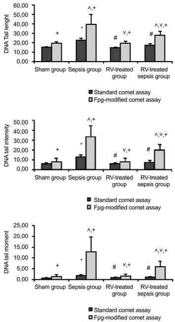
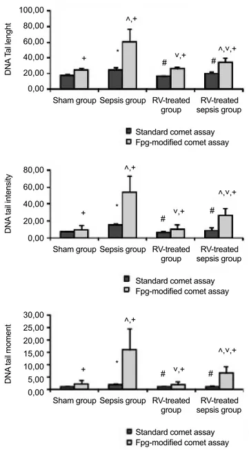
DNA damage in the liver and kidney tissues
DNA damage in the liver and kidney cells are shown in Figures 2a-c and 3a-c, respectively. In the standard comet as-say, there were no statistically significant differences in terms of DNA tail length (TL), DNA tail intensity (TI), and DNA tail moment (TM) in the liver and kidney cells between the sham and the RV-treated groups. All DNA damage parame-ters in the sepsis group were significantly higher than in the sham group (p<0.05) and RV treatment significantly reduced DNA damage induced by sepsis in these tissue cells (24.1%, 45.2%, and 45.0% for TL, TI, and TM in the liver, respec-tively, and 18.5%, 39.1%, and 43.4% for TL, TI, ad TM in
the kidney, respectively) (p<0.05). In the Fpg-modified comet assay, there were no statistically significant differences in all DNA damage parameters of the liver and kidney cells between the sham and the RV-treated groups. These parameters in the septic rats were found to be significantly higher than in the sham group (p<0.05) and RV treatment significantly reduced sepsis-induced DNA strand breakages in the liver and kidney cells (28.9%, 40.6%, and 53.3% for TL, TI, and TM in the liver, respectively, and 43.7%, 50.4%, and 58.8% for TL, TI, and TM in the kidney, respectively) (p<0.05). The enzyme-sensitive sites, more prone for the strand breaks and indicat-ing DNA damage, were found to be significantly higher in the Fpg-modified comet assay than in the standard comet assay. Fpg-sensitive sites increased significantly in the septic rats (p<0.05) and RV significantly reduced Fpg-sensitive sites in-duced by sepsis in all tissue cells (35.2%, 37.6%, and 54.7% for TL, TI, and TM in the liver, respectively, and 61.1%, 54.7%, and 61.1% for TL, TI, TM in the kidney, respectively) (p<0.05).
DISCUSSION
Resveratrol treatment reduces the sepsis-induced oxidative stress, inflammation and DNA damage in the main target tis-sues in Wistar albino rats. Under normal physiological condi-tions, there is a balance between ROS formation and removal by endogenous antioxidant scavenging compounds. Oxidative stress can occur due to imbalance between the excessive pro-duction of ROS and the antioxidant defenses (22). The anti-oxidant defense system is impaired in sepsis (15). The reduc-tion of GSH is supposed to reflect the intracellular oxidareduc-tion impairing cellular functions and the strongest alteration in the antioxidant defense (23,24). The decreases in GSH and increases in MDA levels were determined in the hepatic and renal tissues of septic rats (25). In our study, as for our previ-ous data (13-15), the increases in MDA levels and decreases in GSH levels and SOD and GPX activities in the liver and kid-ney were shown in the septic rats compared to the sham group.
An increasing protective and immunity response charac-terized by the increased nitric oxide (NO), ROS, and inflam-matory cytokines such as TNF-alpha, cause sepsis
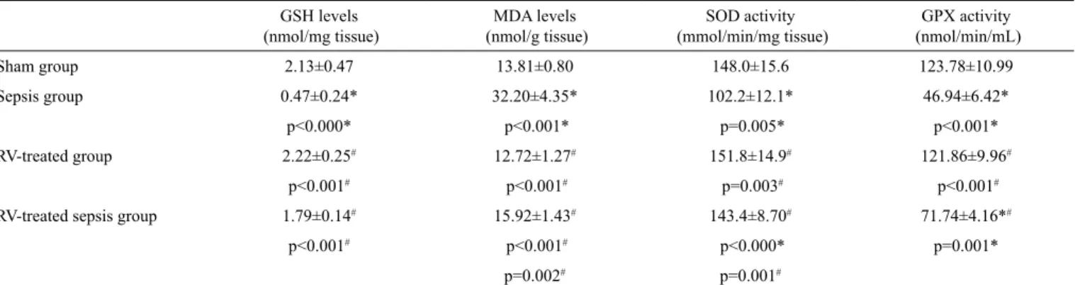
progres-TABLE 2. Oxidative stress parameters in kidney tissues
GSH levels
(nmol/mg tissue) (nmol/g tissue)MDA levels (mmol/min/mg tissue)SOD activity (nmol/min/mL)GPX activity
Sham group 2.13±0.47 13.81±0.80 148.0±15.6 123.78±10.99
Sepsis group 0.47±0.24* 32.20±4.35* 102.2±12.1* 46.94±6.42*
p<0.000* p<0.001* p=0.005* p<0.001*
RV-treated group 2.22±0.25# 12.72±1.27# 151.8±14.9# 121.86±9.96#
p<0.001# p<0.001# p=0.003# p<0.001#
RV-treated sepsis group 1.79±0.14# 15.92±1.43# 143.4±8.70# 71.74±4.16*#
p<0.001# p<0.001# p<0.000* p=0.001*
p=0.002# p=0.001# *p<0.05, compared to the sham group, #p<0.05, compared to the sepsis group. The results were given as the mean±standard deviation. RV: resveratrol; GSH: reduced glutathione; MDA, malondialdehyde; SOD: superoxide dismutase; GPX: glutathione peroxide
TABLE 1. Oxidative stress parameters in the liver tissues
GSH levels
(nmol/mg tissue) (nmol/g tissue)MDA levels (mmol/min/mg tissue)SOD activity (nmol/min/mL)GPX activity
Sham group 3.52±0.40 11.32±2.73 164.6±6.91 86.58±4.69
Sepsis group 1.05±0.30* 24.80±4.36* 131.6±5.86* 20.34±3.31*
p<0.001* p=0.004* p<0.001* p<0.001*
RV-treated group 3.72±0.44# 10.50±1.95# 161.0±10.54# 78.40±2.77#
p<0.001# p=0.004# p=0.007# p<0.001#
RV-treated sepsis group 2.22±0.53*# 15.40±2.80# 147.6±6.27*# 54.30±4.16*#
p=0.014* p=0.026# p=0.019* p<0.001*
p<0.022# p=0.017# p<0.001#
*p<0.05, compared to the sham group, #p<0.05, compared to the sepsis group. The results were given as the mean±standard deviation. RV: resveratrol; GSH: reduced glutathione; MDA: malondialdehyde; SOD: superoxide dismutase; GPX: glutathione peroxide
sion. TNF-alpha, a proinflammatory cytokine, manifests a considerably amplifying effect in the inflammatory response, and leads to severe tissue damages (1,2,15). The severity of tissue injury induced by sepsis is correlated with TNF-alpha level and there is a relation between inflammation and oxida-tive stress (26). In our study, as in our previous data (13-15), plasma TNF-alpha levels significantly increased in the septic rats, indicating an inflammatory response to sepsis.
Single and double DNA strand breaks, base modifications, fragmentation of deoxyribose, formation of DNA-protein cross-links as well as abasic sites may occur as a result of oxidative DNA damage. The alkaline single cell gel electro-phoresis using Fpg, lesion-specific enzyme was used to detect oxidized purines showing the oxidative stress-induced DNA damage (8). In our study, Fpg-sensitive DNA lesions in the he-patic and renal cells significantly increased in the septic rats, referring to the oxidization of purines in the development of sepsis.
Resveratrol, a polyphenolic compound, is well known for its antioxidant feature. There is an increasing interest in RV, since RV may prevent or delay the ischemic and chemically-induced injuries, multi-organ dysfunctions, inflammations, infections,and heart disorders by inhibiting oxidation and the expression of inflammatory mediators and also by regulat-ing microcirculation (3-6,9,27). Dalaklioglu et al. (28) found that RV treatment (20 mg/kg/day, ip) significantly prevented methotrexate-induced hepatotoxicity, as indicated by alanine transaminase, aspartate aminotransferase, and alkaline phos-phatase levels and liver histopathology. RV significantly re-duced the elevated levels of TBARS and activities of catalase and glutathione-S transferase in the hepatic tissue compared to the methotrexate-treated group. RV (5 mg/kg bw for 30 days) found a reduction of the levels of blood glucose, glyco-sylated hemoglobin, TNF-alpha, interleukin-1-beta, interleu-kin-6, NF-kappaB p65 unit and NO with increase in plasma
insulin in diabetic rats (29). Bi et al. (30) found that RV treat-ment significantly inhibited the release of TNF-alpha and ni-trite oxide induced by lipopolysaccharide (LPS) in rat cortical microglia and a mouse microglial cell line N9 in vitro. RV in-hibited the degradation of IkappaB alpha, expression of iNOS and phosphorylation of p38 mitogen-activated protein kinases in N9 microglial cells treated with LPS. In sepsis progression, high-mobility group box 1 (HMGB1) protein, a nuclear
pro-FIG. 1. Plasma TNF-alpha levels in the study group. TNF-alpha: tumor
necrosis factor alpha; RV: resveratrol. *p<0.05, compared to sham group; #p<0.05, compared to sepsis group. The results were given as the mean ± standard deviation. Sham group 250 200 150 100 50 0 Sepsis group * # *,# RV-treated
group sepsis groupRV-treated
TNF-alpha (pg/mL)
FIG. 2. a-c. DNA damage in the liver cells of the study groups. DNA
damage was expressed as (a) DNA tail length; (b) DNA tail intensity; (c) DNA tail moment. RV: resveratrol. *p<0.05, compared to the sham group for the standard comet assay; #p<0.05, compared to the sepsis group for the standard comet assay; ˄p<0.05, compared to the sham group for the Fpg-modified comet assay; ˅p<0.05, compared to the sepsis group for the Fpg-modified comet assay; +p<0.05, standard comet assay was compared to Fpg-modified comet assay. The results were given as the mean ± standard deviation.
Sham group + + + * * * ˄,+ ˄,+ ˄,+ ˄,˅,+ ˄,˅,+ ˄,˅,+ ˅,+ ˅,+ ˅,+ # # # # # # Sham group Sham group Sepsis group Sepsis group Sepsis group RV-treated group Standard comet assay Fpg-modified comet assay
Standard comet assay Fpg-modified comet assay
Standard comet assay Fpg-modified comet assay
RV-treated group RV-treated group RV-treated sepsis group RV-treated sepsis group RV-treated sepsis group 60,00 50,00 40,00 30,00 20,00 10,00 0,00 50,00 40,00 30,00 20,00 10,00 0,00 25,00 20,00 15,00 10,00 5,00 0,00 DNA T ail lenght DNA tail intensity DNA tail moment
tein binding to DNA molecule, promotes the activation of in-nate immune cells, which is suggested to be associated with liver injury. In liver cells, HMGB1 translocation was reported to be related to sirtuin 1 (SIRT1) upregulation. In the septic rats pretreated with RV (60 mg/kg/day for 3 days), serum ami-notransferase activities, proinflammatory cytokines were re-ported to be reduced, thus liver histology was also improved. RV was suggested to prevent sepsis-induced liver damage
via SIRT 1-mediated HMGB1 (31). RV was found to modu-late the expression of genes associated with proinflammatory cytokines by up-regulating the nuclear factor of kappa light chain gene enhancer in IkappaBalpha and down-regulating NFkappaBL1. RV increased the pro-inflammatory cytokines interleukin-6 and TNF-alpha in sepsis (32). Consistent with these studies, we observed that RV treatment significantly in-creased GSH levels and dein-creased MDA levels in the liver and kidney of septic rats. RV also significantly increased SOD and GPX activities in the liver and kidney of sepsis-induced rats. Plasma TNF-alpha level in the RV-treated septic rats was significantly lower than in the septic rats, indicating the pro-tective effects of RV on tissue injury might be mediated by the suppression of the excessive inflammatory response.
In several models, it has been demonstrated that RV has anti-mutagenic and anti-carcinogenic activity (33). The che-mopreventive effect of RV is possibly due to its ability to pre-vent DNA damage and to increase DNA repair (34-36). In the previous study with the peripheral lymphocytes of Wistar rats, RV (100 mg/kg bw, ip) was found to reduce the sepsis-induced oxidative DNA damage using the standard and formamidopy-rimidine DNA glycosylase (Fpg)-modified comet assays (9).
In the study of Kolgazi et al. (37), RV was found to reduce sepsis-induced organ injury. RV was also found to induce premature senescence via decreasing SIRT1 and SIRT2 lev-els in human dermal fibroblasts. The decline in SIRT1/2 ex-pression in response to RV treatment showed that DNA dam-age was regulated by RV (38). Kao et al. (39) reported that RV treatment reduced oxidative stress in human endothelial cells via induction of SIRT1 activation in endothelial cells. RV (100 mg/kg/day) was shown to protect radiation induced chromo-some aberrations in mice (40). RV pretreatment (50 and 100 mg/kg/day, for 7 and 14 days) was found to reduce 8-OHdG, an oxidative DNA damage marker in urinary induced by cispl-atin, via reducing oxidative DNA damage (41). Win et al. (42) showed that RV inhibited ROS-mediated strand breaks in Phi X-174 plasmid DNA. Consistent with these findings, we also found that RV treatment significantly decreased DNA damage induced by sepsis in the liver and kidney tissues of rats. The increases in DNA damage in the liver and kidney cells of RV treated-septic rats using the standard comet and Fpg-modified comet assay were much lower than in septic rats, showing that RV might prevent oxidative DNA strand breakages and increase DNA repair processes.
Our study has some limitations. The results of our study only give the protective effect of a single ip dose of RV (100 mg/kg) after 24 h of sepsis in Wistar albino rats. Due to the survival time of the septic rats being short, we only observed the rats for a short time after the induction of sepsis. We had some financial and technical limitations for this experiment.
FIG. 3. a-c. DNA damage in the kidney cells of the study groups. DNA
damage was expressed as (a) DNA tail length; (b) DNA tail intensity; (c) DNA tail moment. RV: resveratrol. *p<0.05, compared to the sham group for the standard comet assay; #p<0.05, compared to the sepsis group for the standard comet assay; ˄p<0.05, compared to the sham group for the Fpg-modified comet assay; ˅p<0.05, compared to the sepsis group for the Fpg-modified comet assay; +p<0.05, standard comet assay was compared to Fpg-modified comet assay. The results were given as the mean ± standard deviation.
Sham group Sham group Sham group + + + * * * ˄,+ ˄,+ ˄,+ ˄,˅,+ ˄,˅,+ ˄,˅,+ ˅,+ ˅,+ ˅,+ # # # # # # Sepsis group Sepsis group Sepsis group RV-treated group RV-treated group RV-treated group Standard comet assay Fpg-modified comet assay
Standard comet assay Fpg-modified comet assay
Standard comet assay Fpg-modified comet assay
RV-treated sepsis group RV-treated sepsis group RV-treated sepsis group 100,00 80,00 60,00 40,00 20,00 0,00 80,00 60,00 40,00 20,00 0,00 30,00 25,00 20,00 15,00 10,00 5,00 0,00 DNA T al lenght DNA tail intensity DNA tail moment
More parameters such as DNA adduct formation, micronucle-us formation and changes in gene expression related to DNA damage and inflammation would be investigated. Different experimental design at different treatment doses, route, and time and in different animal species would be performed. To determine the pretreatment effects of RV before the progres-sion of sepsis, the rats would be pretreated with RV in the long-term before sepsis induction. The numbers of animals would be increased to achieve more accurate statistical re-sults. The study would be repeated to show the reproducibil-ity of our results. The level of RV and its metabolic products in biological samples would also be determined to reveal the dose-response relationship of RV.
In conclusion, DNA damage might be initiated in sepsis due to the induction of oxidative stress and it seems that RV via increasing the antioxidant status and DNA repair capacity in
vivo has protective effects against the sepsis-induced oxidative
stress and DNA damage. The uses of RV in clinical settings might be beneficial against the progression of tissue injuries induced by sepsis. It is suggested that RV treatment might re-duce the oxidative DNA damage in sepsis-related multi-organ failures, however more investigations are required to clarify the act of protective mechanisms of RV against sepsis. We are planning to determine the pretreatment effects of RV at differ-ent oral doses for long-term before the progression of sepsis
in vivo.
Ethics Committee Approval: Ethics committee approval was
re-ceived for this study from the ethics committee of Hacettepe Univer-sity School of Medicine.
Informed Consent: N/A.
Peer-review: Externally peer-reviewed.
Author contributions: Concept - S.A., N.B., T.T.Ş., A.A.B.;
De-sign - S.A., N.B., M.B., G.T.; Supervision - S.A., N.B., T.T.Ş., A.A.B., M.B., G.T., M.A.; Resource - S.A., N.B., T.T.Ş., A.A.B., M.B., G.T., M.A.; Materials - S.A., N.B., T.T.Ş., A.A.B., M.B., G.T., M.A.; Data Collection and/or Processing - S.A., N.B., T.T.Ş., A.A.B., M.B., G.T., M.A.; Analysis and/or Interpretation - S.S.A., N.B., T.T.Ş., A.A.B., M.B., G.T., M.A.; Literature Search - S.A., N.B., T.T.Ş., A.A.B., M.B., G.T., M.A.; Writing - S.A., N.B., T.T.Ş., A.A.B., M.B., G.T., M.A.; Critical Reviews - S.A., N.B., T.T.Ş., A.A.B., M.A.
Conflict of Interest: No conflict of interests was declared by the
authors.
Financial Disclosure: The authors declared that this study has
re-ceived no financial support.
REFERENCES
1. Andrades M, Ritter C, Moreira JDF, Dal-Pizzol F. Oxidative pa-rameters differences during non-lethal and lethal sepsis develop-ment. J Surg Res 2005;125:68-72. [CrossRef]
2. Andrades ME, Ritter C, Dal-Pizzol F. The role of free radicals in sepsis development. Front Biosci 2009;1:277-87.
3. Bishayee A, Barnes KF, Bhatia D, Darvesh AS, Carroll RT. Res-veratrol suppresses oxidative stress and inflammatory response in diethylnitrosamine-initiated rat hepatocarcinogenesis. Cancer
Prev Res (Phila) 2010;3:753-63. [CrossRef]
4. Aggarwal BB, Bhardwaj A, Aggarwal RS, Seeram NP, Shisho-da S, TakaShisho-da Y. Role of resveratrol in prevention and therapy of cancer: preclinical and clinical studies. Anticancer Res 2004;24:2783-840.
5. Eybl V, Kotyzová D, Cerná P, Koutensky J. Effect of melatonin, curcumin, quercetin, and resveratrol on acute ferric nitrilotriac-etate (Fe-NTA)-induced renal oxidative damage in rats. Hum
Exp Toxicol 2008;27:347-53. [CrossRef]
6. Bujanda L, Garcı´a-Barcina M, Gutie´rrez-de Juan V, Bidaur-razaga J, de Luco MF, Gutie´rrez-Stampa M, et al. Effect of res-veratrol on alcohol-induced mortality and liver lesions in mice.
BMC Gastroenterol 2006;6:35. [CrossRef]
7. Sener G, Toklu HZ, Sehirli AO, Velioğlu-Öğünç¸ A, Cetinel S, Gedik N. Protective effects of resveratrol against acetamin-ophen-induced toxicity in mice. Hepatol Res 2006;35:62-8. [CrossRef]
8. Collins AR, Duthie SJ, Dobson VL. Direct enzymic detection of endogenous oxidative base damage in human lymphocyte DNA. Carcinogenesis 1993;14:1733-5. [CrossRef]
9. Aydin S, Bacanli M, Taner G, Şahin T, Başaran AA, Başaran N. Protective effects of resveratrol on sepsis-induced DNA damage in the lymphocytes of rats. Hum Exp Toxicol 2013;32:1048-57. [CrossRef]
10. Parker SJ, Watkins PE. Experimental models of gram negative sepsis. Br J Surg 2001;88:22-30. [CrossRef]
11. Ritter C, Andrades M, Frota ML, Pinho R, Polydoro M, Klamt F, et al. Oxidative parameters and mortality in sepsis induced by ce-cal ligation and perforation. Intensive Care Med 2003;29:1782-9. [CrossRef]
12. Della-Morte D, Dave KR, DeFazio RA, Bao YC, Raval AP, Per-ez-Pinzon MA. Resveratrol pretreatment protects rat brain from cerebral ischemic damage via a sirtuin 1-uncoupling protein 2 pathway. Neuroscience 2009;159:993-1002. [CrossRef] 13. Taner G, Aydin S, Bacanli M, Sarıgöl Z, Sahin T, Basaran AA,
et al. Modulating Effects of Pycnogenol® on Oxidative Stress and DNA Damage Induced by Sepsis in Rats. Phytother Res 2014;28:1692-700. [CrossRef]
14. Bacanlı M, Aydın S, Taner G, Göktaş HG, Sahin T, Başaran AA, et al. The protective role of ferulic acid on sepsis-induced oxida-tive damage in Wistar albino rats. Environ Toxicol Pharmacol 2014:38:774-82. [CrossRef]
15. Bacanlı M, Aydın S, Taner G, Göktaş HG, Şahin T, Başaran AA, et al. Does rosmarinic acid treatment have protective role
against sepsis-induced oxidative damage in Wistar Albino rats? Hum Exp Toxicol 2015; pii: 0960327115607971.
16. Russell WMS, Burch, RL. The Principles of Humane Experi-mental Technique, London: Methuen & Co., Ltd.; 1959. 17. Graham JM. Homogenization of mammalian tissues. Scientific
World J 2002;2:1626-9. [CrossRef]
18. Mihara M, Uchiyama M. Determination of malonaldehyde precursor in tissues by thiobarbituric acid test. Anal Biochem 1978;86:271-8. [CrossRef]
19. Singh NP, McCoy MT, Tice RR, Schneider EL. A simple tech-nique for quantitation of low levels of DNA damage in indi-vidual cells. Exp Cell Res 1988;175:184-91. [CrossRef] 20. Anderson D, Yu TW, Phillips BJ, Schmezer P. The effect of
vari-ous antioxidants and other modifying agents on oxygen-radical-generated DNA damage in human lymphocytes in the comet as-say. Mutat Res 1994;307:261-71. [CrossRef]
21. Bakare AA, Patel S, Pandey AK, Bajpayee M, Dhawan A. DNA and oxidative damage induced in somatic organs and tissues of mouse by municipal sludge leachate. Toxicol Ind Health 2012;28:614-23. [CrossRef]
22. Gutteridge JMC, Mitchell J. Redox imbalance in the critically ill. Br Med Bull 1999;55:49-75. [CrossRef]
23. Himmelfarb J, McMonagle E, Freedman S, Klenzak J, Mc-Menamin E, Le P, et al. Oxidative stress is increased in criti-cally ill patients with acute renal failure. J Am Soc Nephrol 2004;15:2449-56. [CrossRef]
24. Ciriolo MR, Palamara AT, Incerpi S, Lafavia E, Buè MC, De Vito P, et al. Loss of GSH, oxidative stress, and decrease of in-tracellular pH as sequential steps in viral infection. J Biol Chem 1997;272:2700-8. [CrossRef]
25. Sener G, Toklu H, Kapucu C, Ercan F, Erkanli G, Kaçmaz A, et al. Melatonin protects against oxidative organ injury in a rat model of sepsis. Surg Today 2005;35:52-9. [CrossRef]
26. Waage A, Halstensen A, Espevik T. Association between tumor necrosis factor in serum and fatal outcome in patients with me-ningococcal disease. Lancet 1987;1:355-7. [CrossRef]
27. Hascalik S, Celik O, Turkoz Y, Hascalik M, Cigremis Y, Mizrak B, et al. Resveratrol, a red wine constituent polyphenol, protects from ischemia-reperfusion damage of the ovaries. Gynecol
Ob-stet Invest 2004;57:218-23. [CrossRef]
28. Dalaklioglu S, Genc GE, Aksoy NH, Akcit F, Gumuslu S. Resveratrol ameliorates methotrexate-induced hepatotoxicity in rats via inhibition of lipid peroxidation. Hum Exp Toxicol 2013;32:662-71. [CrossRef]
29. Palsamy P, Subramanian S. Ameliorative potential of resvera-trol on proinflammatory cytokines, hyperglycemia mediated oxidative stress, and pancreatic beta-cell dysfunction in strep-tozotocin-nicotinamide-induced diabetic rats. J Cell Physiol 2010;224:423-32. [CrossRef]
30. Bi XL, Yang JY, Dong YX, Wang JM, Cui YH, Ikeshima T, et al. Resveratrol inhibits nitric oxide and TNF-alpha production by lipopolysaccharide-activated microglia. Int Immunopharmacol 2005;5:185-93. [CrossRef]
31. Chen Z, Wang G, Jing H, Zhang X, Li M, Peng J, Tian X. Novel role of resveratrol: suppression of high-mobility group protein box 1 nucleocytoplasmic translocation by the upregulation of sirtuin 1 in sepsis-induced liver injury. Shock 2014;42:440-7. [CrossRef]
32. Leiro J, Arranz JA, Fraiz N, Sanmartín ML, Quezada E, Orallo F. Effect of cis-resveratrol on genes involved in nuclear fac-tor kappa B signaling. Int Immunopharmacol 2005;5:393-406. [CrossRef]
33. Uenobe F, Nakamura S, Miyazawa M. Antimutagenic effect of resveratrol against Trp-P-1. Mutat Res 1997;373:1997-2000. [CrossRef]
34. Chakraborty S, Roy M, Bhattacharya RK. Prevention and repair of DNA damage by selected phytochemicals as measured by single cell gel electrophoresis. J Environ Pathol Toxicol Oncol 2004;23:215-26. [CrossRef]
35. Yen GC, Duh PD, Lin CW. Effects of resveratrol and 4-hexyl-resorcinol on hydrogen peroxide-induced oxidative DNA dam-age in human lymphocytes. Free Radic Res 2003;37:509-14. [CrossRef]
36. Alturfan AA, Tozan-Beceren A, Sehirli AO, Demiralp E, Sener G, Omurtag GZ. Resveratrol ameliorates oxidative DNA dam-age and protects against acrylamide-induced oxidative stress in rats. Mol Biol Rep 2012;39:4589-96. [CrossRef]
37. Kolgazi M, Sener G, Cetinel S, Gedik N, Alican I. Resveratrol reduces renal and lung injury caused by sepsis in rats. J Surg Res 2006;134:315-21. [CrossRef]
38. Kilic Eren M, Kilincli A, Eren Ö. Resveratrol induced premature senescence is associated with DNA damage mediated SIRT1 and SIRT2 down-regulation. PLoS One 2015;10:e0124837. [CrossRef]
39. Kao CL, Chen LK, Chang YL, Yung MC, Hsu CC, Chen YC, et al. Resveratrol protects human endothelium from H(2)O(2)-induced oxidative stress and senescence via SirT1 activation. J
Atheroscler Thromb 2010;17:970-9. [CrossRef]
40. Carsten RE, Bachand AM, Bailey SM, Ullrich RL. Resveratrol reduces radiation-induced chromosome aberration frequen-cies in mouse bone marrow cells. Radiat Res 2008;169:633-8. [CrossRef]
41. Attia SM. Influence of resveratrol on oxidative damage in ge-nomic DNA and apoptosis induced by cisplatin. Mutat Res 2012;741:22-31. [CrossRef]
42. Win W, Cao Z, Peng X, Trush MA, Li Y. Different effects of ge-nistein and resveratrol on oxidative DNA damage in vitro. Mutat