Skewed X inactivation in an X linked nystagmus
family resulted from a novel, p.R229G, missense
mutation in the FRMD7 gene
Y Kaplan,
1I Vargel,
2T Kansu,
3B Akin,
4E Rohmann,
5,6S Kamaci,
7E Uz,
8T Ozcelik,
8B Wollnik,
5,6N A Akarsu
4,9 cThe supplementary table ispublished online only at http:// bjo.bmj.com/content/vol92/ issue1
1
Department of Neurology, Gaziosmanpas¸a University, Medical Faculty, Tokat, Turkey;
2
Department of Plastic and Reconstructive Surgery, Hacettepe University Medical Faculty, Ankara, Turkey;
3
Department of Neurology, Hacettepe University Medical Faculty, Ankara, Turkey;4Gene Mapping Laboratory, Pediatric Hematology Unit, Hacettepe University Medical Faculty, Ankara, Turkey;5Center for
Molecular Medicine Cologne (CMMC), University of Cologne, Cologne, Germany;6Institute of Human Genetics, University of Cologne, Cologne, Germany;
7Department of Orthodontics,
Hacettepe University, Faculty of Dentistry, Ankara, Turkey;
8Department of Molecular
Biology and Genetics, Bilkent University, Faculty of Science, Ankara, Turkey;9Department of Medical Genetics, Hacettepe University Medical Faculty, Ankara, Turkey
Correspondence to: N A Akarsu, Hacettepe University Medical Faculty, Department of Pediatrics, Pediatric Hematology Unit, Gene Mapping Laboratory, Room No. 24, Sihhiye, 06100, Ankara, Turkey; nakarsu@hacettepe. edu.tr
Accepted 6 October 2007 Published Online First 25 October 2007
ABSTRACT
Aims: This study aimed to identify the underlying genetic defect of a large Turkish X linked nystagmus (NYS) family. Methods: Both Xp11 and Xq26 loci were tested by linkage analysis. The 12 exons and intron–exon junctions of the FRMD7 gene were screened by direct sequencing. X chromosome inactivation analysis was performed by enzymatic predigestion of DNA with a methylation-sensitive enzyme, followed by PCR of the polymorphic CAG repeat of the androgen receptor gene.
Results: The family contained 162 individuals, among whom 28 had NYS. Linkage analysis confirmed the Xq26 locus. A novel missense c.686C.G mutation, which causes the substitution of a conserved arginine at amino acid position 229 by glycine (p.R229G) in exon 8 of the FRMD7 gene, was observed. This change was not documented in 120 control individuals. The clinical findings in a female who was homozygous for the mutation were not different from those of affected heterozygous females. Skewed X inactivation was remarkable in the affected females of the family. Conclusions: A novel p.R229G mutation in the FRMD7 gene causes the NYS phenotype, and skewed X inactivation influences the manifestation of the disease in X linked NYS females.
Congenital nystagmus (NYS) is an ocular oscilla-tory movement disorder caused by a motor instability that can manifest with or without afferent visual system dysfunction. This entity is defined both clinically and by electronystagmogra-phy. Certain clinical features usually differentiate congenital NYS from other oscillations. NYS may be irregular, but is always conjugate and horizon-tal, though very rarely vertical. There may be a torsional component. Some patients with this abnormality also show head oscillations, which tend to increase when the patient attends to an object, an effort that also increases the NYS. Therefore, it seems probable that both the head tremor and the ocular oscillations are the conse-quence of a common disordered neural mechan-ism.1
The underlying defect in congenital NYS remains elusive. The Eye Movement Abnormalities and Strabismus Working Group proposed the term, infantile nystagmus syndrome, for the NYS that is known as congenital NYS. Although there is some controversy concerning classification of NYS in infancy, it has been suggested that NYS with an onset before 6 months of age can be divided into three categories:2 (1)
congenital idiopathic NYS in which no visual or associated neurological impairment can be found; (2) sensory deficit NYS in which there is a visual abnormality, such as ocular albinism, optic nerve hypoplasia, congenital stationary night blindness and blue cone monochromatism; (3) neurological NYS, which is associated with a neurological disorder. Category 1 is also called congenital motor NYS, which is presumed to have a primary defect in the part of the brain responsible for ocular motor control.3
Congenital motor NYS is a genetically hetero-geneous disorder. Autosomal dominant (MIM 164100), recessive (MIM 257400) and X linked (MIM 310700) patterns of inheritance are described for this disorder. X linked inheritance is the most common form of NYS (NYS1). An irregular dominant pattern of X linked inheritance has been frequently reported, although some pedigrees support X linked recessive inheritance.4
The penetrance is full in males and approximately 50% to 29% in females.5 6
Large intrafamilial variance in waveforms can be observed. Two distinct loci, one on the Xq26–q273
and the other on the Xp11.47regions have been reported as the
likely loci for X linked dominant NYS. The locus on the long arm (Xq26) has been confirmed by subsequent reports of various ethnic populations.8– 10To the best of our knowledge, there are only two
reports supporting the Xp11.4 locus.7 11
The pat-tern of inheritance and clinical profile of Xp11-linked families are not different from Xq26-Xp11-linked pedigrees. X linked recessive NYS has also been mapped to the Xq26 region, which harbours the X linked dominant NYS locus.10Recently, mutations
in the FRMD7 (FERM domain-containing 7) gene have been reported as a molecular cause in Xq26-linked families.6
Little is known about the function of the FRMD7 gene; however, the restricted expression pattern of this gene in the human embryonic brain and developing neural retina suggests a role in eye movement and gaze stability.6
Herein, we report an extensive NYS pedigree, including 162 individuals across six generations from southeastern Turkey. The mode of inheri-tance is clearly X linked, demonstrating a reduced penetrance in female obligate gene carriers. Genetic linkage analysis confirmed the Xq26–q27 locus, and further mutation analysis identified a novel p.R229G missense mutation in the FRMD7 gene that causes this disorder. We also detected a predisposition to skewed X inactivation in affected
females, suggesting that the X inactivation mechanism might have a role in manifestation of the disease in females.
METHODS Clinical evaluation
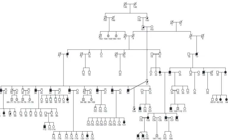
The family was identified during fieldwork to study a large craniosynostosis pedigree in Antakya, Turkey. The complete pedigree structure contains over 427 individuals, including a separate branch of congenital idiopathic NYS. Only the NYS branch is included in this study, and the pedigree structure is shown in fig 1. Pedigree formation was completed in the field by NA, IV and SK. Neurological evaluation of 23 individuals was conducted by YK, a neurologist and member of the family. The recruitment criteria were NYS noted at birth or during the first 3 months of life, and no abnormalities of the ocular or neural visual pathway. Visual acuity of all the patients was measured by Snellen card. Eye movements were recorded by a video camera in 16 patients. Video recordings were further reviewed by a neuro-ophthalmologist (TK). Peripheral blood samples were collected from 48 individuals with informed consent for further molecular studies. The Hacettepe University Ethics Committee approved the study (FON 02/6-13)
Genetic linkage analysis
The family was tested for linkage to two reported loci on chromosome regions Xp117and Xq26.3Map order and physical
positions (Mb) of the polymorphic markers were obtained from the University of California Santa Cruz (USCS), Genome Bioinformatics Center (http://genome.ucsc.edu). Oligonucleotide samples were purchased from MWG-Biotechnology (Ebersberg,
Germany). Site-specific PCR, 7% polyacrylamide gel electrophor-esis and silver staining technique were used in genotyping the individuals. Gels were manually pictured. The Cyrillic program was used to generate haplotype and input files for linkage analysis. Two-point linkage was performed with the MLINK component of the LINKAGE package, assuming X linked dominant inheritance with 0.36 penetrance in females. Mutant allele frequencies were kept as 0.0001. Equal marker allele frequency of DNA markers was assumed.
Mutation analysis
We designed primers to amplify all 12 protein-coding exons and adjacent intronic sequences of the FMRD7 gene by PCR using standard conditions (supplementary table). Due to its size, exon 12 was amplified in three overlapping fragments. Subsequent to amplification, PCR fragments were purified, with QIAquick spin columns (Qiagen), and directly sequenced using the corresponding forward primers with the ABI PRISMH BigDyeTMTerminator v2.0 cycle sequencing ready reaction kit and an ABI PRISMH 3100 genetic analyser (Applied Biosystems). We re-sequenced all identified mutations in independent experiments, tested for co-segregation within the families, and screened 120 Turkish control individuals for the c.686C.G mutation by PCR and subsequent restriction digestion using BccI (MBI Fermentas, Germany).
Evaluation of X chromosome inactivation (XCI)
Genotyping of a highly polymorphic CAG repeat in the androgen-receptor (AR) gene was performed to assess the XCI patterns.12
DNA was divided into two identical aliquots, one of
Figure 1 Complete pedigree structure of X linked idiopathic congenital NYS. Full shaded symbols indicate the NYS phenotype. Quarter shaded symbols demonstrate individuals with craniosynostosis. The craniosynostosis branch (relatives of individuals 150, 151 and 51) was not included in this study.
which was incubated overnight at 37uC with the methylation-sensitive restriction enzyme HpaII (MBI Fermentas, Vilnius, Lithuania), for the digestion of unmethylated (or active) alleles. A second restriction enzyme, RsaI (MBI Fermentas, Vilnius, Lithuania), which recognises a four-base-pair sequence not present in the amplified region of the AR locus, was also included in the reaction to facilitate the HpaII digestion process. Male DNA with a cytogenetically verified 46,XY karyotype was used as the control for complete digestion. The other half of the DNA was treated similarly, but without HpaII. After restric-tion-enzyme digestion, residual DNA was amplified using the primers 59-GTC CAA GAC CTA CCG AGG AG-39 and 59-CCA GGA CCA GGT AGC CTG TG-39. PCR products were
separated on 10% denaturing 29:1 acrylamide-bisacrylamide gel for 5 h at 20 W. Gels were stained with ethidium bromide and visualised under UV light. Densitometric analysis of the alleles was performed at least twice for each sample using the appropriate software (MultiAnalyst v1.1; Bio-Rad, Hercules, CA). A corrected ratio (CrR) was calculated by dividing the ratio of the predigested sample (upper/lower allele) by the ratio of the non-predigested sample for normalisation of the ratios that were obtained from the densitometric analyses. The use of a CrR compensates for preferential amplification of the shorter allele when the number of PCR cycles increases.13A skewed population
is defined as a cell population with .80% expression of one of the AR alleles. This corresponds to CrR values of ,0.25 or .4.0. Table 1 Examination findings of the family members
PID Gender DOB Nystagmus Head oscillations DM type II Obesity Other findings
23 F 1939 Pendular + – – –
24 M 1929 Pendular + + + Asthma, corneal opacity on the left, hypertension
32 M 1961 Pendular+ jerk + + + Diabetic neuropathy, cataract, hypertension
38 M 1950 Pendular – + + Diabetic neuropathy, amyloid lichenosis
40 M 1945 Pendular + + + –
42 M 1943 Pendular + + + Diabetic neuropathy, psoriasis, hypertension
46 M 1939 Pendular + + + Diabetic neuropathy, ptosis on the left
48 M 1930 Pendular + + + Diabetic neuropathy, hypertension, ptosis on the right
65 F 1981 Pendular+ jerk + – – –
66 F 1982 Pendular+ jerk – – – Strabismus
93 F 1980 Pendular – – – –
105 M 1986 Pendular+ jerk + – + Febrile convulsion
108 M 1985 Pendular + – – – 112 M 1998 Pendular – – – – 121 F 1971 Pendular + – + – 131 F 1965 Pendular + – – – 133 F 1969 Pendular – – – – 136 M 1968 Pendular + – + – 138 M 1993 Pendular – – – – 139 M 1993 Pendular – – – – 36 M 1957 – – + + – 39 F 1951 – – – + – 43 F 1951 – – – + – 53 F 1959 – – + + Psoriasis, hypertension 54 F 1960 – – – + – 69 M 1978 – – – + – 79 M 1971 – – – + – 80 M 1974 – – – + – 81 M 1976 – – – + – 82 M 1989 – – – + – 87 F 1970 – – – + – 91 M 1969 – – – + – 94 M 1989 – – – + Allergic conjuctivitis 95 M 1976 – – – + – 107 F 1983 – – – + – 115 M 1975 – – – + – 116 M 1979 – – – + – 117 M 1980 – – – + Asthma bronchiale 118 F 1970 – – – + – 119 F 1978 – – – + Asthma bronchiale 120 F 1984 – – – + – 122 F 1986 – – – + – 123 M 1967 – – – + – 124 M 1968 – – – + – 125 M 1969 – – – + – 126 M 1960 – – – + – 134 M 1968 – – – + –
RESULTS
Clinical presentation and formal genetics of the family
The most striking findings of this pedigree were, 1, NYS and, 2, obesity and type 2 diabetes in some family members (table 1). Other findings included craniosynostosis and allergic com-plaints, such as bronchial asthma. In all, 20 patients had rhythmic pendular type NYS, with varying frequencies and amplitudes. Video recordings of 16 individuals supported the clinical observations. On lateral gaze, four individuals had horizontal jerk NYS, whereas six patients had spontaneous head oscillations, and 13 had oscillations during fixation (table 1). Visual acuity among the patients varied from 20/20 to 20/100. Direct ophthalmoscopic findings were unremarkable. In all, seven NYS patients had diabetes, and 10 were obese. Among the non-NYS family members, 27 were obese (two of them with diabetes) (table 1) suggesting independent segregation of the two disorders (NYS and obesity and/or type 2 diabetes) in the same family. Regarding the NYS phenotype, male-to-male transmission was not observed. This finding supported X linked inheritance. There were 33 obligate gene carrier females in this family, and only 12 of them developed NYS (fig 1); therefore, the penetrance of the NYS phenotype in females was estimated to be approximately 36% for this family.
Linkage studies
The family was tested for a linkage to both Xp11.4-p11.37and
Xq26–q273 loci. The NYS phenotype was previously mapped
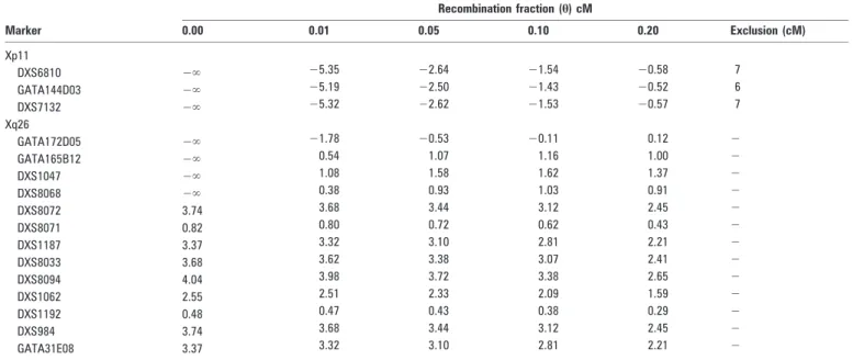
between the DNA markers DXS8015 and DXS1003 on the Xp11.4 locus. The DNA markers DXS6810, GATA144D04 and DXS7132 were used in this study, and the physical positions of these markers are: DXS8015 (39.44 DXS6810, (42.57 Mb)-GATA144D04 and (44.67 Mb)-DXS1003 (46.19 Mb)-DXS7132 (64.33 Mb). Four NYS patients were recombinant for the entire region (data not shown). Negative LOD scores were also obtained. The excluded region was 6–7 cM for each DNA marker (table 2).
Significant LOD scores were obtained for the entire region, with a maximum of 4.04 at h = 0 cM for the DNA marker DXS8094 (table 2) in the Xq26–q27 region (fig 2). A single
cross-over event in an NYS patient (person 40) positioned the disease gene telomeric to the DNA marker DXS8068. We also observed the first homozygote female case of the NYS phenotype (fig 1, person 133). No phenotypic differences were observed among the females, in terms of homozygote and heterozygote states (table 1).
Mutation results
We sequenced all 12 coding exons of the recently described X linked NYS gene FRMD7, which was located in the critical region in two affected male individuals of the family, and identified the c.686C.G mutation in exon 8 of the gene (fig 3A). The mutation co-segregated with the disease in the family (fig 3B) and was not observed in 120 healthy individuals of the same ethnic background. The novel c.686C.G mutation predicted a substitution of a conserved arginine at amino acid position 229 in the functionally important FERM-C domain of the protein by glycine (p.R229G). Interestingly, three other missense mutations in the FERM-C domain of FRMD7 have been described in the original gene identification paper6(fig 3C),
suggesting an important role for this domain in the pathogen-esis of congenital NYS.
X chromosome inactivation results
Since at least seven females in the pedigree had NYS, we analysed the XCI patterns to verify if skewed XCI could be responsible for the clinical manifestation of the disease. Skewed patterns with ratios of 81:19 per cent in individual 65, 85:15 per cent in 121, and 80:20 per cent in 131 and 133 was apparent, while individual 23 was not informative, and only individuals 93 and 66 displayed random XCI with ratios of 57:43 per cent and 62:38 percent, respectively. Among the non-NYS females, XCI status was analysed in 18 individuals. With the exception of individual 35, who had a skewed XCI (93:7) pattern, and four more non-informative females (individuals 41, 43, 54 and 86), all women displayed random XCI profiles (table 3). Skewed X inactivation was significantly increased in the NYS females when compared with the non-NYS females in the family (odds ratio was 26:1; 95% CI = 1.83 to 367.7).
Table 2 Two point LOD scores with the DNA markers selected from Xp11 and Xq26 regions
Recombination fraction (h) cM Marker 0.00 0.01 0.05 0.10 0.20 Exclusion (cM) Xp11 DXS6810 2‘ 25.35 22.64 21.54 20.58 7 GATA144D03 2‘ 25.19 22.50 21.43 20.52 6 DXS7132 2‘ 25.32 22.62 21.53 20.57 7 Xq26 GATA172D05 2‘ 21.78 20.53 20.11 0.12 2 GATA165B12 2‘ 0.54 1.07 1.16 1.00 2 DXS1047 2‘ 1.08 1.58 1.62 1.37 2 DXS8068 2‘ 0.38 0.93 1.03 0.91 2 DXS8072 3.74 3.68 3.44 3.12 2.45 2 DXS8071 0.82 0.80 0.72 0.62 0.43 2 DXS1187 3.37 3.32 3.10 2.81 2.21 2 DXS8033 3.68 3.62 3.38 3.07 2.41 2 DXS8094 4.04 3.98 3.72 3.38 2.65 2 DXS1062 2.55 2.51 2.33 2.09 1.59 2 DXS1192 0.48 0.47 0.43 0.38 0.29 2 DXS984 3.74 3.68 3.44 3.12 2.45 2 GATA31E08 3.37 3.32 3.10 2.81 2.21 2
DISCUSSION
In this study, we described a large family with X linked congenital NYS. The disorder was completely penetrant in males; however, reduced penetrance was observed in females. Various penetrance rates, ranging from 54% to 29%, are suggested to be due to family size.3 5 6Our study also supported
36% penetrance in females. This extremely low penetrance rate in females might challenge the mode of inheritance estimations. For genetic counselling purposes, it might be difficult to distinguish X linked recessive and dominant patterns in small family trees. Genetic linkage analysis provided strong evidence of linkage to the previously established locus on Xq26–27,
which is the major locus for X linked NYS (NYS1). A recombination event in a single affected male positioned the disease gene telomeric to the DNA marker DXS8068 (fig 2). Combining these data with previous studies, the 8 Mb region residing between DNA markers DXS8068 and DXS1211 containing the recently reported FRMD7 gene6was critical for
this family.
We identified the novel p.R229G missense mutation in the FRMD7 gene in the study family. The following points support the disease-causing nature of this mutation: (i) p.R229G co-segregated with the disease in this family; (ii) our control studies excluded p.R229G as a non-synonymous polymorphism Figure 2 Haplotype structure between the NYS phenotype and the DNA markers selected from Xq26. The order and the physical locations (Mb) of the DNA markers are shown to the upper left. A single recombination event in affected individual 40 positioned the disease gene telomeric to DXS8068 containing the FRMD7 gene (130.93 Mb).
Figure 3 Identification of a novel missense mutation in the FRMD7 gene. (A) Representative sequence chromatograms of the identified c.686C.G (p.R229G) mutation in an affected male (mutant), a carrier female (carrier) and an unaffected male (wild-type). (B) Co-segregation of the mutation in the family. Results of BccI restriction fragment analysis presenting an undigested 234 bp PCR-fragment in affected male individuals, a fully digested PCR fragment in unaffected males (two fragments 131 bp and 101 bp in size), and one undigested and one digested allele in carrier females (three fragments 234 bp, 131 bp and 101 bp in size). A non-digested sample, labelled as C, was included as a control (C) schematic view of conserved FRMD7 domains and locations of described mutations located in the FERM-C domain.
Table 3 Distribution of skewed x-inactivation between nystagmus and normal members of the family
PID DNA no. Status Skewing degree
Skewed 1 35 N42 Normal 93:7 2 121 N46 Affected 85:15 3 65 N31 Affected 81:19 4 131 N40 Affected 80:20 5 133 N21 Affected 80:20 Random 1 33 N18 Normal 72:28 2 45 N11 Normal 71:29 3 107 N28 Normal 71:29 4 72 N41 Normal 69:31 5 15 N30 Normal 67:33 6 11 N37 Normal 67:33 7 53 N27 Normal 63:37 8 66 N16 Affected 62:38 9 93 N26 Affected 57:43 10 118 N13 Normal 56:44 12 99 N10 Normal 54:46 13 122 N14 Normal 54:46 14 119 N43 Normal 53:47 15 120 N12 Normal 51:49 16 Spouse of 129 N7 normal 50:50
PID reflects personal identification numbers on fig 1. A total of five individuals (fig 1, individuals 23, 41, 43, 54 and 86) were not informative. The disease status of non-informative individuals was normal except person 23.
in the Turkish population; (iii) the mutation is located in the FERM-C domain in the surrounding of previously described missense mutations in families with congenital NYS.6 14
The functional role of FRMD7 remains to be elucidated. The FRMD7 transcript is abundantly expressed in most tissues, and a localised and restricted expression was found to be involved in embryonic development in regions affecting motor control of eye development.6Homologies of the B41 and FERM-C domains
of FRMD7 to other proteins, such as FARP1 and FARP2, which are involved in neurite branching of cortical neurons, led to the hypothesis that FRMD7 could also be involved in the develop-ment of similar neuronal pathways.
Skewed X inactivation has consistently been suggested as a mechanism that may influence the penetrance of X linked NYS in females;3 7 8 9 however, except for only one study,7 an X
inactivation pattern was not previously studied in NYS families. No correlation between X inactivation patterns and the NYS phenotype was observed in this sole study.7
Nevertheless, NYS in the family used in that study was linked to the Xp11 region rather than the major NYS locus on Xq26–q27. Our Xq26-linked NYS family implies that skewed XCI could be a factor that influences the clinical manifestation of NYS in females. It is well established that skewed XCI is a rare event in a diverse group of control females.15 16 In agreement with this prior observation,
we only observed skewed X inactivation in a single healthy spouse (individual 35). None of the obligate gene carrier females and none of the remaining healthy spouses demonstrated skewed X inactivation (table 3); however, increased suscept-ibility to skewed X inactivation was apparent in the clinically affected females. On the other hand, we observed random X inactivation in two affected females, individuals 66 and 93, with XCI scores of 62:38 and 57:43, respectively. This finding may be a reflection of tissue mosaicism, which has been clearly shown in women.16
To the best of our knowledge, inactivation status of the FRMD7 gene has not been studied. Further investigations of the X inactivation status of FRMD7 might help contribute to an understanding of the irregular pattern of inheritance of NYS1.
Acknowledgements: We are grateful to the family members for their participation in the study. We thank the Hacettepe University Craniofacial Surgery Study Group members: Yucel Erk, Emin Mavili, and Gokhan Tuncbilek (Plastic and Reconstructive
Surgery), Kemal Benli (Neurosurgery), Aysenur Cila (Radiology), and Sevim Balci (Genetics) for their evaluation of the family members with craniosynostosis. This work was presented in the 8th European Neuro-Ophthalmology Society (EUNOS) Meeting, 26–29 May 2007, Istanbul, Turkey.
Funding: This study was supported by The Hacettepe University Research Foundation (number 00-01-101-010), The Scientific and Technological Research Council of Turkey (number TUBITAK-SBAG 3334) and The International Centre for Genetic Engineering and Biotechnology (ICGEB-CRP/TUR04-01).
Competing interests: None.
REFERENCES
1. Leigh RJ, Averbuch-Heller L. Nystagmus and related ocular motility disorders. In: Walsh & Hoyt’s clinical neuro-ophthalmology, Vol. 1. 5th edn. In: N R Miller, NJ Newman, eds. Williams & Wilkins, Baltimore, 1998:1483.
2. Hoyt C. Clinical congenital nystagmus. Evaluation and treatment. North American Neuro-Ophthalmology Society 31st Annual Meeting Syllabus, 2005:319.
3. Kerrison JB, Vagefi MR, Barmada MM, et al. Congenital motor nystagmus linked to Xq26–q27. Am J Hum Genet 1999;64:600–7.
4. Forrsman B, Ringer B. Prevalence and inheritance of congenital nystagmus in a Swedish population. Ann Hum Genet 1971;35:139–47.
5. Kerrison JB, Giorda R, Lenart TD, et al. Clinical and genetic analysis of a family with X linked congenital nystagmus (NYS1). Ophthalmic Genet 2001;22:241–8. 6. Tarpey P, Thomas S, Sarvananthan UM, et al. Mutations in FRMD7, a newly
identified member of the FERM family, cause X linked idiopathic congenital nystagmus. Nat Genet 2006;38:1242–4.
7. Cabot A, Rozet JM, Gerber S, et al. A gene for x-linked idiopathic congenital nystagmus (NYS1) maps to chromosome Xp11.4–p11.3. Am J Hum Genet 1999;64:1141–6.
8. Mellot M, Brown J, Fingert JH, et al. Clinical characterization and linkage analysis of a family with congenital X linked nystagmus and deuteranomaly. Arch Ophthalmol 1999;117:1630–3.
9. Zhang B, Xia K, Ding M, et al. Confirmation and refinement of a genetic locus of congenital motor nystagmus in Xq26.3–q27.1 in a Chinese family. Hum Genet 2005;116:128–31.
10. Guo X, Li S, Jia X, et al. Linkage analysis of two families with x-linked recessive congenital motor nystagmus. J Hum Genet 2006;51:76–80.
11. Oetting WS, Armstrong CM, Holleschau AM, et al. Evidence for genetic heterogeneity in families with congenital motor nystagmus (CN). Ophthalmic Genet 2000;21:227–33.
12. Allen RC, Zoghbi HY, Moseley AB, et al. Methylation of HpaII and HhaI sites near the polymorphic CAG repeat in the human androgen-receptor gene correlates with X chromosome inactivation. Am J Hum Genet 1992;51:1229–39.
13. Delforge M, Demuynck H, Vandenberghe P, et al. Polyclonal primitive hematopoietic progenitors can be detected in mobilized peripheral blood from patients with high-risk myelodysplastic syndromes. Blood 1995;86:3660–7.
14. Schorderet DF, Tiab L, Gaillard MC, et al. Novel mutations in FRMD7 in X linked congenital nystagmus. Hum Mutat 2007;28:525.
15. Busque L, Mio R, Mattioli J, et al. Nonrandom X-inactivation patterns in normal females: lyonization ratios vary with age. Blood 1996;88:59–65.
16. Sharp A, Robinson D, Jacobs P. Age- and tissue-specific variation of X chromosome inactivation ratios in normal women. Hum Genet 2000;107:343–9.