* Corresponding Author
Received: 18 August 2018 Accepted: 31 May 2019
Determination of 1H and 13C Nuclear Magnetic Resonance Chemical Shift Values of Glyoxime Molecule with Experimental and Theoretical Methods
Halil Uğur TAŞDEMİR 1,*, Fatih SEVGİ 2, Ercan TÜRKKAN 1
1Necmettin Erbakan University, A. K. Education Faculty, Physics Education Department, Konya, Türkiye [email protected] , ORCID Address: http://orcid.org/0000-0002-6205-0092
[email protected] , ORCID Address: http://orcid.org/0000-0003-4365-5544 2Selçuk University, Vocational School of Health Sciences Department, Konya, Türkiye
[email protected] , ORCID Address: http://orcid.org/0000-0002-5049-2053
Abstract
In this study, the conformational analysis was performed by the semi-empirical PM3 method to determine the molecular structure of the glyoxime molecule. Each of conformer was optimized using the Density Functionals Theory (DFT) with DFT / B3LYP / 6-311G ++ (d, p) method basis set combination. As a result of the optimization, the most stable structure was determined according to the energy order. The chemical shift values of 1H and 13C, which were Nuclear Magnetic Resonance (NMR) parameters of this stable structure, were calculated in liquid phase and gas phase using DFT method and six different basis sets.Furthermore, the effect of intermolecular hydrogen bonding on 1H chemical shift values was investigated by dimer molecular modeling at the level of B3LYP / 6-31G ++ (d, p) in the DFT method. Also, the 1H and 13C chemical shift values of the glyoxime molecule were determined experimentally. Structural analyzes of the glyoxime molecule were made by comparing the calculated NMR parameters with the experimental NMR parameters.
Keywords: Glyoxime, Nuclear magnetic resonance, Density functional theory,
Hydrogen bonding.
Adıyaman University Journal of Science
dergipark.org.tr/adyusci
ADYUSCI
9 (1) (2019) 99-112
100
Glioksim Molekülünün 1H ve 13C NMR Kimyasal Kayma Değerlerinin Deneysel ve Teorik Metotlar ile Belirlenmesi
Özet
Bu çalışmada glioksim molekülünün moleküler yapısını belirlemek için konformasyon analizi yarı deneysel PM3 metodu ile yapılmıştır. Elde edilen konformasyonlar Yoğunluk Fonksiyonelleri Metodu(DFT) kullanılarak DFT/B3LYP/6-311G++(d,p) metot baz seti kombinasyonu ile optimize edilmiştir. Optimizasyon sonucunda enerji durumuna göre en kararlı yapı bulunmuştur. Bu kararlı yapıya ait Nükleer Manyetik Rezonans (NMR) parametreleri olan 1H ve 13C kimyasal kayma
değerleri, DFT metodu ve 6 farklı baz seti kullanılarak sıvı fazda ve gaz fazında hesaplanmıştır. Ayrıca moleküller arası hidrojen bağının, 1H kimyasal kayma
değerlerine etkisi DFT metodunda B3LYP/ 6-31G++(d,p) seviyesinde dimer moleküler modellemesi yapılarak incelenmiştir. Aynı zamanda glioksim molekülünün 1H ve 13C
kimyasal kayma değerleri deneysel olarak da tespit edilmiştir. Hesaplanan NMR parametreleri ile deneysel NMR parametreleri karşılaştırılarak glioksim molekülünün yapı analizleri yapılmıştır.
Anahtar Kelimeler: Glioksim, Nükleer manyetik rezonans, Yoğunluk
fonksiyonelleri teorisi, Hidrojen bağı.
1. Introduction
The Nuclear Magnetic Resonance (NMR) technique was a spectroscopic method used in natural sciences. The NMR technique helps explain the structures of the molecules by giving information about the electrons around nuclei that have non-zero nuclear spin [1,2]. Ab-Initio calculations give reliable results to define the molecular structures and NMR coupling constants for small and medium-sized molecules [3-11].
Some of the oximes have local anesthetic effects, while others have antimicrobial properties. Oxime derivatives with antimicrobial properties were used as
101
antibiotics in the health field [12-17]. Furthermore, some oxime derivatives have parasiticidal properties [18,19]. In addition, oximes were used in arrhythmia, intraocular pressure reduction, and in the treatment of some psychiatric disorders [20-22].
In this study, the most stable structure of the glyoxime molecule was determined by the theoretical calculations. The 1H and 13C chemical shift values of the most stable structure were calculated. The 1H and 13C chemical shift values of the glyoxime molecule were determined experimentally. Calculated chemical shift values were compared with experimental values. The effect of intermolecular hydrogen bonds on 1H chemical shift values was investigated by theoretical calculations. As a result, the structure of the glyoxime molecule had been determined using the theoretical and experimental values.
2. Materials and Methods
2.1. Synthesis
The glyoxime sample used in the study was synthesized in chemistry department of Selcuk University Science Faculty. The synthesis method was carried out according to the literature [23]. Accordingly, 55 grams (1.37 mol) of NaOH are dissolved in 150 ml of water in ice bath and 139 gram (2 mol) of NH2OHHCl is added. 1 mol 40%
aqueous Glyoxal was slowly added to this cooled solution with ice bath and stirred for 15 minutes. The mixture, kept at room conditions overnight, was cooled to about 0 ° C and the resulting white precipitates were filtered, washed with cold water and dried in vacum.
2.2. The measured 13 C and 1H Chemical Shift Values of the Glyoxime Molecule
The 1H and 13C NMR spectra of the glyoxime molecule were recorded at room temperature in DMSO-d 6 solution using Varian 400 MHz NMR spectrometer in chemistry department of Selcuk University Science Faculty. Tetra methyl silane (TMS) was used as a reference sample in NMR measurements. The experimental chemical shift values were found with reference to the coupling constant values of the TMS molecule.
102 2.3.Computational Details
The correct determination of the NMR parameters of the glyoxime molecule depends on the knowledge of the molecular structure of the glyoxime molecule. Conformational analysis may be considered as the first step in the process of determining the molecular structure. Conformational space scanning of the glycoxomic molecule was performed using the Spartan02 program and the PM3 semi-emperical method [24]. Conformational analysis of the glyoxime molecule was performed by giving 10o degree rotations to the single bonds. Optimization and harmonic frequency calculations for each of the 56 conformers obtained as a result of the conformational analysis were made using the DFT/B3LYP/6-311++G(d,p) method and basis sets [25]. According to the result of the harmonic frequency calculations, all points correspond to the local minima. Accordingly, the points found as a result of harmonic frequency calculations show stable regions on the potential energy surface. According to the results of these calculations, the most stable structure was determined by ordering the energies of the conformers. The 1H and 13C experimental chemical shift values of the glyoxime molecule were determined relative to the 1H coupling constant values of the
Tetra methyl silan (TMS) molecule. In the theoretical calculations, the 1H and 13C
chemical shift values of the glyoxime molecule were also calculated relative to the 1H
and 13C coupling constant values of the TMS molecule. 1H and 13C chemical shift
values calculed as
In these equations : The chemical shift value of a hydrogen atom, : The
coupling constant for the hydrogen atoms in the TMS molecule : the coupling
constants of the hydrogen atoms in the sample, : The chemical shift value of a carbon
atom, : The coupling constant for the carbon atoms in the TMS molecule ,
: the coupling constants of the carbon atoms in the sample.
Molecular coupling constants were calculated using DFT / B3LYP method and six different basis sets, for the determination of 1H and 13C NMR chemical shifts of the
103
most stable structure of the glyoxime molecule in gas and solution phases. The 1H and 13C coupling constants of the TMS molecule were calculated for six basis sets and the
chemical shift values were theoretically determined as described above. In addition, a dimer model of the glyoxime molecule was performed to study the effect of hydrogen bonding on 1H chemical shift values. The DFT / B3LYP / 6-31 ++ G (d, p) method and basis set combination was used for dimer modeling. Both optimization and harmonic frequency calculations were performed with Gaussian03 program. 1H and 13C chemical shift values were calculated with the same program [26].
3. Results and Discussions
The property to be examined by a spectroscopic method in a molecule is closely related to the molecular structure. For this reason, while a spectroscopic property of a molecule is calculated, it is first necessary to know the molecular structure of the molecule.
The schematic representation of the most stable structure obtained as a result of the optimization process performed with the combination of the DFT / B3LYP/6-311++G(d,p) method base set was given in Figure 1. When the structure of the glyoxime molecule was examined, it can be said that the molecule was symmetrical and had C2h symmetry.
Figure 1 Schematic representation of the most stable structure of the glyoxime
104
Some bond lengths and bond angle values calculated in this work and X-ray study results were shown in Table 1 for the glyoxime molecule.
Table 1 Some bond length and bond angle values of molecule for X-ray structure and
the calculated most stable structure [27]
Bond length Calculated (Ao) X-ray structure (Ao)
R(1,2) 1.4517 1.4528 R(1,3) 1.29086 1.2849 R(1,9) 1.09067 1.0926 R(2,10) 1.09067 1.0926 R(3,5) 1.39187 1.3854 R(4,7) 1.39187 1.3854 R(5,6) 0.96 0.9947 Bondangles (degree) (o) (o) A(2,1,3) 117.635 118.00 A(2,1,9) 120.270 120.51 A(3,1,9) 122.095 121.49 A(1,2,10) 120.270 120.51 A(4,2,10) 122.095 121.49 A(2,4,7) 110.650 112.31 A(3,5,6) 102.448 104.76 D(3,1,2,4) 179.965 179.63 D(9,1,2,10) 179.907 179.16 D(2,1,3,5) 179.981 178.79 D(1,2,4,7) 179.982 178.79 D(1,3,5,6) 179.986 164.24
The calculated bond length and bond angle values for the glyoxime molecule were in good agreement with the values obtained from the X-ray spectrum. Therefore, the modeled structure that is theoretically determined could be used to calculate the chemical shift values of 1H and 13C of the glyoxime molecule. The chemical shift values of 1H and 13C of the glyoxime molecule were calculated in gas phase and liquid phase with 6 different basis sets in order to examine the effect of basis set and calculated environment in the calculation of chemical shift values.
The 1H and 13C NMR spectra of the glyoxime molecule were recorded in the DMSO-d6 solution at room temperature for evaluating which basis sets were more
105
effective in chemical shift calculations in the glyoxime molecule. The recorded NMR spectra were given in Figure 2 and Figure 3.
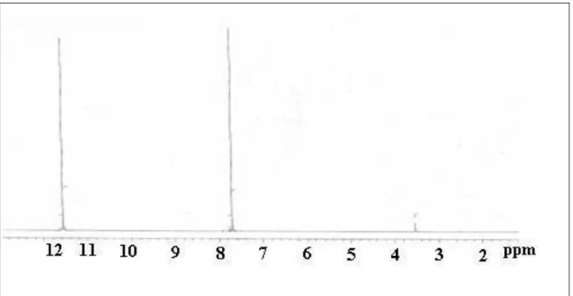
Figure 2 The 1H chemical shift spectrum of glyoxime molecule in the DMSO-d6
solution [27]
Figure 3 The 13C chemical shift spectrum of glyoxime molecule in the DMSO-d 6
106
Experimentally measured chemical shift values and chemical shift values calculated with 6 different basis sets in gas phase and liquid phase were given in Table 2.
Table 2 The calculated and measured chemical shift values of 1H and 13C for the glycoside molecule in gas phase and liquid phase [27]
Gas phase H9 and H10
(ppm) H6 and H8 (ppm) C1 and C2 (ppm) 6-31G(d,p) 7.92 6.66 143.08 6-31+G(d,p) 8.16 6.69 146.30 6-311+G(d,p) 8.18 6.74 157.58 6-311G(d,p) 8.10 6.53 155.16 6-31++G(d,p) 8.16 6.69 146.30 6-311++G(d,p) 8.21 6.74 157.58
Liquid Phase (DMSO-d6)
6-31G(d,p) 8.16 8.32 144.69 6-31+G(d,p) 8.45 8.62 148.72 6-311+G(d,p) 8.50 8.40 160.29 6-311G(d,p) 8.36 8.21 157.18 6-31++G(d,p) 8.48 8.67 148.72 6-311++G(d,p) 8.47 8.47 160.29 Experimental (ppm) 7.7 11.5 146.07
The experimentally determined 13C chemical shift values of the glyoxime molecule were 146.07 ppm as seen in Table 2. Considering the molecular structure of the glyoxime molecule, it was expected that carbon atoms will give a single peak. The chemical shift values of the carbon atom 1 and carbon atom 2 given in Figure 1 calculated with three base sets of 6-31G(d,p), 6-31+G(d,p) ve 6-31++G(d,p) were well matched to the experimental values both in gas phase and liquid phase. When the experimental 1H NMR spectrum of the glyoxime molecule is examined, two distinct
singlet peaks were observed.
These peaks were observed at 11.5 ppm and 7.7 ppm. The chemical shift corresponding to 11.5 ppm is thought to be the OH hydrogen of the molecule and the chemical shift corresponding to 7.7 ppm is thought to be the hydrogen bound to the carbon atoms. The difference between experimental values with calculated chemical shift values for 6 different basis set in solution and gas phase of H9 and H10 atoms was
107
less than 1 ppm. Therefore, the calculated chemical shift values and the experimental chemical shift values were in good agreement for H9 and H10 atoms. The difference between the experimental chemical shift values for H6 and H8 atoms and the chemical shift values calculated with 6 different base sets was about 5 ppm in the gas phase and about 3 ppm in the solution phase. According to these results, the theoretical chemical shift values were not in good agreement with experimental chemical shift values, for H8 and H6 atoms.
According to the literature research, chemical shift values of hydrogen atoms with hydrogen bonding were larger than chemical shift values of hydrogen atoms without hydrogen bonding [29,30]. Theoretical calculations show that the chemical shift values of OH hydrogens did not match the experimental chemical shift values since a single molecule approach was employed in solution. The glyoxim molecule was modeled as a dimer in solution to calculate the effect of the hydrogen bond on the 1H
chemical shift value, which was theoretically calculated. A schematic representation of the dimer molecular model was shown in Figure 4.
108
The stable structure of the dimer model was calculated by the DFT / B3LYP / 6-311 ++ G (d, p) method basis set combination. The 1H chemical shift values of the molecular dimer model were calculated in solution phase using the DFT/B3LYO/6-31++G(d,p) method and basis set combination. When Table 2 was examined, it appears that this method and basis set combination were suitable for the calculation. According to the results of the calculation, the chemical shift value of the hydrogen-bonding H atom was calculated as 11.3 ppm . This theoretical value was in very good agreement with the experimental value. Accordingly, the intermolecular hydrogen bonding significantly affect the 1H chemical shift values, for the glyoxime molecule and this situation had been supported by the theoretical calculations.
Conclusions
In this study, the 1H and 13C chemical shift values of the glyoxime molecule were determined theoretically and experimentally. The experimental 13C chemical shift
values of the glyoxime molecule were consistent with theoretical calculated in the solution phase. However, it had been observed that the 1H chemical shift values for the
hydrogen atoms, which were formed hydrogen bonding, do not agree with the experimental values. Theoretical calculations had been carried out by using a dimer molecule approach instead of a single molecule approach for resolve this disagreement. It had been seen that theoretical and experimental values agree with each other as a result of the dimer molecular approach.
Accordingly, instead of the single molecule approach for 1H chemical shift calculations in oxime molecules, at least a dimer molecule approach should be made and the hydrogen bond effect should be taken into account. If the calculations were performed in solution phase, both 1H and 13C chemical shift calculations would agree with the experimental values for oxime molecules.
109 References
[1] Kaupp, M., Bühl, M., Malkin, V. G., Calculation of NMR and EPR parameters, Wiley-Vch, Weinheim, 2004.
[2] Wu, A., Zhang, Y., Xu X., Yan, Y., Systematic Studies on the Computation
of Nuclear Magnetic Resonance Shielding Constants and Chemical Shifts: The Density Functional Models, Journal of Computational Chemistry, 28(15), 2431-2442, 2007.
[3] Hehre, W. J., Radom, L., Schleyer, Pvon R., Pople, J. A., Ab Initio Molecular Orbital Theory, John Wiley & Sons, New York, 1986.
[4] Foresman, J. B., Frisch, A., Exploring Chemistry with Electronic Structure Methods, Gaussian, Pittsburgh, 1996.
[5] Frisch, M. J., Frisch, A., Foresman, J. B., Gaussian 94 Users Reference, Gaussian, Pittsburgh, 1995.
[6] Ruud, K., Helgaker, T., Kobayashi, R., Jorgensen, P., Bak, K. L., Jensen, H. J. A., Multiconfigurational self-consistent field calculations of nuclear shieldings using
London atomic orbitals, Journal of Chemical Physics, 100(11), 8178-8185, 1994.
[7] Chesnut, D. B., Ab Initio Calculations of NMR Chemical Shielding, Annual, Reports on NMR Spectroscopy, 29, 71-122, 1994.
[8] de Dios, A. C., Ab initio calculations of the NMR chemical shift, Progress in Nuclear Magnetic Resonance Spectroscopy, 29(3-4), 229-278, 1996
[9] Barszczewicz, A., Jaszu´nski, M., Jackowski, K., Ab initio calculations of the
oxygen atom NMR shielding in the carbonyl group, Chemical Physical Letters, 203(4),
404-408, 1993.
[10] Cheeseman, J. R., Trucks, G. W., Keith, T. A., Frisch, M. J., A comparison
of models for calculating nuclear magnetic resonance shielding tensors, Journal of
Chemical Physics, 104(14), 5497-5509, 1996.
[11] Kupka, T., Koaski, M., Pasterna, G., Ruud, K., Towards more reliable
prediction of formaldehyde multinuclear NMR parameters and harmonic vibrations in the gas phase and solution, Journal of Molecular Structure (THEOCHEM), 467(1),
110
[12] DeHaven-Hudkins, D. L., Komer, K. M., Peterson, J. A., Mavunkel, B. J., Rzeszotarski, W. J., Opioid agonist properties of two oxime derivatives of naltrexone,
NPC 831 and NPC 836, Pharmacology Biochemistry and Behavior, 44(1), 45-50, 1993.
[13] DeHaven-Hudkins, D. L., Brostrom, P. A., Allen, J. T., Lesko, L. J., Ferkany, J. W., Kaplita, P. V., Mavunkel, B. J., Rzeszotarski, W. J., Steranka, L. R.,
Pharmacologic Profile of NPC 168 (naltrexone phenyl oxime), A Novel Compound With Activity At Opioid Receptors, Pharmacology Biochemistry and Behavior, 37(3),
497-504, 1990.
[14] Schenone, S., Bruno, O., Ranise, A., Bondavalli, F., D'Amico, M., Parrillo, C., Lampa, E., Rossi, F., N-Substituted 0-(3-amino-2- hydroxypropyl) Oximes of
1,3,3-trimethyl-5-endo-(1-piperidinyl or 4- morpholinyl)-2-oxabicyclo (2,2,2)-octan-6-ones With Platelet Antiaggregating and Local Anesthetic Activities, Farmaco, 47(10),
1249-1262, 1992.
[15] Ranise, A., Bondavalli, F., Bruno, O, Schenone, P, Faillace, G., Coluccino, A., Filippelli, W., Di Sarno, A., Marmo, E., Omega-dialkylaminoalkyl Ethers of
3-EXO-dialkylamino-(z)-camphoroximes With Anitarrhythmic and Local Anesthetic Activities,
Farmaco, 45(2), 187-202, 1990.
[16] Gasc, J. C., d'Ambrieres, S. G., Lutz, A., Chantot, J. F., New Ether Oxime
Derivates of Erythromycin A. A Structure-Activity Relationship Study, The Journal of
Antibiotics (Tokyo), 44(3), 313-330, 1991.
[17] Cooper, C. S., Klock, P. L., Chu, D. T. W., Hardy, D. J., Swanson, R. N., Plattner, J. J., Preparation and in vitro and in vivo evaluation of quinolones with
selective activity against Gram-positive organisms, Journal of Medicinal Chemistry,
35(8), 1392-1398, 1992.
[18] Tsukamoto, Y., Sato, K., Mio, S., Sugai, S., Yanai, T., Kitano, N., Muramatsu, S., Nakada, Y., Ide, J., Synthesis of 5-keto-5-oxime Derivates of
Milbemycins and Their Activities Against Microfilariae, Agricultural and Biological Chemistry, 55(10), 2615-2621, 1991.
[19] Bowman, D. D., Darigrand, R. A., Frongillo, M. K., Barr, S. C., Flanders, J.A., Carbone, L.G., Treatment of experimentally induced trichinosis in dogs and cats, American Journal of Veterinary Research, 54(8), 1303-1305, 1993.
111
[20] Abdalla, S., Khalili, F., Effects of Dichloroglyoxime on Isolated
Guinea-Pig Smooth Muscle and Atrium, Drug and Chemical Toxicology, 15(2), 145-159, 1992.
[21] Bodor, N., Elkoussi, A., Improved Delivery Through Biological
Membranes.LVI.Pharmocological Evaluation of Alprenoxime-A New Potential Aniglaucoma Agent, Pharmaceutical Research, 8(11), 1389-1395, 1991.
[22] Ballantyne, B., Ophtalmic Effects of Oximes: A Review, Veterinary and Human Toxicology, 33(2), 151-154, 1991.
[23] Olofson, R., Michelman, J., Furuzan, Journal of Organic Chemistry, 30, 1854-1859, 1965.
[24] Spartan 08 Programme, Wavefunction Inc., Irvine, CA 92612, USA, 2008. [25] Lee, C., Yang, W., Par,r R.G., Development of the Colle–Salvetti
correlation- energy formula into a functional of the electron density, Physical Review B
37(2), 785–789, 1988.
[26] Gaussian 03 Programme, Revision E.01, Frisch, M. J., Trucks, G. W., Schlegel, H. B., Scuseria, G. E., Robb, M. A., Cheeseman, J. R., Montgomery, Jr., J. A., Vreven, T., Kudin, K. N., Burant, J. C., Millam, J. M., Iyengar, S. S., Tomasi, J., Barone, V., Mennucci, B., Cossi, M., Scalmani, G., Rega, N., Petersson, G. A., Nakatsuji, H., Hada, M., Ehara, M., Toyota, K., Fukuda, R., Hasegawa, J., Ishida, M., Nakajima, T., Honda, Y., Kitao, O., Nakai, H., Klene, M., Li, X., Knox, J. E., Hratchian, H. P., Cross, J. B., Adamo, C., Jaramillo, J., Gomperts, R., Stratmann, R. E., Yazyev, O., Austin, A. J., Cammi, R., Pomelli, C., Ochterski, J. W., Ayala, P. Y., Morokuma, K., Voth, G. A., Salvador, P., Dannenberg, J. J., Zakrzewski, V. G., Dapprich, S., Daniels, A. D., Strain, M. C., Farkas, O., Malick, D. K., Rabuck, A. D., Raghavachari, K., Foresman, J. B., Ortiz, J. V., Cui, Q., Baboul, A. G., Clifford, S., Cioslowski, J., Stefanov, B. B., Liu, G., Liashenko, A., Piskorz, P., Komaromi, I., Martin, R.L., Fox, D.J., Keith, T., Al- Laham, M. A., Peng, C. Y., Nanayakkara, A., Challacombe, M., Gill, P. M. W., Johnson, B., Chen, W., Wong, M. W., Gonzalez, C., Pople, J. A., 2003. Gaussian, Inc., Pittsburgh P.A.
[27] Taşdemir H. U., Calculation of NMR Parameters of Some Molecules Using HF, Post HF and DFT Methods Selçuk University, Master Thesis, Konya, 2010.
112
[28] Jeffrey, G. A., Ruble, J.R., Pople, J. A., Neutron Diffraction at 9 K and ab
initio Molecular Orbital Studies of the Molecular Structure of Glyoxime, Acta
Crystallography, B38: 1975-1980, 1982.
[29] Hori, S., Yamauchi, K., Kuroki, S., Ando, I., Proton NMR Chemical Shift
Behavior of Hydrogen-Bonded Amide Proton of Glycine-Containing Peptides and Polypeptides as Studied by ab initio MO Calculation, International Journal of Molecular
Sciences, 3(8), 907-913, 2002.
[30] Malek, K., Vala, M., Kozlowski, H., Proniewicz, L. M., Experimental and
theoretical NMR study of selected oxocarboxylic acid oximes, Magnetic Resonance in
![Figure 1 Schematic representation of the most stable structure of the glyoxime molecule [27]](https://thumb-eu.123doks.com/thumbv2/9libnet/4484495.78650/5.892.279.652.813.1054/figure-schematic-representation-stable-structure-glyoxime-molecule.webp)
![Table 1 Some bond length and bond angle values of molecule for X-ray structure and the calculated most stable structure [27]](https://thumb-eu.123doks.com/thumbv2/9libnet/4484495.78650/6.892.188.706.314.800/table-length-values-molecule-structure-calculated-stable-structure.webp)
![Table 2 The calculated and measured chemical shift values of 1 H and 13 C for the glycoside molecule in gas phase and liquid phase [27]](https://thumb-eu.123doks.com/thumbv2/9libnet/4484495.78650/8.892.196.694.299.683/table-calculated-measured-chemical-values-glycoside-molecule-liquid.webp)
![Figure 4 Schematic representation of the dimer modeling for glyoxime molecule [27]](https://thumb-eu.123doks.com/thumbv2/9libnet/4484495.78650/9.892.132.765.621.1069/figure-schematic-representation-dimer-modeling-glyoxime-molecule.webp)
