GENETIC AND EPIGENETIC ANALYSIS OF
IMMORTAL AND SENESCENCE ARRESTED
LIVER CANCER CELLS
A THESIS SUBMITTED TO
THE DEPARTMENT OF MOLECULAR BIOLOGY AND
GENETICS
AND THE INSTITUTE OF ENGINEERING AND SCIENCE
OF
BİLKENT UNIVERSITY
IN PARTIAL FULFILLMENT OF THE REQUIREMENTS
FOR THE DEGREE OF DOCTOR OF PHILOSOPHY
By
G. SEVGİ BAĞIŞLAR
August 2009
I certify that I have read this thesis and that in my opinion it is fully adequate, in scope and in quality, as a thesis for the degree of Doctor of Philosophy.
________________________________ Prof. Dr. Mehmet Öztürk (Supervisor) I certify that I have read this thesis and that in my opinion it is fully adequate, in
scope and in quality, as a thesis for the degree of Doctor of Philosophy.
________________________________ Asst. Prof. Dr. Özlen Konu
I certify that I have read this thesis and that in my opinion it is fully adequate, in scope and in quality, as a thesis for the degree of Doctor of Philosophy.
________________________________ Asst. Prof. Dr. Cengiz Yakıcıer
I certify that I have read this thesis and that in my opinion it is fully adequate, in scope and in quality, as a thesis for the degree of Doctor of Philosophy.
________________________________ Prof. Dr. Funda Yılmaz-Barbet I certify that I have read this thesis and that in my opinion it is fully adequate, in scope and in quality, as a thesis for the degree of Doctor of Philosophy.
________________________________ Doç. Dr. Esra Erdal
Approved for the Institute of Engineering and Science
_______________________________ Prof. Dr. Mehmet Baray
To my mother…..
Anneme…..
ABSTRACT
G. SEVGİ BAĞIŞLAR
PhD in Molecular Biology and Genetics Supervisor: Prof. Dr. Mehmet Öztürk
August 2009, 123 Pages
Genetic and epigenetic aspects of cellular senescence and immortality in hepatocellular carcinoma (HCC) are poorly elucidated. The aim of our thesis was to characterize senescence and immortality gene network (SIGN) involved in these cancers. We also wished to explore epigenetic changes associated with senescence and immortality of HCC cells. First, we identified differentially expressed genes in immortal, pre-senescent and senesce-arrested Huh7 clones. Our microarray analysis revealed 6390 probesets significantly changing among groups. Moreover, the significant gene signature could successfully discriminate both replicative senescent cells, and oncogene-induced senescent cells from their immortalized counterparts. E2F1 targets, stem-cell related genes, DNA repair, RNA splicing and cell cycle related gene sets were enriched specifically in immortal cells, whereas immune function, stress response, electron transporter activity, protein modification, metabolism, chromatin biogenesis related gene groups were significantly up-regulated in senescent clones. Next, we integrated gene expression data from senescence-programmed and immortal HCC cells with the data from cirrhosis and HCC tissues to generate a SIGN signature. This signature identified several HCC classes, including one “normal-like”, and two with increased expression of immortality genes. Senescence-to-immortality transition was accompanied by hepatic dedifferentiation and increased expression of cell proliferation, chromosome modification and DNA damage response genes. Finally, we identified a large set of upregulated DNA damage checkpoint and DNA repair genes that showed significant associations with some SIGN classes of HCC tumors. As retinoblastoma/E2F pathway plays a key role in cellular senescence, we also analyzed E2F and DP family members in senescent and immortal hepatocellular carcinoma cells. E2F1, E2F5, E2F7, E2F8 and DP1 were up-regulated in immortal hepatocellular carcinoma (HCC) cell lines as compared to senescent cells, whereas E2F3a and DP-2 expressions were downregulated. Upregulation of DP2
expression in senescent cells correlated with increased DP2 protein expression, as tested with TGF-beta induced senescence models. Finally, we demonstrated important epigenetic changes associated with hepatocellular immortality and senescence. Among histone methyltransferases and demethylases, MLL3, FBXL11, SUV420H1, UTX, SMYD2, SETD2, JMJD2B, JMJD3, JARID1B and ASH1L genes were up-regulated, and EZH2 was down-regulated in senescent cells. These changes were accompanied with changes in histone methylation patterns. Of particular interest, H3K27me1, H3K27me3, H4K20me3, H3R2me2a and H4R3me2a forms of methylated histones displayed increased expression in both Huh7 and MRC5 senescent cells, as compared to their immortal forms. Finally, H3K27me3, H4K20me3, H3K36me3, H3R17me2a, H4R3me2a also showed decreased expression in some cirrhotic liver and primary HCC tumors. In conclusion, we demonstrated that a large set of senescence and immortailty genes were dysregulated in HCC. This profound change in gene expression was associated with differential expression of histone modifying enzymes, as well as histone methylation status. Thus, the immortalization of hepatocytes during hepatocellular carcinogenesis is accompanied with global gene expression changes probably mediated by a major modification of their epigenetic program via histone demethylation.
ÖZET
İMMORTAL VE HÜCRE YAŞLANMASI PROGRAMLI KARACİĞER KANSER
HÜCRELERİNDE GENETİK VE EPİGENETİK ANALİZİ
G. SEVGİ BAĞIŞLAR
Doktora Tezi, Moleküler Biyoloji ve Genetik Bölümü Danışman: Prof. Dr. Mehmet ÖZTÜRK
Ağustos 2009, 123 Sayfa
Hepatoselüler karsinomda (HCC), hücre yaşlanması ve immortalitesinin genetik ve epigenetik özellikleri henüz zayıf olarak açıklanmıştır. Tezimizin amacı; karaciğer kanserinde rol alan hücre yaşlanması ve immortalite gen ağını karakterize etmektir. Ayrıca, hücre yaşlanması ve immotral HCC hücrelerinin epigenetik değişikliklerini de incelemek istedik. İlk önce, immortal, erken hücre yaşlanması ve hücre yaşlanması Huh7 klonlarında gen ifade farklılıklarını bulduk. Mikroarray çip çalışmamız sonucunda 6390 prob seti, bu gruplar arasında değişiklik gösterdi. Daha fazlası, bu anlamlı gen seti; hem replikatif hücre yaşlanması hem de oncogen indüklenmiş hücre yaşlanması modellerini, bunların immortalize edilmiş karşılıklarından başarı ile ayırdı. Biyolojik fonksiyon gruplama çalışmaları; E2F1 hedef genlerinin, kök hücre ilişikli genlerin, DNA tamir, RNA splicing ve hücre döngüsü ilişik gen gruplarının özellikle immortal hücrelerde ifadesinin arttığını gösterdi. Öte yandan, immün sistem, stres cevabı, elektron taşınımı, protein modifikasyonu, metabolizma ve kromatin biyogenezi ile ilgili grupları yaşlanmış hücre klonlarında ifade gösteriyordu. Ayrıca, immortal ve hücre yaşlanması programlı hücre datasıyla siroz ve HCC doku datasını birleştirerek, ortak değişen “hücre yaşlanması ve immortal gen ağı (SIGN) listesini elde ettik. SIGN gen seti, farklı HCC alt gruplarını birbirinden ayırmayı başardı. SIGN gen setinde hücre yaşlanmasından immortale doğru anlamlı olarak değişen gruplar; hepatic de-differensiye genleri, hücre döngüsü, kromozom modifikasyonu ve DNA hasarına cevap veren gen grupları idi. Ayrıca, geniş bir sayida DNA tamir genlerinin tümor oluşumu ve ilerlemesi ile alakalı olduğunu belirledik.
Retinoblastoma/E2F yolağı hücre yaşlanmasında önemli bir rol oynamaktadır, biz de bu yüzden E2F ve DP protein ailesi üyelerinin yaşlanmış ve immortal programlı hepatoselüler karsinomlarında analiz ettik. E2F1, E2F5, E2F7, E2F8 ve DP1 genlerinin
ifadesi immortal hücrelerde artış gösterdi. Öte yandan E2F3a ve DP2 genlerinin ifadesi azalmıştı. DP-2 ifadesinin senesens Huh7 hücrelerde artışı; DP2 protein artışı ile ilintilendirildi ve aynı artış TGF-beta indüklenmiş hücre yaşlanmasında da gözlendi. Son olarak, hepatoselüler immortalite ve yaşlanmış hücrelerinde önemli epigenetik değişiklikler gösterdik. Histone metiltransferaz ve demetilazlar arasında; MLL3, FBXL11, SUV420H1, UTX, SMYD2, SETD2, JMJD2B, JMJD3, JARID1B ve ASH1L genlerinin hücre yaşlanmasında artış gösterdiğini ve EZH2 geninin azalış gösterdiğini gözlemledik. Bu değişiklikler eşliğinde, histon metilasyon seviyeleri de değişikliğe uğruyordu. Yaşlanmış Huh7 ve MRC5 hücrelerde immortal eşdeğerlerine göre, H3K27me1- and 3, H4K20me3, H3R2me2a ve H4R3me2a rezidülerinin artış gösterdiğini gözledik. H3K27me3, H4K20me3, H3K36me3, H3R17me2a, ve H4R3me2a rezidüleri bazı sirotik karaciğer ve HCC tümörlerinde azalan bir ifade gösterdi.
Sonuç olarak, geniş sayıda immortal ve hücre yaşlanması genlerinin HCC de de değiştiğini göstermiş olduk. Histon modifiye eden genleri ve histon metilasyon seviyesi de değişikliğe uğruyordu. Yani, hepatositlerin immortalizasyon süreçleri global gen ifade değişikliği ile oluyor ve, büyük ihtimalle, bu değişim epigenetik program tarafından yönlendiriliyor.
ACKNOWLEDGEMENTS
I would like to express my gratitude to Prof. Mehmet Öztürk for his supervision, endless support and valuable suggestions throughout the course of my studies. It has always been a privilege for me to be accepted in his team, work with him and being educated by him.
I would like to thank to the jury members for evaluating my thesis.
I would like to express my special thanks to Assist. Prof. Özlen Konu for sharing her excellent experiences on bioinformatics, her support, her patience, and being such a kind person all the time.
I would like to attend my very special thanks to Assist. Prof. Tamer Yağcı for his support, for scientific discussions and for paying attention to my scientific ideas, even if they were foolish. I would like to thank to Assist. Prof. Cengiz Yakıcıer for his support, his friendship, his kindness. I would like to thank to Assist. Prof. Uygar Tazebay for his kindness and patience. For three of you, thank you for being there.
I would like to thank to Prof. Dr. Funda Yılmaz-Barbet and Prof. Dr. Hakan Bozkaya for their supports in my projects.
I would like to thank to my previous supervisor Prof. Dr. Tayfun Özçelik for teaching me, and all PI’s in MBG for educating me and being patient to me. I would like to thank to Prof. Dr. Stéfan Dimitrov for his very kind supports.
I would like to thank to my project partners Ayça Arslan-Ergül and Haluk Yüzügüllü for working and being with me, and all current and past group friends Şerif Şentürk, Mine Mumcuoğlu, Nuri Öztürk for teaching me a lot, and Nilgün Taşdemir, Pelin Gülay, Eylül and Gökhan.
I would like to express all my gratitude to my friends Bàlà Gür-Dedeoğlu, Elif Uz, Ceren Çıracı and Elif Yaman for the happy days and being with me. I thank to Tolga Acun, Hani Al-Otaibi, Ender Avcı and Emin BeyAbi for being there with smiley faces when I need anything. I would like to thank other MBG graduate students that I can not list here for making the PhD life easier.
I would like to thank to “ma p’tite” Sophie Barral for being her, being my friend or more than a friend. I would like to thank to Angeline Eymery for being very lovely to me everytime. Thanks god, I came to France and I met you. I would like to express my
deepest feelings to my dear friends Nicolas Reynoird, Sébastien Cadau, Leila Todeschini, Aurelia Vavasseur, Faycal Boussouar, Hong Lien and Xavier Camous. I feel lucky to know you. Thank you for being my friends.
I would like to thank to Mrs. Sevim Baran, Mrs. Füsun Elvan, Mr. Abdullah Ünnü, Mr. Turan Daştandır, Mrs. Bilge Özbayoğlu, Miss. Tülay Arayıcı for their very kind helps to make my life easier. This thesis could not be completed without your helps. I would like to thank to Mrs. Denise Leardini, Mrs. Sanie Claraz, Mr. Michail Gidopoulos and Mrs. Dalenda Benmedjahed for helping me and for loving me like a family.
Lastly, I wish to thank to my parents and my sister, Betül, and brother, Ümit, for their endless support during my life and loving me in every condition. I dedicate this thesis to my mother, my endless love source, who believed in me, trusted in me, who is absolutely unique and a perfect mother.
TABLE OF CONTENTS SIGNATURE PAGE ii ABSTRACT iv ÖZET vi ACKNOWLEDGEMENTS viii TABLE OF CONTENTS x
LIST OF FIGURES xvii
LIST OF TABLES xix
CHAPTER 1. INTRODUCTION 1
1.1 Hepatocellular malignancy 1
1.2 Pathogenesis of hepatocellular carcinoma 2
1.2.1 Viral induced hepatocarcinogenesis 2
1.2.2 Alcohol-induced hepatocarcinogenesis 4
1.2.3 Aflatoxin-B1-induced hepatocarcinogenesis 5
1.3 Genetic and epigenetic events in HCC 5
1.3.1 The p53 tumor suppressor 5
1.3.2 β-Catenin and AXIN1 6
1.3.3 ErbB receptor family 6
1.3.4 MET ad HGF 7
1.3.5 Methylation of cancer-relevant genes 7
1.3.6 c-myc 7
1.4 Liver cirrhosis and senescence 8
1.4.1 Cellular senescence 8
1.4.1.1 Replicative senescence 9
1.4.1.2 Oncogene and ROS-induced senescence 10
1.4.2 Senescence as an anti-tumor mechanism in hepatocellular carcinoma 10
1.4.3 Cyclin-dependent inhibitors 11
1.5 Expression profiling using Affymetrix GeneChip Microarrays 12
1.6 Rb/E2F pathway 13
1.6.1 DP-2 14
1.7 Chromatin midifications and hepatocellular carcinoma 15
1.7.1 DNA methylation 15
1.7.1.1 DNA methylation and cancer 16
1.7.2 Histone modifications 18
1.7.2.1 Histone modifying enzymes 19
1.7.2.2 Histone methylation and cancer 21
1.7.2.2.1 H3K27 methylation 22
1.7.2.2.1.1 EZH2 22
1.7.2.2.2 H3K9 methylation 23
1.7.2.2.2.1 HP1-a H3K9 methylation “reader” 23
1.7.2.2.2.2 SUV39H-RIZ1-H3K9 methylation “writers” 23 1.7.2.2.2.3 JMJD2C- H3K9 methylation “eraser” 24
1.7.2.2.3 H3K4 methylation 24
1.7.2.2.3.1 MLL-SMYD3- H3K4 methylation “writers” 24 1.7.2.2.3.2 ING proteins- H3K9 methylation “readers” 25
1.7.2.2.4 H3K36, H4K20 and H3K79 methylation 25
1.7.2.2.4.1 H3K36, H4K20, H3K79methylation‘writers’ 25 1.7.2.3 Histone lysine acetylation and cancer 25
1.7.2.4 Histone methylation and senescence 26
1.7.2.5 Histone variants 27
CHAPTER 2. OBJECTIVES AND RATIONALE 28
CHAPTER 3. MATERIALS AND METHODS 30
3.1 Materials 30
3.1.1 Reagents 30
3.1.2 Nucleic acids and proteins 30
3.1.3 Oligonucleotides 30
3.1.4 Enzymes 30
3.1.5 Electrophoresis 30
3.1.6 Protein transfer materials 31
3.1.7 Tissue culture reagents and cell lines 31
3.1.8 Antibodies and chemiluminescence 31
3.1.9 Kits 32
3.2 Solutions and media 32
3.2.1 General solutions 32
3.2.2 Tissue culture solutions 32
3.2.3 RNA solutions 33
3.2.4 Protein extraction and western blotting solutions 34
3.2.5 Immunofluoroscence and immunoperoxidase solutions 35
3.2.7 Immunohistochemistry solutions 36
3.3 Methods 36
3.3.1 Tissue culture techniques 36
3.3.1.1 Cell lines 36
3.3.1.2 Cell lines for microarray study 37
3.3.1.3 Thawing cell lines 37
3.3.1.4 Cryopreservation of cell lines 37
3.3.1.5 TGF-β and LiCL treatment 38
3.3.2 RNA extraction 38
3.3.2.1 Extraction of total RNA from tissue culture cells 38
3.3.2.2 Extraction of total RNA from tissue samples 38
3.3.3 Expression microarray analysis 39
3.3.3.1 Microarray experiments 39
3.3.3.2 Data processing and quality controls 40
3.3.3.3 Determination of differentially expressed gene sets 40
3.3.3.4 Visualization of dataset 42
3.3.3.5 Data integration 42
3.3.3.6 Functional gene annotation cluster analysis 42
3.3.3.6.1 DAVID 42
3.3.3.6.2 GSEA 43
3.3.3.6.3 Onto express 43
3.3.3.6.4 Ingenuity Pathway Analysis 43
3.3.3.7 Meta Data 43
3.3.3.7.1 HPEC replicative senescence data 43
3.3.3.7.2 IMR90 Oncogene-induced senescence data 44
3.3.3.7.3 HCC molecular classification data 44
3.3.3.8 BRB Array Tools 44
3.3.4 Quantification of nucleic acids 44
3.3.4.1 Horizantal agarose gels of DNA samples 44
3.3.4.2 Gel electrophoresis of of total RNA 45
3.3.6 Polymerase chain reaction (PCR) 45
3.3.6.1 Primer design for expression analysis for SQ and Q PCR 46
3.3.6.2 Expression analysis of a gene by semi-quantitative PCR 49
3.3.6.2.1 Determination of optimal cycle of a gene for 49
semi-quantitative PCR 3.3.6.2.2 GAPDH normalization 49
3.3.6.2.3 PCR amplification of target region 49
3.3.6.3 Quantitative real-time PCR 50
3.3.6.3.1 Amplification efficiency calculations 50
3.3.7 Total protein isolation 51
3.3.7.1 Protein isolation from tissue culture cells 51
3.3.7.2 Protein isolation tissue samples 51
3.3.7.3 Histone extraction 51
3.3.8 Quantification of proteins 52
3.3.9 Western blotting 52
3.3.10 Immunofluorescence 53
3.3.11 Immunoperoxidase 53
3.3.12 Immunohistochemistry on paraffin-embedded tissue sections 54
3.3.13 SABG assay 55
CHAPTER 4. RESUTS 56
4.1 Identification of senescence and immortality gene network and its role in 56
hepatocellular carcinoma 4.1.1 Expression analysis of immortal and reprogrammed senescent cells of 56
hepatocellular carcinoma 4.1.2 Differentially expressed gene set between immortal, pre- and senescent 57
clones 4.1.3 Confirmation of microarray data 59
4.1.4 Analysis of significant gene set on other senescence microarray data 61
4.1.5 Functional classification of senescence and immortality genes 63
4.1.6 Identification of the role of senescence and immortality genes in HCC 68
4.1.6.2 DNA damage response genes as potential therapeutic targets 76
4.2 Expression analysis of E2F/DP family in senesence and immortality,senescence 78
association of DP-2 4.2.1 Differential expression of E2F/DP family genes in 78
senescent/immortal hepatocellular carcinoma cells 4.2.2 Expression analysis of differentially regulated E2F/DP genes in 80
HCC cell lines 4.2.3 DP2 protein is abundant in senescent clone but lost in immortal clone 81
4.2.4 Examination of the DP-2 isoforms in different cell cycle conditions 83
and upon TGF-β treatment 4.3 Epigenetic changes in immortality and senescence of liver in vitro and in vivo 86
4.3.1 Differential expression of histone modifying enzyme genes in senescent 86 and immortal hepatocellular carcinoma cells 4.3.2 Histone methylation changes between immortal and senescent Huh7 87
clones 4.3.3 Does histone methylation levels differ in-vivo? 93
4.3.4 Histone variant differences in immortal and senescent Huh7 cells 96
CHAPTER 5. DISCUSSION 98
5.1 Global expression analysis of immortality and senescence in liver cancer 98
5.1.1 Identification of differentially expressed genes between immortal, 98
pre- and senescent Huh7 clones 5.1.2 Establishing a senescence and immortality gene network signature 99
for cirrhosis and hepatocellular carcinoma 5.2 DP-2 is associated with senescence 101
5.3 Histone methylation levels of some H3 and H4 residues change in immortality 102
and senescence of liver in vitro and in vivo CHAPTER 6. FUTURE PERSPECTIVES 105
REFERENCES 107
APPENDIX A 116
APPENDIX C 122
APPENDIX D 127
LIST OF FIGURES
Figure 1.1 Multistage process of hepatocarcinogenesis 2
Figure 1.2 Mechanisms of hepatocarcinogenesis 5
Figure 1.3 Senescence controlled by the p53 and p16-Rb pathways 9
Figure 1.4 Senescence pathways 10
Figure 1.5 E2F/DP family 15
Figure 1.6 Epigenetic alterations in tumor progression 16 Figure 1.7 DNA methylation differences in some cancers 17
Figure 1.8 Histone modifications 19
Figure 1.9 Histone modifying enzymes 20
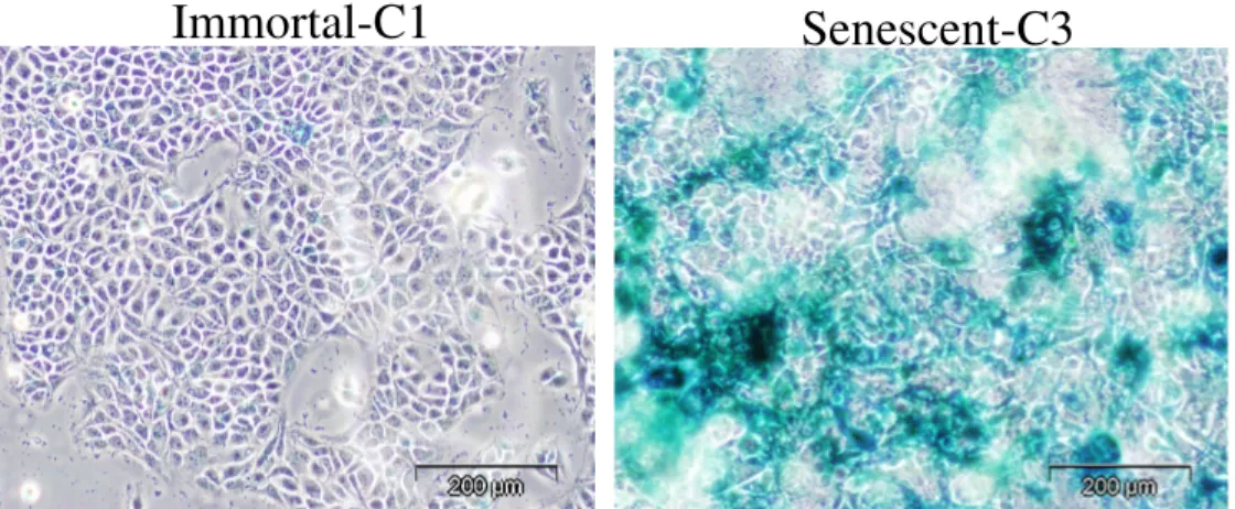
Figure 4.1.1 Senescence associated B-galactosidase (SABG) staining of 56 immortal and senescent cells
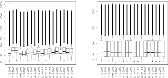
Figure 4.1.2 Quality assesment of microarray data 57
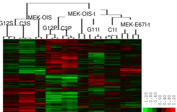
Figure 4.1.3 Plots of microarray data before and after normalization 58 Figure 4.1.4 Huh7 isogenic clones significant gene list 58
unsupervised hierarchical cluster analysis
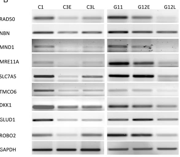
Figure 4.1.5 Confirmation of microarray data 61
Figure 4.1.6 Meta-analysis with previously published senescence data 63 Figure 4.1.7 Enrichement plots and cluster dendograms of 3 enriched gene sets 66
Figure 4.1.8 Biological functions 67
Figure 4.1.9 Venn diagram 68
Figure 4.1.10 Hierarchical clustering of senescence-programmed and 70 immortal cell lines, and cirrhosis and hepatocellular carcinoma (HCC) tissues
Figure 4.1.11 The SIGN signature separates hepatocellular carcinomas 71 (HCCs) into distinct subclusters
Figure 4.1.12 Binary analysis and senescence-immortality associated gene 72 rate of Boyault groups
Figure 4.1.13 The protein networks generated through the use of Ingenuity 74 Pathway Analysis
presenescent and senescent cells
Figure 4.2.2 mRNA expression levels of differentially regulated E2F/DP genes 80 in HCC cell lines
Figure 4.2.3 DP-2 immunostaining 82
Figure 4.2.4 Expression analysis of DP-2 protein 83 Figure 4.2.5 Western blot analysis of DP-2 in LiCl and TGF- β treated cells 84 Figure 4.3.1 Expression analysis of histone methyltransferases and 86
de-methylases in immortal and senescence Huh7 clones
Figure 4.3.2 Histone methylation status of H3 and H4 tail modifications in 88 immortal, pre- and senescent cells
Figure 4.3.3 Immunoperoxidase analysis of histone methylation residues 92 in replicative senescence model of MRC5
Figure 4.3.4 Histone mehtylation in in-vivo 95
Figure 4.3.5 Histone variant levels of immortal, pre- and senescent cells 96
LIST OF TABLES
Table 3.1 Antibodies 31
Table 3.2 Required R packages 40
Table 3.3 Experiment file of in-vitro data 41
Table 3.4 Primers and their sequences 46
Table 3.5 A standard curve preparation with BSA dilution 52 Table 3.6 Protein sample preparation for Bradford assay 52 Table 4.1.1 The numerical report of significant probes and genes 59 Table 4.1.2 Examples of significantly upregulated GSEA gene sets 64 Table 4.1.3 Biological pathways affected in hepatocellular carcinoma 76
classes ccording to SIGN signature
ABBREVIATIONS
APC Adeno poliposis coli
APS Ammonium persulphate
bp Base Pairs
BSA Bovine serum albumin
cDNA Complementary DNA
CDKI Cycline dependent kinase inhibitor
Ct Cycle Threshold
ddH2O Double distilled water
DMEM Dulbecco’s Modified Eagle’s Medium
DMSO Dimethyl Sulfoxide
DNA Deoxyribonucleic Acid
dNTP Deoxyribonucleotide triphosphate
DNMT DNA methyltransferase
DP Dimerization partner
ds Double strand
EGFR Epidermal growth factor receptor
EtBr Ethidium Bromide
ER Endoplasmic reticulum
FBS Fetal Bovine Serum
H Histone
HAT Histone acetyl transferase
HCC Hepatocellular carcinoma
HB Hepatoblastoma
HBcAg Hepatitis B core antigen
HBV Hepatitis B virus
HBx Hepatitis B protein x
HCV Hepatitis C virus
HDAC Histone deacetylase
HGF Hepatocyte growth factor
HRP Horseradish peroxidase
LCD Large cell dysplasia
µg Microgram mg Miligram min Minute µl Microliter ml Mililiter µm Micrometer µM Micromolar mM Milimolar
mRNA Messenger RNA
Oligo(dT) Oligodeoxythymidylic acid
PBS Phosphate Buffered Saline
PDL Population doubling
pRB Retinoblastoma protein
pmol Picomole
q-PCR Quantitative real time RT-PCR
Rpm Revolutions Per Minute
RMA Robust multichip average
RT PCR Reverse Transcription PCR
ROS Reactive oxygen species
SABG senescence-associated b-galactosidase
SAHF Senescence associated heterochromatin loci
SCD Small cell dysplasia
Sec Second
TAE Tris-Acetate-EDTA buffer
TBS Tris buffered saline
TERC Telomerase RNA
TERT Telomerase reverse transcriptase
Tm Melting Temperature
Tris Tris (Hydroxymethyl)- Methylamine
UV Ultraviolet
v/v volume/volume
CHAPTER 1. INTRODUCTION 1.1 Hepatocellular malignancy
The liver is a vital organ present in vertebrates and some other animals; it has a wide range of functions, a few of which are detoxification, protein synthesis, and production of biochemicals necessary for digestion. The liver plays a major role in metabolism and has a number of functions in the body, including glycogen storage, decomposition of red blood cells, plasma protein synthesis, hormone production, and detoxification. It lies below the diaphragm in the thoracic region of the abdomen. It produces bile, an alkaline compound which aids in digestion, via the emulsification of lipids. It also performs and regulates a wide variety of high-volume biochemical reactions requiring highly specialized tissues, including the synthesis and breakdown of small and complex molecules, many of which are necessary for normal vital functions (Anthea et al, 1993).
Primary cancer of the liver is the fourth most common cause of death from cancer (estimated mortality is more than 600,000 deaths per year) and the third most common malignancy in human (Ferlay et al, 2001), (1). The lethality of liver cancer stems in part from its resistance to existing anticancer agents, a lack of biomarkers that can detect surgically resectable incipient disease, and underlying liver disease that limits the use of chemotherapeutic drugs (2). There are two main kinds of primary liver cancer, hepatoma and cholangiocarcinoma. Hepatoma is cancer of the hepatocytes, the main functioning liver cells. Cholangiocarcinoma originates in the bile ducts. Hepatoblastoma (HB) is the most common pediatric liver malignancy, comprising approximately 1% of all pediatric cancers (3). Liver angiosarcoma is a very rare type of primary liver cancer developing from the cells of blood vessels within the liver. Hepatocellular carcinoma (HCC) is one of the most common malignant liver tumors (83% of all cases) in the world with a high prevalence in Asia and sub-Saharan Africa (4). Recent studies have shown that the incidence of HCC has substantially increased in the USA as well as in other areas including Japan and Europe (El-Serag and Mason, 1999),(Taylor-Robinsonet al., 1997). HCC is one of the few human cancers in which an underlying etiology can often be identified in most cases. Hepatocarcinogenesis nearly always develops in the setting of chronic hepatitis or cirrhosis; conditions in which many hepatocytes are killed, inflammatory cells invade the liver and connective tissue (5).
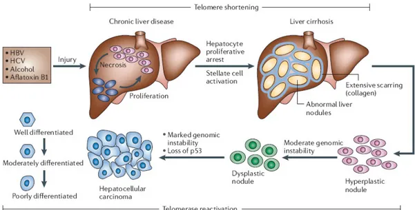
Development of HCC is a multistep process and slow. The sequential events leading to HCC may be summarized in five steps: chronic liver injury that produces inflammation, cell death, cirrhosis and regeneration, DNA damage, dysplasia, and finally HCC (Figure 1.1) (6), (2).
Figure 1.1: Multistage process of hepatocarcinogenesis (2).
1.2 Pathogenesis of hepatocellular carcinoma
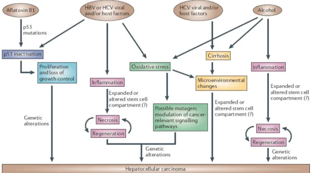
HCC affects all segments of the world population, although significant differences in HCC incidence in various countries reflect the regional differences in the prevalence of specific etiological factors as well as ethnicity (American Cancer Society. Cancer Facts and FIGS 2005. American Cancer Society [online], http://www.cancer.org/docroot/home/index.asp (2005)). The most prominent factors associated with HCC include chronic hepatitis B and C viral infection, chronic alcohol consumption, aflatoxin-B1-contaminated food and virtually all cirrhosis-inducing conditions (Figure 1.2) (7). Other etiological factors have also been
proposed to lead to HCC, albeit at a lower frequency; such as certain metabolic disorders, diabetes, non-alcoholic fatty liver disorders. In addition, gender can also influence the risk and behavior of HCC, with males accounting for a larger fraction of cases (8).
1.2.1 Viral-induced hepatocarcinogenesis
Hepatitis B virus (HBV) infects approximately 2 billion individuals worldwide and causes an estimated 320,000 deaths annually. Approximately 30–50% of HBV-related deaths are attributable to HCC (9). The impact of HBV infection on HCC development is reflected by the correlation between increased incidences of HCC in patients with increasing levels of HBV DNA in serum (10). Hepatitis C virus (HCV) infects approximately 170 million individuals worldwide (11). Approximately 20% of chronic HCV cases develop liver
cirrhosis, and 2.5% develop HCC (12). The viral-associated mechanisms driving hepatocarcinogenesis are complex and involve both host and viral factors.
HBV is a non-cytopathic, partially double-stranded hepatotropic DNA virus classified as a member of the hepadnaviridae family. The HBV genome encodes several viral proteins essential to its life cycle, including a reverse transcriptase/DNA polymerase (pol), the capsid protein known as hepatitis B core antigen (HBcAg), and the L, M and S envelope proteins that associate with the endoplasmic reticulum (ER) membrane as part of their replication process. HBV also encodes a number of proteins whose functions are not fully understood, such as protein x (HBx) (13).
Several lines of evidence support the direct involvement of HBV in the transformation process. First, HBV genome integration has been associated with host DNA microdeletions (14) that can target cancer-relevant genes including telomerase reverse transcriptase (TERT), platelet-derived-growth-factor receptor-β (PDGFRβ ), PDGFβ and mitogen activated protein kinase 1 (MAPK1), among others (15). Second, HBx transcriptional activation activity can alter the expression of growth-control genes, such as SRC tyrosine kinases, Ras, Raf, MAPK, ERK, JNK and others (16), (17). Finally, HBx can bind and inactivate the tumor suppressor p53 in vitro, therefore increasing cellular proliferation and survival and compromising DNA-damage checkpoints (18), (19). The hepatocarcinogenic potential of HBx has been genetically validated in HBx transgenic mice, of which 90% develop HCC (20).
Host–viral interactions seem to contribute to hepatocarcinogenesis in several ways. A robust T-cell immune response is presumably elicited to combat viral infection, however, this response contributes to hepatocyte necrosis, inflammation and consequently regeneration, leading to carcinogenesis (13). Such continuous replication of hepatocytes might enable the
propagation of oncogenic lesions and telomere erosion with consequent genomic instability. Another proposed mechanism of HBV-induced hepatocarcinogenesis might stem from viral– ER physical interactions, that provoke ER stress and ultimately the induction of oxidative stress (21), which can stimulate growth- and survival-signaling pathways, cause mutations through the generation of free radicals and activate stellate cells (22).
HCV is a non-cytopathic virus of the flaviviridae family. The HCV positive-stranded RNA genome encodes non-structural proteins (NS2, NS3, NS4A, NS5A and NS5B), which associate with the ER membrane to form the viral replicase and viral envelope proteins (E1 and E2). An important recent advance has been the establishment of a cell-culture model supporting efficient HCV replication and infectious particle production (23) (24), enabling the molecular dissection of these processes for the first time. 5–10% of HCV-infected patients
develop liver cirrhosis after 10 years of infection, a frequency that is approximately 10–20-fold higher than HBV, a highly relevant association as cirrhosis is a significant correlate of HCC development (25).
Both viral and host factors are thought to contribute to HCC development in the setting of HCV infection, analogous to HBV (13). One theory for HCV-induced hepatocarcinogenesis posits that the continuous cycles of hepatocyte death caused by the immune response to the virus and subsequent regeneration provide a context for the accumulation and propagation of mutations. HCV RNA and/or core proteins have been suggested to impair dendritic cell functions that are important for T-cell activation (26). In
addition, HCV core proteins have been shown to interact with components of the MAPK signaling pathway (such as ERK, MEK and Raf) and therefore modulate cell proliferation (27). NS5A has also been shown to interact with and inactivate p53 by sequestration to the
perinuclear membrane, thereby affecting the p53-regulated pathways that control cell-cycle progression, cellular survival, response to hypoxic and genotypic stresses, and tumor angiogenesis (28).
1.2.2 Alcohol-induced hepatocarcinogenesis
Chronic alcohol intake has been implicated in causing the production of proinflammatory cytokines through monocyte activation and provoking increased concentrations of circulating endotoxin, activating Küpffer cells which release many chemokines and cytokines (including TNFα, interleukin-1β (IL1β), IL6 and prostaglandin E2) with adverse effects on hepatocyte survival (29). In the setting of chronic ethanol exposure, hepatocytes show increased sensitivity to the cytotoxic effects of TNFα, which sets the stage for chronic hepatocyte destruction–regeneration, stellate cell activation, cirrhosis and ultimately HCC (29).
Alcohol also damages the liver through oxidative stress mechanisms. Alcoholic hepatitis shows increased isoprostane, a marker of lipid peroxidation (30). Oxidative stress might contribute to hepatocarcinogenesis in several ways; promoting fibrosis and cirrhosis via activation of stellate cells, actin on HCC-relevant signalling pathways, such as the documented reduction in tyrosine phosphorylation of STAT1, and causing accumulation of oncogenic mutations.
Figure 1.2 Mechanisms of hepatocarcinogenesis (2). 1.2.3 Aflatoxin-B1-induced hepatocarcinogenesis
Ingestion of the fungal toxin, aflatoxin B1, also poses an increased risk for the development of HCC. Aflatoxin B1 seems to function as a mutagen, and is associated with a specific p53 mutation (codon 249, G to T mutation) (31).
1.3 Genetic and epigenetic events in HCC
The neoplastic evolution of HCC proceeds through a multi-step histological process that is less well defined than that of other cancer types (Figure 1.2). The molecular analysis of human HCC has shown many genetic and epigenetic alterations that result in the deregulation of key oncogenes and tumor-suppressor genes including TP53, β-catenin, ErbB receptor family members, MET and its ligand hepatocyte growth factor (HGF), p16(INK4a), E-cadherin and cyclooxygenase 2 (COX2).
1.3.1 The p53 tumour suppressor
Although it is widely accepted that p53 deficiency participates in the development of HCC, whether p53 mutation contributes to cancer initiation, progression or both remains an area of active investigation. In humans, analyses of HBV- and HCV-related HCCs have shown a greater frequency of p53 mutations in advanced malignancies (43%) than in regenerative nodules (~7%) (32). In the context of aflatoxin B1, regions of high aflatoxin B1 exposure
show frequent p53 mutations in early-stage HCC lesions, whereas regions of low aflatoxin B1 exposure show p53 mutations in much later stages of HCC (33).
1.3.2 β-catenin and AXIN1
β-catenin is a crucial downstream component of the Wnt signalling pathway. When Wnt signalling is engaged, the adenomatosis polyposis coli (APC) and Axin proteins no longer bind β-catenin, with consequent β-catenin stabilization and translocation to the nucleus where it associates with the Tcf family of transcription factors. This transcription factor complex trans-activates a host of target genes governing cancer-relevant processes, including MYC, cyclin D1, COX2, and matrix metalloproteinase 7 (MMP7) (34).
β-catenin mutations and increased nuclear expression have been detected in human HCC (35). In some reports, β-catenin over-expression and mutations have been related to early-stage HCCs (5) and in others to HCC progression (36). Over-expression and mutations of β-catenin occur more frequently in HCV-related HCCs compared with HBV-related HCCs (37).
Aberrant accumulation of beta-catenin is observed at high frequency in many cancers (38). This accumulation correlates with either mutational activation of CTNNB1 (beta-catenin) or mutational inactivation of APC and Axin1 genes in some tumors. In addition to mutations in the beta-catenin gene, mutations in the Axin1 and Axin2 genes may alter the Wnt signaling pathway, resulting in accumulation of beta-catenin in HCC. In literature; there are studies indicating that Axin1 mutation is observed in a specific portion of HCC cases (10%-25% in different studies) (39), (40). Somatic mutations of exon 3-5 of AXIN1 have been observed in 25% of HCC patients. Moreover, reduced or absent expression of axin was seen in 66.7% HCCs tested. The abnormal expression of beta-catenin and axin proteins was closely correlated with mutations of AXIN1 and beta-catenin (P < 0.0001 and P = 0.008, respectively). The researchers suggest that mutation of AXIN1 gene is a frequent and late event for HCC, associated with cirrhosis, and is correlated significantly with abnormal expression of axin and beta-catenin. Transduction of the wild-type Axin gene (AXIN1) induces apoptosis in HCC cells as well as in colon cancer cells.
1.3.3 ErbB receptor family
The examination of these receptor tyrosine kinases has documented the overexpression of ERBB1 (also known as epidermal growth factor receptor (EGFR)) in 68% of HCC cases,
ERBB3 in 84%, ERBB2 (also known as HER2) in 21% and ERBB4 in 61% (but at a lower level) (41).
1.3.4 MET and HGF
Overexpression of the MET receptor has been reported in advanced human HCCs (42). The role of MET signalling in HCC development has been confirmed in mouse models, whereby mice transgenic for the MET ligand HGF, one of the most potent hepatocyte mitogens, develop HCCs by 1.5 years of age (43).
1.3.5 Methylation of cancer-relevant genes
Aberrant DNA methylation patterns have been reported in human HCC (44). Methylation has been detected in the earliest stages of hepatocarcinogenesis, and to a greater extent in tumor progression (45). Specific hypermethylation events in HCC have targeted p16(INK4a),
E-cadherin, COX2, apoptosis-associated speck-like protein (ASC) and deleted in liver cancer 1 (DLC1), among others (18).
Studies have reported methylation at p15, SOCS1, RIZ1 and CASP8 in HCC (46). Methylation status of RASSF1A, SOCS1 and CASP8 in 97 tumors found to be hypermethylated in 30.9, 33.0 and 15.5%, respectively. Moreover, methylation status of RASSF1A but not the other 2 genes predicted the outcome of HCC (47). In an other study, Okochi et. al. detected that aberrant methylation of the SOCS-1 gene in 30 of 50 (60%) HCC specimens. No corresponding nontumorous liver tissues have showed SOCS-1 methylation. Subsequent Northern analysis proved that methylation of the SOCS-1 promoter inactivated translation and diminished expression of SOCS-1 mRNA. They analyzed the correlation between the clinicopathological data and SOCS-1 aberrant methylation and found that HCC derived from liver cirrhosis had a significant relationship with SOCS-1 methylation (P = 0.0207) (48).
1.3.6 c-Myc
Recently, Kaposi-Novak et. al. identified that the MYC oncogene as a plausible driver gene for malignant conversion of the dysplastic nodules. They showed that induction of MYC target genes occurred ubiquitously during malignant conversion (49).
1.4 Liver cirrhosis and senescence
Liver cirrhosis, the irreversible terminal stage of chronic liver disease, characterized by widespread fibrous scarring, serious complications of liver cirrhosis includes those: accumulation of fluid in the abdomen (ascites), bleeding disorders (coagulopathy), increased pressure in the blood vessels (portal hypertension), and confusion or a change in the level of consciousness (hepatic encephalopathy). Regenerative nodules are characteristic lesions of the cirrhotic liver. Dysplastic foci, which are smaller than 1 mm, can be found in regenerative nodules. There are two types of dysplastic foci in cirrhotic livers, small cell-dysplasia (SCD) and the large cell-dysplasia (LCD), according to the nuclear/cytoplasmic ratio.
Hepatocyte telomere shortening and senescence are general markers of human liver cirrhosis, and correlates with progression of fibrosis in cirrhosis samples (Wiemann SU.et al., 2002). Additionally, Paradis V et al. observed an increasing percentage of replicative
senescent liver cells from normal liver to chronic hepatitis and HCC (Paradis V. et al., 2002).
1.4.1 Cellular senescence
The term ‘‘cellular senescence” was initially used by Hayflick and colleagues to define cells that ceased to divide in culture (50). Today, cellular senescence is recognized as a response of proliferating somatic cells to stress and damage from exogenous and endogenous sources. It is characterized by permanent cell cycle arrest. Senescent cells also display altered morphology and an altered pattern of gene expression, and can be recognized by the presence of senescence markers such as senescence-associated b-galactosidase (SABG), p16INK4A, senescence-associated DNA-damage foci and senescence-associated heterochromatin foci (51). This cellular response has both beneficial (anti-cancer) and probably deleterious (such as tissue aging) effects on the organism.
Upstream checkpoint kinases, such as ATM or ATR are activated in response to DNA damage in the form of double-stand breaks. These kinases phosphorylate downstream factors including CHK1 and CHK2 that in turn phosphorylate p53. Phosphorylation of p53 results in its activation by the displacement of the MDM2 protein. Critical involvement of this p53 activating pathway has been reported for both telomere-dependent, and oncogene-induced senescence (52). Most cells senesce owing to engagement of the p53 pathway, p16–pRB pathway, or both (Figure 1.3).
Figure 1.3 Senescence controlled by the p53 and p16–pRB pathways (51). 1.4.1.1 Replicative senescence
Human chromosome telomere ends which are composed of TTAGGG repeats (5–20 kb) in a DNA-protein complex formed by six telomere-specific proteins, called ‘‘shelterin” (53)
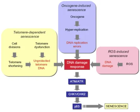
prevent genomic instability and the loss of essential genetic information by ‘‘capping” chromosome ends. They are also indispensable for proper recombination and chromosomal segregation during cell division. Telomeres become shorter with every cell division in somatic cells, because of replication complex’s inability to copy the ends of linear DNA, which also makes them a ‘‘cell cycle counter” for the cell (54). Telomeres are added to the end of chromosomes with a complex containing the RNA template TERC and the reverse transcriptase TERT (55). Most somatic cells lack telomerase activity because the expression of TERT is repressed, in contrast to TERC expression. It is now well known that telomere-dependent senescence is induced by a change in the protected status of shortened telomeres, whereby the loss of telomere DNA contributes to this change (56). At least two other forms of telomere-independent senescence are presently known: (1) oncogene-induced senescence; and (2) reactive oxygen species (ROS)-induced senescence (Figure 1.4).
Figure 1.4 Senescence pathways (52).
1.4.1.2 Oncogene and ROS-induced senescence
Oncogene-induced senescence had initially been identified as a response to expression of Ras oncogene in normal cells, accompanied by accumulation of p53 and p16INK4a. In addition to Ras, other oncogenes including Raf, Mos, Mek, Myc and Cyclin E also induce senescence (57). Similar to telomere-dependent senescence, oncogene-induced senescence is also primarily a DNA damage response (Figure 1.3).
ROS-induced senescence, the other telomere-independent senescence pathway is gaining importance, also. Experimental induction of ROS accumulation in cells (for example by mild H2O2 treatment or glutathione depletion) induces senescence-like growth arrest in different cell types, whereas anti-oxidant treatment can inhibit senescence (58). More importantly, ROS have been identified as critical mediators of both telomere-dependent and oncogene-induced senescence (52).
1.4.2 Senescence as an anti-tumor mechanism in hepatocellular carcinoma
Recent findings indicate that senescence induction is a powerful mechanism of HCC regression. Xue et al. expressed H-ras oncogene and suppressed endogenous p53 expression in mouse hepatoblasts which produced massive HCCs upon implantation into livers of athymic mice (59). However, these tumors regressed rapidly upon restoration of p53 expression. Tumor regression was due to differentiation and massive senescence induction,
followed by immune-mediated clearance of senescent cells. These observations may indicate that oncogene-induced senescence is also involved in HCC. On the other hand, HCCs induced by tet-regulated c-Myc activation in mouse liver cells differentiate into mature hepatocytes and biliary cells or undergo senescence (60). Thus, senescence induction may also be relevant to oncogene inactivation in HCC. So far, all the reported examples of senescence induction in HCC cells are in the form of a telomere-independent permanent cell cycle arrest. Until recently, it was unknown whether replicative senescence could also be induced in immortal cancer cells. Our group reported recently that immortal HCC cells can revert spontaneously to a replicative senescence phenotype (61). HCC cells generated progeny that behaved, in vitro, similar to normal somatic cells. Such senescence-programmed progeny (C3 and G12 clones) lacked telomerase activity due to TERT repression (probably mediated by SIP1 gene), and displayed progressive telomere shortening in cell culture, resulting in senescence arrest. On the other hand, immortal clones (C1 and G11) had indefinite proliferation capacity with high tumorigenic capacity.
1.4.3 Cyclin-dependent inhibitors as common mediators of senescence arrest
Most if not all senescence pathways result in the activation of cyclin-dependent kinase inhibitors (CDKIs) in order to induce permanent cell cycle arrest. Senescent cells accumulate at G1 phase of the cell cycle due to an inability to enter into S phase in order to initiate DNA synthesis. The transition of proliferating cells from G1 to S phase requires the release of E2F factors from their inhibitory partner retinoblastoma protein (pRb) following phosphorylation by cyclin-dependent kinases (CDKs), in particular by CDK4/CDK6 and CDK2 at this stage of the cycle (62). The senescence arrest is mediated by inhibition of pRb phosphorylation by CDK4 and CDK2. The activities of these enzymes are controlled by different mechanisms, but the major proteins involved in the control of senescence arrest are CDKIs. Almost all known CDKIs have been reported to be implicated in senescence arrest, but three of them are best characterized: p16INK4a and p15INK4b which inhibit CDK4/CDK6, and p21Cip1 which inhibits CDK2 (Fig. 2). p21Cip1 is one of the main targets of p53 for the induction of cell cycle arrest following DNA damage (63). Pathways that generate DNA damage response and p53 activation use p21Cip1 as a major mediator of cellular senescence to control pRb protein (64). Exceptionally, p21Cip1 can be activated by p53-independent pathways to induce senescence (65).
1.5 Expression Profiling Using Affymetrix GeneChip Microarrays
The approximately 25,000 genes in mammalian genomes can be transcribed at different levels. Measurements of gene expression for ten thousands of genes in parallel give the most comprehensive picture of steady-state levels of transcripts and is used in basic and applied research. Microarrays are the most frequently used technology for genome-wide expression profiling; from the various available microarray platforms, Affymetrix GeneChips are most frequently used for expression profiling and over 3,000 scientific publications describe results of this technology. In medical research, expression profiling by microarrays holds great promises for better understanding of diseases, identification of new therapeutic targets, and subclassification of diseases to identify individualized treatment strategies (66). Microarray studies provide evidence that a large set of growth control genes is deregulated in HCC (1).
A typical microarray experiment involves the hybridization of an mRNA molecule to the DNA template from which it is originated. Many DNA samples are used to construct an array. The amount of mRNA bound to each site on the array indicates the expression level of the various genes. This number may run in thousands. All the data is collected and a profile is generated for gene expression in the cell (67).
Affymetrix microarray technology uses oligonucleotides consisting of 25 bases. A special technique called photolithographical array production is applied to sequence or synthesize the oligonucleotides on a glass support. Each gene (or transcript) is represented by 22 different oligonucleotide fragments that are attached to a tiny section of the chip (density of up to 500 000 sections per 1.6 cm2). Each section carries 22 different oligonucleotide sequences (11 matches and 11 mismatches). Thus, the expression of a gene is given by 11 signal intensities (compared to 1 signal for cDNA microarrays). The 11 mismatch strands serve to determine the specificity of measured signal. The production of target DNS includes some further steps. After the isolation of mRNA and the reverse transcription to cDNA, the cDNA strands are converted to double stranded DNA molecules. After this step, the DNA is converted to cRNA molecules using a special polymerase (T7 polymerase) and fluorescently labelled. This cRNA is hybridised to the microarray. Unbound cRNA are washed away and signal intensities are scanned. The Affymetrix GeneChip Human Genome U133 set is made up of over 1,000,000 unique oligonucleotide features covering over 39,000 transcript variants which represent 33,000 of the best characterized human genes. Sequences used in the design of array were selected from GeneBank, dbEST, and RefSeq.
1.6 Rb/E2F pathway
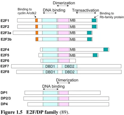
The Rb-E2F pathway links growth-regulatory pathways to a transcription program involved in DNA synthesis, cell-cycle progression, cell division, apoptosis, DNA repair, differentiation and senescence (68).This transcription program is repressed when hypophosphorylated pRb is bound to E2F proteins or recruit co-repressors to the proteins, and active when pRb is phosphorylated by CDK4/6, which frees E2F proteins to function as transcription factors via forming heterodimers with DP proteins. Eight E2F genes encode nine major proteins that share a related DNA-binding domain and are classified as either ‘activating’ or ‘repressor’ E2Fs (69).E2F1, E2F2 and E2F3a localize to the promoters of target genes in the G1/S phase
and activate transcription of these genes; whereas E2F4, E2F5 and, most probably, E2F3b bind their target promoters in association with Rb family members in the G0/G1 phase
coincident with their repression (70). E2Fs 1-5 have pocket-protein binding domains that enable them to interact with pRb and its homologs p107 and p130. E2Fs 6-8 lack this domain, and repress transcription via other mechanisms; for example, E2F6 exerts its effect by binding to the Polycomb group’s transcriptional repressors. The E2F proteins, except E2F7 and 8,
form heterodimers with DP (or TFDP), which enhances their DNA binding, transactivation and pRb-binding activities DP1 and DP2 (which is the human equivalent to murine DP3) proteins are 70% homologous and each form functional dimers with any E2F protein (71). The
other DP family protein, DP4, which has been recently characterized, shares 90% homology with DP1 and heterodimerizes with E2F (72).DP proteins have a DNA-binding domain that shares sequence homology with the E2F DNA-binding domain (73). As pRb protein is the key player in cell-cycle progression, it is not surprising that it and its upstream regulators, such as Cyclin D1, cyclin-dependent kinase 4 and p16INK4a are frequently mutated in numerous types
of human tumours (73).And thus, attention has focused on the involvement of downstream regulators of pRb in tumourigenesis, of which E2F/DP transcription factors are the best characterized set. Increased levels of E2F1 have been associated with tumorigenesis and a poorer outcome of melanoma, small-cell lung carcinoma, breast and pancreas carcinomas both in vitro and in vivo, whereas decreased E2F1 expression has resulted in more aggressive disease progression in colon cancer, bladder cancer and diffuse large B-cell lymphoma (74), (75), (76), (77).
It is becoming apparent that the function of any given E2F/DP family protein in regulating cell proliferation, apoptosis, quiescence or senescence is more complex than previously thought and most probably dependent on cell type and context (78). The Rb pathway is among the pathways most frequently disrupted in HCC.RB1 is inactivated by
direct mutation or loss of the RB1 gene, and phosphorylation of the pRb protein is deregulated through aberrant cdk activity (via loss of p16INK4a expression or cyclin-D1 amplification) (5).
Up-regulation of pRb-free E2F1-DP1 and E2F2-DP1 heterodimers is found to be a major cause of aberrant G1-S transition in c-myc/TGF-β-dependent HCC models (79). Also,
Hepatitis B viral core protein (HBc) has been found to interact with E2F1 and reduce the DNA-binding ability of E2F1 to the p53 promoter (80).Mutations of E2F4 have been detected in HCC (81). Yasui et al. showed that the DP1 gene was located at 13q34, a frequent amplification site in HCC, and that over-expression of this gene was correlated with large tumour size (82). Nonetheless, the involvement of different DP subunits (including splice variants) and different E2F-DP heterodimers in the oncogenic and tumor suppressor mechanisms (such as immortality and senescence) in HCC are not completely understood. 1.6.1 DP-2 (TFDP2)
Human cells express at least three DP2 isoforms (of 55, 48 and 43 kDa) (83). Despite being identified more than 13 years ago (84),the DP2 gene remains underexplored, particularly in terms of its role in tumorigenesis.
An increase in DP2expression has been reported in a smal set of HCC tissues by RT-PCR (85).
It has been reported that the expression of DP2 is controlled during tissue development (86), (87) and cell cycle (88). Moreover, the expression of DP2 in adult tissues is highly variable, detected in some tissues - including liver - but not in others.Differential expression of DP2 was also observed in cancer cell lines (71).
Figure 1.5 E2F/DP family (89).
1.7 Chromatin modifications and hepatocellular carcinoma
The term epigenetics refers to changes in gene expression or phenotype caused by mechanisms other than changes in the underlying DNA sequence, hence the name epi- (Greek: over; above) -genetics. These changes may remain through cell divisions for the remainder of the cell's life and may also last for multiple generations.
1.7.1 DNA methylation
The 5' cytosine of CpG dinucleotides within mammalian genomes can be methylated by denovo DNA methyltransferases such as DNMT3A and DNMT3B. Maintenance of DNA methylation is performed by DNMT1, utilizing hemimethylated DNA as a substrate. This provides a mechanism to propagate the epigenetic mark following DNA replication. The methyl groups serve as docking sites for gene silencing proteins. In general, DNA methylation correlates with increased chromatin condensation and gene silencing (90).
There are several ways in which altered patterns of DNA methylation lead to disease. Alterations in methylation patterns are responsible for several congenital diseases that affect growth through the misregulation of imprinted genes. In addition to alterations in the patterns of DNA methylation, loss of DNA methyl transferase also leads to disease. Given that mutations within DNA methyltransferase genes are associated with disease, it follows that mutations within genes encoding proteins that bind to methylated cytosines also result in
disease. MeCP2 (methyl-CpG-binding protein 2) is a member of a class of DNA methyl binding proteins (MBDs) that specifically recognize methylated cytosine residues. These binding proteins function by recruiting histone deacetylases (HDACs) to silencetarget genes. Mutations in the X-linked gene encoding MeCP2 are responsible for approximately 95% of classic Rett syndrome cases (RTT, OMIM 312750) (91).
1.7.1.1 DNA methylation and cancer
CpG dinucleotides are generally methylated in normal cells, with the exception of hypomethylation at CpG “islands” located upstream of many active genes. In contrast, cancer cells exhibit a global hypomethylation and CpG island hypermethylation (92) (Fig. 1.6 and 1.7). This shift in the pattern of DNA methylation frequently results in inappropriate silencing of genes. Global DNA hypomethylation (also known as demethylation) is associated with activation of protooncogenes, such as c-JUN, c-MYC, and c-Ha-Ras, and generation of genomic instability. Hypermethylation on CpG islands located in the promoter regions of tumor suppressor genes results in transcriptional silencing and genomic instability. For example, expression of the serine protease inhibitor family member maspin is reduced due to methylation of promoter sequences in many advanced forms of cancer (93).
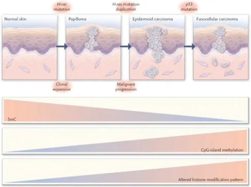
Figure 1.6 Epigenetic Alterations in Tumor Progression, skin tumor as a model (94). A growing number of genes undergoing aberrant CpG island hypermethylation in HCC have been discovered, suggesting that de novo methylation is an important mechanism underlying malignant transformation in the liver. Epigenetic silenced genes are involved in
important molecular pathways of carcinogenesis e.g., cell cycle regulation, apoptosis, DNA repair or cell adhesion (45). The genes frequently found to be methylated in HCC are APC, GSTP1, RASSF1A, p16, COX-2, CCND2, SPINT2, RUNX3, CFTR, HINT1, RIZ1 and E-cadherin (45). Furthermore, a combination of RASSF1A, CCND2 and SPINT2 showed 89-95% sensitivity, 91-100% specificity and 89-97% accuracy in discriminating between HCC and non-HCC tissues, and correctly diagnosed all early HCCs (95). Also, it was shown that one of the regulation mechanisms of hTERT promoter was DNA methylation (96). Some miRNA’s were also shown to be silenced by DNA methylation and play a role in hepatocellular carcinogenesis, for example micro-RNA 1 (97). Also, methylation of Tip30 promoter was associated with poor prognosis in human hepatocellular carcinoma (98). Yan et. al. associated the promoter methylation and reduced T-cadherin expression (40% HCC) with the development and progression of hepatocellular carcinoma.
Calvisi et. al. showed that the extent of global DNA hypomethylation and CpG hypermethylation correlates with biologic features and clinical outcomes of HCC. They suggested that aberrant methylation is a major event in both early and late stages of liver malignant transformation and might constitute a critical target for cancer risk assessment, treatment, and chemoprevention of HCC (99).
Figure 1.7 DNA methylation differences in some cancers (Esteller M, Epigenetics in cancer, NEJM, 2008).
1.7.2 Histone modifications
The nucleosome is the fundamental unit of chromatin and it is composed of an octamer of the four core histones (H3, H4, H2A, H2B) around which 147 base pairs of DNA are wrapped. The core histones are predominantly globular except for their N-terminal ‘‘tails,’’ which are unstructured. A striking feature of histones, and particularly of their tails, is the large number and type of modified residues they possess. These modifications serve to alter charge interactions of the histone tails with DNA, thereby influencing chromatin packaging. In addition, these modifications serve as binding sites for specific factors that “read” a proposed histone code. In most cases, specific modifications correlate with biological functions such as chromatin condensation, transcriptional regulation and DNA replication. Generally speaking, two kinds of enzymatic activities impinge on chromatin structure. One family involves mainly ATP hydrolysing enzymes that can re-model chromatin by ‘shuffling’ nucleosomes. Another family includes a set of enzymes that are able to modify histones covalently, at specific residues, located most commonly at the histone tails (100). There are at least eight distinct types of modifications found on histones. We have the most information regarding the modifications include acetylation, phosphorylation, methylation and ubiquination. Although these modifications have been known for many years to occur on histones, their function is only recently being recognised. There are over 60 different residues on histones where modifications have been detected either by specific antibodies or by mass spectrometry. However, this represents a huge underestimate of the number of modifications that can take place on histones. Extra complexity comes partly from the fact that methylation at lysines or arginines may be one of three different forms: mono-, di-, or trimethyl for lysines and mono- or di- (asymmetric or symmetric) for arginines (101). The timing of the appearance of a modification will depend on the signaling conditions within the cell.
Whereas lysine acetylation almost always correlates with chromatin accessibility and transcriptional activity, lysine methylation can have different effects depending on which residue is modified. Methylation of histone H3 lysine 4 (H3K4) and H3 lysine 36 is associated with transcribed chromatin. In contrast, methylation of H3 lysine 9 (H3K9), H3 lysine 27 (H3K27), and H4 lysine 20 (H4K20) generally correlate with repression. Distinct histone modifications can influence each other and may also interact with DNA methylation,
in part through the activities of protein complexes that bind modified histones or methylated cytosines (102). (Fig. 1.8).
1.7.2.1 Histone modifying enzymes
Most modifications have been found to be dynamic, and enzymes that remove the modification have been identified (Fig. 1.9).
Figure 1.8 Histone modifications (Upstate)
Of all the enzymes that modify histones, the methyltransferases and kinases are the most specific. This is perhaps the reason why methylation is the most characterized modification to date. In some cases, the specificity of enzymes that modify histones can be influenced by other factors: complexes in which enzymes are found (103); proteins that associate with the enzyme may affect its selection of residue to modify (104), or the degree of methylation (mono-, di-, or tri-) at a specific site (105). Proteins are recruited to modifications
and bind via specific domains. Methylation is recognized by chromo-like domains of the Royal family (chromo, tudor, MBT) and nonrelated PHD domains, acetylation is recognized by bromodomains, and phosphorylation is recognized by a domain within 14-3-3 proteins.
Also, recently Chang et. al. identified that the Jumonji domain-containing 6 protein (JMJD6) was a JmjC-containing iron- and 2-oxoglutarate-dependent dioxygenase that demethylates histone H3 at arginine 2 (H3R2) and histone H4 at arginine 3 (H4R3) in both biochemical and cell-based assays (106).
The abundance of modifications on the histone tail makes ‘‘crosstalk’’ between modifications very likely. Mechanistically such communication between modifications may occur at several different levels. Firstly, many different types of modification occur on lysine residues. This will undoubtedly result in some form of antagonism since distinct types of modifications on lysines are mutually exclusive. Secondly, the binding of a protein could be disrupted by an adjacent modification. The best example of this is that of phosphorylation of H3S10 affecting the binding of HP1 to methylated H3K9 (107). Thirdly, the catalytic activity of an enzyme could be compromised by modification of its substrate recognition site; for example, isomerization of H3P38 affects methylation of H3K36 by Set2 (108). Fourthly, an enzyme could recognize its substrate more effectively in the context of a second modification; the example here is the GCN5 acetyltransferase, which may recognize H3 more effectively when it is phosphorylated at H3S10 (109).
1.7.2.2 Histone methylation and cancer
Hypermethylation of the CpG islands in the promoter regions of tumor-suppressor genes in cancer cells is associated with a particular combination of histone markers: deacetylation of histones H3 and H4, loss of H3K4 trimethylation, and gain of H3K9 methylation and H3K27 trimethylation. The presence of the hypo-acetylated and hypermethylated histones H3 and H465 silences certain genes with tumor-suppressor–like properties, such as p21WAF1, despite the absence of hypermethylation of the CpG island (94). In human tumors generally, modifications of histone H4 entail a loss of monoacetylated and trimethylated forms (110). These changes appear early and accumulate during the development of the tumor. The losses occur predominantly at the monoacetylated Lys16 and trimethylated Lys20 residues of histone H4 in association with hypomethylated repetitive DNA sequences. They have been found in breast and liver cancer (111); (112). In prostate cancer, weak immunohistochemical
staining of two histone modifications (the dimethylation of lysine 4 and the acetylation of lysine 18 of histone H3) has been proposed as a marker of a high risk of recurrence. Expression patterns of histone modifying enzymes distinguish cancer tissues from their normal counterparts, and they differ according to tumor type. In leukemias and sarcomas, chromosomal translocations that involve histone-modifier genes, such as histone