| Journal of Clinical and Analytical Medicine
1
A Case Report: Primary Epiploic Apendagitis
A Rare Cause of Abdominal Pain:
Primary Epiploic Appendagitis (PEA)
Karın Ağrısının Nadir Bir Sebebi:
Primer Epiploik Apendajitis
DOI: 10.4328/JCAM.4436 Received: 24.02.2016 Accepted: 15.03.2016 Printed: 01.07.2016 J Clin Anal Med 2016;7(4): 567-9 Corresponding Author: Gulbanu Erkan, Istanbul Medipol Universitesi Tıp Fakultesi Hastanesi (Medipol Mega) TEM Avrupa Oto Yolu Göztepe Çıkışı No:1, 34214, Istanbul, Turkey. E-Mail: [email protected]
Özet
Primer epiploik appendajitis (PEA), epiploik apendiksleri drene eden santral venin torsiyon yada spontan trombozu sonucu gelişen nadir bir hastalıktır. Sıklıkla ken-dini sınırlar ve cerrahi tedavi gerektirmez, ancak cerrahi girişim yada agresif me-dikal tedavi gerektiren appandisit, divertikülit yada kolesistit gibi tabloları taklit edebilir. Akut karın ağrısıyla başvuran hastalarda gereksiz cerrahi girişimi önlemek adına PEA akılda bulundurulmalıdır. Biz de karın ağrısıyla başvuran ve PEA tanısı alan bir olgumuzu sunacağız.
Anahtar Kelimeler
Karın Ağrısı; Primer Epiploik Apendajitis; Tomografi
Abstract
Primary epiploic appendagitis (PEA) is a rare disease caused by torsion or spon-taneous thrombosis of the central vein that drains epiploic appendages (EA). Pri-mary Epiploic Appendagitis (PEA) is an ischemic infarction. Although PEA is a self-limiting disease and does not require surgical intervention in most cases, it may mimic diseases that require surgical intervention or aggressive medical therapy, such as appendicitis, diverticulitis, or cholecystitis. In order to avoid un-necessary surgical intervention, PEA should be kept in mind when patients present with acute abdominal pain. In this report, we present a PEA case admitted with abdominal pain.
Keywords
Abdominal Pain; Primary Epiploic Apendagitis; Computed Tomography Gulbanu Erkan1, Cem Gezen2, Sabriye Sennur Bılgın3 1Department of Internal Diseases, Gastroenterology, 2Department of General Surgery, 3Department of Radiology, İstanbul Medipol University Medical School, İstanbul, Turkey
This case report was presented at the 32th National Gastroenterology week in 25-29 November 2015, Antalya, Turkey as a poster presentation. This case report has not been previously published full text, and it is not under consideration for publication elsewhere.
| Journal of Clinical and Analytical Medicine A Case Report: Primary Epiploic Apendagitis
2
Introduction
Epiploic appendages (EA) are pedunculated structures filled with fat and situated along the external aspect of the colon. EA occur all along the entire colon but are mainly present in the transverse and sigmoid parts of the colon. On average, approxi-mately 50 to 100 appendages can be present in an adult colon. Epiploic Appendagitis is a rare, self-limiting, inflammatory dis-ease of EA. Primary Epiploic Appendagitis (PEA) is an ischemic infarction caused by torsion or spontaneous thrombosis of the central vein that drains EA (1). Secondary epiploic appendagi-tis (SEA) occurs when the epiploic appendage is inflamed due to another abdominal inflammatory process, such as acute pendicitis, diverticulitis, or cholecystitis. In primary epiploic ap-pendagitis (PEA), no inflammation is present in other abdominal organs (1). Keeping PEA in mind in patients presenting with acute abdominal pain is of prime importance for avoiding un-necessary laparotomy and surgical intervention.
Case Report
A 67-year-old woman presented with a sudden severe pain in the left lower quadrant. Arrhythmia was detected in the physical examination, and defense and rebound tenderness were positive in the left lower quadrant. Patient history revealed that the pa-tient had had atrial fibrillation and had been hospitalized in the intensive care unit due to an acute thrombotic cerebrovascular event three weeks earlier. The patient was using a proton pump inhibitor and rivaroxaban. Whole blood count revealed a normal white blood cell (WBC) count, sedimentation of 39 mm/h, and a CRP of 26 mg/L. Direct abdominal X-ray revealed no air-fluid level or perforation. Due to the suspicion of mesenteric isch-emia or acute diverticulitis, oral intake was discontinued and the patient was given intravenous fluid and antibiotic therapy (ciprofloxacin and metronidazole). An emergency abdominal CT scan revealed a circular wall thickening of the distal descend-ing colon, hypodense central area surrounded by a hyperdense ring in the paracolic fatty tissue, and an impression of fatty tissue inflammation in the most external part, consistent with PEA (Figures 1 and 2). No signs of acute mesenteric ischemia
or acute diverticulitis were detected. Following clinical improve-ment, the patient was discharged and outpatient clinic follow-up was recommended. Colonoscopy revealed normal findings 4 weeks following the initial abdominal findings.
Discussion
Appendicitis epiploica (AE), also known as epiploic appendagi-tis, hemorrhagic epiploiappendagi-tis, or epiplopericoliappendagi-tis, is a self-limiting rare clinical condition (1). Primary epiploic appendagitis (PEA) results from torsion or spontaneous thrombosis of the central vein that drains EA (1). The exact prevalence of PEA remains unknown since the disease is highly rare and self-limiting. PEA may affect any age group but is more commonly seen in the fourth and fifth decades. PEA is more common in men than in women (1).
PEA patients commonly present with acute-onset and well-localized abdominal pain. The pain is mostly located in the left lower quadrant (60-80%) but may also be present in the right lower quadrant (2,3). Some patients may also present with subferile fever. Abdominal physical examination mostly reveals palpation, localized tenderness, and defense. WBC count, sedi-mentation, and CRP may be normal or slightly increased (4). The diagnosis of PEA is mostly by chance, commonly estab-lished during the imaging-based diagnosis of patients present-ing with an acute abdominal pain (1). It should also be con-sidered when explorative laparotomy fails to reveal any other common cause of acute abdomen. Abdominal CT is the gold
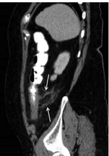
Figure 1. An oval lesion (arrow) with an impression of peripheral rim which ap-pears hypodense due to central fat density and hyperdense due to serosal inflam-mation is seen which suggests the presence of epiploic appendigitis in the poste-rior of the descending colon. Linear density suggestive of thrombotic structure in the central part of the lesion and wall thickening caused by inflammation in the adjacent peritoneum are visible (arrow head).
Figure 2. An oval lesion with an impression of peripheral rim which appears hy-podense due to central fat density and hyperdense due to serosal inflammation is seen which suggests the presence of epiploic appendicitis in the posterior of the descending colon, and staining is viewed in the fat planes around the lesion.
| Journal of Clinical and Analytical Medicine
568
| Journal of Clinical and Analytical Medicine A Case Report: Primary Epiploic Apendagitis
3
standard for the diagnosis of PEA and also rules out patholo-gies in other abdominal organs (1). On CT, EA is normally not detected, but an inflamed EA is viewed as an oval or circular pedunculated pericolonic lesion with fat density. The lesion de-tected on CT is surrounded by a high-density peripheral rim and represents the inflammation in the serosal layer. A linear or fo-cal density corresponding to the thrombotic vein may appear in the middle of the lesion. Moreover, inflamed lines may appear in the pericolonic fat tissue (1,4,5). PEA may be confused with any acute abdominal disease but mostly mimics acute diverticulitis and acute appendicitis (3).
Knowledge about the treatment of PEA is highly limited and relies solely on case reports or case series. The most common treatment method is the conservative approach with anti-in-flammatory drugs or short-term opiates (acetaminophen/co-deine) (3,4,6). Antibiotic use and hospitalization is usually not required in uncomplicated cases (7). Surgical treatment is per-formed in patients not responding to the conservative approach or in patients developing complications, such as intussuscep-tion, abscess, or intestinal obstruction (8).
In conclusion, PEA is a rare cause of abdominal pain. In patients presenting with acute abdominal pain, PEA should be kept in mind during the differential diagnosis in order to prevent un-necessary laparotomy and surgical intervention.
Competing interests
The authors declare that they have no competing interests.
References
1. Mollà E, Ripollés T, Martínez MJ, Morote V and Roselló-Sastre E. Primary epi-ploic appendagitis: US and CT findings. Eur Radiol 1998;8:435-8.
2. Sandrasegaran K, Maglinte DD, Rajesh A and Akisik FM. Primary epiploic ap-pendagitis: CT diagnosis. Emerg Radiol 2004;11:9-14.
3. Hwang JA, Kim SM, Song HJ, et al. Differential diagnosis of left-sided abdominal pain: primary epiploic appendagitis vs colonic diverticulitis. World J Gastroenterol. 2013;19(40):6842-8.
4. Rioux M, Langis P. Primary epiploic appendagitis: clinical, US, and CT findings in 14 cases. Radiology 1994;191(2):523-6.
5. Singh AK, Gervais DA, Hahn PF, Rhea J, Mueller PR. CT appearance of acute ap-pendagitis. AJR Am J Roentgenol 2004;183(5):1303-7.
6. Vinson DR. Epiploic apendagitis: a new diagnosis for the emergency physician. Two case reports and a review. J Emerg Med 1999;17:827-32.
7. Sangha S, Soto JA, Becker JM, Farraye FA. Primary epiploic appendagitis: an underappreciated diagnosis. A case series and review of the literature. Dig Dis Sci 2004;49:347-50.
8. Bonnefoy S, Corberand D, Sinayoko L, Harnois F, Mennecier D, Thiolet C. Epi-ploic appendagitis: report of a case. Gastroenterol Clin Biol 2008;32:1092-4. How to cite this article:
Erkan G, Gezen C, Bılgın SS. A Rare Cause of Abdominal Pain: Primary Epiploic Appendagitis (PEA). J Clin Anal Med 2016;7(4): 567-9.
Journal of Clinical and Analytical Medicine | 569