Journal of Gastroenterology and Hepatology (2005) 20, 482 DOI: 10.1111/j.1400-1746.2005.03817.x
Blackwell Science, LtdOxford, UKJGHJournal of Gastroenterology and Hepatology0815-93192005 Blackwell Publishing Asia Pty LtdMarch 2005203482482Images of InterestGastrointestinalGastrointestinal
Contributed by
M Kantarci,* C Duran† and M Sirvanci†
*Department of Radiology, GATA Haydarpasa Education Hospital and †Department of Radiology, Florence Nightingale Hospital, Kadir
Has University School of Medicine, Istanbul, Turkey.
Contributions to the Images of Interest Section are wel-comed and should be submitted to Professor IC Roberts-Thomson, Department of Gastroenterology, The Queen Elizabeth Hospital, Woodville South, South Australia 5011, Australia.
© 2005 Blackwell Publishing Asia Pty Ltd
IMAGES OF INTEREST
Gastrointestinal: Epiploic appendagitis
How did we get the word epiploic appendagitis? The word ‘epiploic’ is synonymous with omental. Appendagitis is more straightforward and means inflammation of a part that is subordinate in size or function to the main structure. Indeed, appendices epiploicae are small fatty projections covered by peritoneum that are scattered over the free surface of most of the large intestine. They are absent from the cecum, the appendix and the rectum. They have no known structural or functional purpose and have only rarely been associated with pathological changes or clinical syndromes.
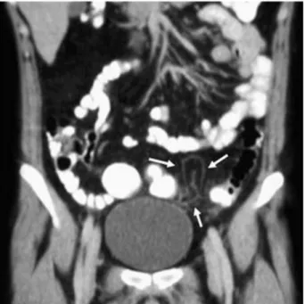
The images illustrated below were from an 18-year-old man who was admitted to hospital with pain in the left iliac fossa that radiated into the region of the umbilicus. Pain was aggravated by deep breathing and coughing. His tem-perature was normal and the only abnormal laboratory finding was a mild leucocytosis. A computed tomography (CT) scan was performed. The axial image (Fig. 1) revealed an oval-shaped lesion (arrows) with a rim of soft tissue that shows increased attenuation and fine stranding consistent with inflammation. The coronal reconstruction con-firmed that the lesion was pericolonic (Fig. 2, arrows). The adjacent colonic wall and nearby segments were normal. The findings were consistent with epiploic appendagitis and his symptoms resolved after bed rest and analgesics for 5 days. The cause of epiploic appendagitis is torsion or venous thrombosis of an appendage causing ischemia or inf-arction. Similar radiological findings may be seen with acute diverticulitis. However, the latter disorder mostly occurs in patients greater than 50 years of age, and CT scans normally show additional features including unaffected diverticula and thickening of the colonic wall. Furthermore, some patients have radiological features of an abscess such as a pericolonic collection with gas bubbles or a fluid level.
Figure 1 Figure 2
Reproduction of color photographs has kindly been sponsored by a grant from AstraZeneca.