Yazışma Adresi/Address for Correspondence: Dr. Ali Murat Sedef, Şanlıurfa Research and Training Hospital, Department of Medical Oncology, Şanlıurfa, Turkey E-mail: [email protected]
ARAŞTIRMA / RESEARCH
Clinicohistopathological features and treatment outcomes of
neuroendocrine tumors: a single center experience
Nöroendokrin tümörlerin klinikohistopatolojik özellikleri ve tedavi sonuçları: tek
merkez deneyimi
Ali Murat Sedef
1, Fatih Köse
2, Ahmet Taner Sümbül
2, Hüseyin Mertsoylu
2, Ali Ayberk Besen
2Ahmet Sezer
2, Özgur Özyılkan
2, Hüseyin Abalı
31Şanlıurfa Research and Training Hospital, Department of Medical Oncology, Şanlıurfa, Turkey 2Baskent University, Department of Medical Oncology, Adana, Turkey
3Acıbadem University, Department of Medical Oncology, Adana, Turkey
Cukurova Medical Journal 2018;43(3):533-538
Abstract Öz
Purpose: Tumor and patient characteristics of
neuroendocrine tumors (NET) significantly change between geographical locations that probably induced by environmental and genetic factors throughout the world. Therefore, reporting single center experience may help clarifying epidemiological view and improving decision-making process.
Materials and Methods: We performed retrospective
analysis of 115 patients of NETs those who followed by Baskent University, department of Medical Oncology and department of General Surgery to record patients and tumors characteristics, treatment modalities, survival rates, and prognostic factors.
Results: Median overall survival (OS) time for all group
and localized NETs were 44 and 24 months, respectively. Most common primary site was found as gastrointestinal system and then pancreatic region. Curative surgical resection rate was 46% and 8.5% of patients presented with carcinoid syndrome. Liver metastasis was far the most common metastatic site compared to lung, bone, and lymph node metastasis. Over 70 percent of patients were treated with chemotherapy and somatostatin analogs.
Conclusion: Patients with higher grade, male gender, and
advanced age (>65 years old) had poor survival rate. However, relatively low number of patients and less usage of (<10%) of new treatment modalities created limitations for producing future directions from our study.
Amaç: Nöroendokrin tümörler (NET) tümör ve hasta
özellikleri, muhtemelen tüm dünyada çevresel ve genetik faktörlerin yol açtığı coğrafi alanlar arasında önemli değişiklik göstermektedir. Bu nedenle, tek merkez deneyimi bildirmek, epidemiyolojik görüşleri açıklığa kavuşturmaya ve karar verme sürecini geliştirmeye yardımcı olabilir
Gereç ve Yöntem: Başkent Üniversitesi Tıbbi Onkoloji
Anabilim Dalına başvuran ve takip edilen, tümör özelliklerini, tedavi yöntemlerini, sağkalım oranlarını ve prognostik faktörlerini kaydettiğimiz 82 NET tanılı hastanın retrospektif analizi yapılmıştır.
Bulgular: Tüm grup ve lokal evredeki NET'lerin genel
sağkalım süresi, sırasıyla 44 ve 24 aydır. En sık görülen primer tümör alanı gastrointestinal sistem ve daha sonra pankreas bölgesi olarak bulundu. Küratif cerrahi rezeksiyon oranı karsinoid sendrom ile başvuran hastaların%46'sında ve %8.5'inde idi. Karaciğer metastazı akciğer, kemik ve lenf nodu metastazı ile karşılaştırıldığında en yaygın metastatik bölge idi. Hastaların %70'inden fazlası kemoterapi ve somatostatin analoğu ile tedavi edildi.
Sonuç: Daha yüksek greydli, erkek cinsiyeti olan ve ileri
yaşta (>65 yaş) hastalarda sağkalım oranı düşüktür. Bununla birlikte, nispeten düşük hasta sayısı ve yeni tedavi yöntemlerinin (<%10) daha az kullanımı, çalışmamızın gelecek için yönermeler üretebilmesi için sınırlamalar getirdi.
Key words: Neuroendocrine tumors, outcomes, single
INTRODUCTION
Neuroendocrine tumors (NETs) are a group of clinically and pathologically distinct heterogeneous family of cancer1. Clinically significant annual
incidence rate of NETs is reported between 2,5-5 per 100,000 people, but its incidence rate increased in the last decades2,3. Moreover, with the relatively
higher survival rates compared to adenocarcinomas, in current state NETs are more prevalent than pancreas and gastric cancers combined1. The
common primary locations were gastrointestinal system and lungs; though NETs may originate from atypical locations like ovary, maxillary sinus, bone, and kidney and they may be presented with clinical syndrome resulted by excessive secretion of peptide hormones4.
Management requires multidisciplinary approach with careful consideration of anatomic and histopathological characteristics of tumors. Total surgical excision is the only curative treatment modality, but more patients presented with advanced disease. The treatment modalities for these patients include cytotoxic chemotherapy, liver and tumor directed therapy, somatostatin analogs, and targeted therapy. However, optimal time and order of these expanding treatment options are not clear and we need to better understand tumor biology and biological behavior of the NETS to clarify to optimization. Using this retrospectively designed protocol, we aim to the study demographic characteristics, the overall, stage and site specific outcomes, prognostic factors, used treatment modalities and outcomes in 82 patients with NETs those who were followed in our center.
MATERIALS AND METHODS
Baskent University patient database was searched for international ICD codes NETs between the years of 2006 and 2013. Consecutive 115 neuroendocrine tumor patients those who were followed at Baskent University Medical Oncology and General Surgery departments were identified after obtaining ethical clearance from ethical committee. Among those, 82 patients with NET were enrolled for this study. The reasons for exclusions (n=43) included primary lung tumors (n=28) and non-follow-up patients15.
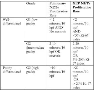
The tumors were graded as G1, G2, or G3 following the WHO (version 2010) classification.
Grade 1 and 2 tumors represent good differential tumors, grade 3 tumors represent poorly differentiated tumors. Ki-67 and proliferation index
were taken into account when classifying the tumors. WHO classification is indicated in Table-1.
Table 1. WHO Grading Systems for gastroenteropancreatic (GEP) and Pulmonary NETs
Grade Pulmonary NETs Proliferative Rate GEP NETs Proliferative Rate
Well-differentiated G1 (low grade) < 2 mitoses/10 hpf AND No necrosis <2 mitoses/10 hpf AND <3% Ki-67 index G2 (intermediate grade) 2-10 mitoses/10 hpf OR necrosis 2-20 mitoses/10 hpf OR 3%-20% Ki-67 index Poorly
differentiated G3 (high grade) >10 mitoses/10 hpf >20 mitoses/10 hpf OR > 20% Ki-67 index
Data collection
The clinical and laboratory information of the patients was transferred safely from the electronic records to the SPSS form. Demographic and clinicopathologic variables of the patients were determined by considering the disease characteristics and follow-up criteria included in the international guidelines. Demographics, clinicopathological characteristics, radiographic data and treatment modalities of patients were extracted from electronic medical records for all 115 patients. In all cases, immunohistochemistry for synaptophysin, chromogranin and Ki-67 was performed in Baskent University Department of Pathology. Conventional tomography PET CT was used for staging of the patients
Statistical analysis
All results were presented as rate for categorical values, or mean and median for continuous variables. Survival curves were estimated by the Kaplan-Meier method and log-rank tests were used for univariate statistical comparisons. Overall survival (OS) was calculated from the date of
histological diagnosis of NETs to the date of the last visit or death. The Cox proportional hazards model was used to determine the variables associated with OS of Neuroendocrine tumors. Adjusted Hazard ratio (HR) and %95 confidence interval (95% CIs) were used for estimation. All data were analyzed using SPSS version 17.0 statistics were made by using SPSS version 17.0 and a p value of <0.05 was considered statistically significant.
RESULTS
Patients characteristics
Of the 82 patients, there were 46 male patients (56%). Median age was 55 years (range 28-78). GEP-NETs, lung and other localizations neuroendocrine tumors were found in 45 (55.6 %), 13(16%), and 23 (28 %) patients, respectively. Though, GEP-NETs in 45 (55.6%) patients constitutes most often localization, rare localizations were present in Table 2. Curative surgery was performed in 38 (46%) patients, including 5 carcinoid tumors; the remaining 44 (54%) of patients were not appropriate for the curative resection. There were 7 (8.5%) patients those who presented with symptoms of excess peptide hormones. Profound diarrhea was most common symptom and seen in 4 (4.9%) patients. Out of the 7 patients, 2 of them treated with curative resection and 5 of them treated symptomatically with somatostatin analogs.
Table 2. Tumor localizations of 82 NETs
Localizations Percentage % Gastric (n:20) 24 Pancreas (n:16) 19 Lung (n:13) 16 Primary unknown (n:10) 12 Appendix (n:5) 6 Small bowel (n:4) 5 Rectum (n:4) 5 Bone (n:2) 2,5 Maxililary sinuses (n:2) 2,5 Colon (n:1) 1 Kidney (n:1) 1 Bladder (n:1) 1 Adrenal gland ( n:1) 1 Ovarian (n:1) 1 Paravertebral area (n:1) 1
Staining for the synaptophysin and chromogranin were positive in 65 (79%) and 65 (79%) patients.
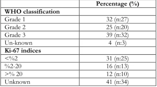
While, 60 patients were positive for both synaptophysin and chromogranin, 5 patients in each group were positive for either synaptophysin or chromogranin only. Grade, Ki 67 and mitotic rate were evaluated in 79 (96.3%), 48 (58.5%), and 29 (35.4%) patients, respectively (Table 3). Metastases to liver were found in 40 (48.8%) patients and liver was the far commonest metastatic site for the whole group.
Table-3. WHO classification and ki-67 indexes of tumor Percentage (%) WHO classification Grade 1 32 (n:27) Grade 2 25 (n:20) Grade 3 39 (n:32) Un-known 4 (n:3) Ki-67 indices <%2 31 (n:25) %2-20 16 (n:13) >% 20 12 (n:10) Unknown 41 (n:34)
Treatments and outcomes
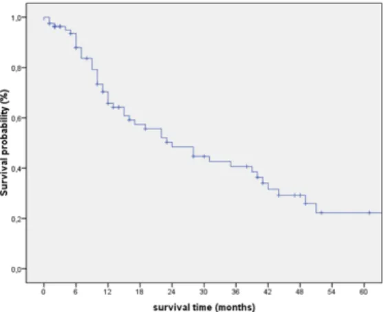
Mean follow-up time was 21.3 months (range 16.9-25.6) and 45 (56%) patients were died during the study period. Out of the 82 patients, 38 (46%) patients were treated with curative surgical resection. There were 10 patients with grade 3 tumor and these patients were treated with adjuvant platin-etoposide combination chemotherapy. There were 17 patients with grade 1 tumor those who showed excellent prognosis with no relapse during surveillance. In addition, there were 8 patients with grade 2 tumor, those 3 of them were treated with the systemic chemotherapy (platin-etoposide), showed also excellent prognosis without disease relapse. Median OS time was estimated 44 months [31.7- 56.3 (95 % CI)] (figure-1). Mitotic index and age showed statistically significant effect on OS in univariate analysis with p=0.007 and p=0.05, respectively. However, in multivariate analysis, WHO grade, age, and sex showed statistically significant effect on OS with p=0.015, p=0.003, and p=0.027, respectively were detailed in Table 4 for the curatively resected patients
For the whole group, treatment modalities were systemic chemotherapy, somatostatin analogs, metastectomy, trans arterial chemoembolization (TACE), radionuclide treatment, everolimus, and
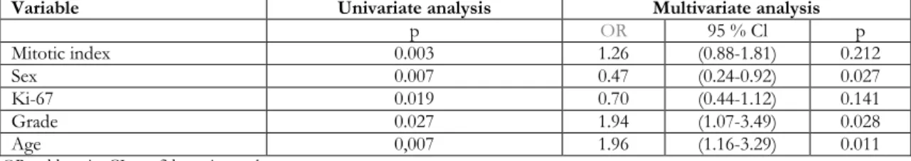
sunitinib in 36 (43%), 34 (41.5%), 1 (1.2%), 7 (8.5%), 9 (11%), 3 (3.7%), and 3 (3.7%) patients, respectively. The most common cytotoxic chemotherapy protocols were cisplatin+etoposid, 5-FU + Dacarbasine + epirubicin, capesitabin +temozolamid and carboplatin + irinotecan combinations.Though, the liver is most common site; liver, bone, and lymph node metastases were found in 40 (48.8%), 8 (9.8%), and 3 (3.7%) patients, respectively. Median OS was estimated as 24 months [13.8- 34.2 (95% CI)] for whole group (figure-2). Mitotic index, sex, Ki-67, and WHO grade showed statistically significant effect on OS in univariate analysis with p=0.003, p=0.007, p=0.019, and p=0.027, respectively. However, in multivariate analysis, WHO grade, sex and age showed statistically significant effect on OS with p=0.028, p=0.027, and p=0.011, respectively (Table 5).
DISCUSSION
NETs are probably one of the most heterogeneous group of cancers and difficult to grasp into a
universally accepted classification system for more than 3 decades5. Indeed, all commonly used
classification systems using proliferation markers, tumor stage, and primary tumor localization [GEP-NET’s (gastric mucosa, the small and large intestine, the rectum and pancreas) and lung] aim to establish clear epidemiologic view and precision in treatment decision 6-8. Recent epidemiological studies showed
increase rate of NETs in Europe and United States and now, prevalence of NETs is higher than all gastrointestinal cancers except colon cancer5.
However, studies also showed tumor and patients characteristics, prognostic factors, treatment modalities, and survival rates significantly differ between countries, even sometimes differ in the same country3,9,10. Here, our study provides valuable
insight and showed that NETs are not benign disease, though have a slower clinical course compared to adenocarcinoma with median OS time of 24 months. Over 50 percent of NETs are GEP-NETs. In addition, WHO grade, sex, and age have significant effect on OS in multivariate analysis.
Figure-1: Median OS time was estimated 44 months [31.7- 56.3 (95 % CI)] who treated with curative surgical resection
Figure 2. Median OS was estimated as 24 months [13.8- 34.2 (95% CI)] for whole group.
Table 4. Multivariate analysis of prognostic factors for overall survival who treated with curative surgical resection.
Variable Univariate analysis Multivariate analysis
p HR 95 % Cl p
Mitotic index 0.007 0.08 (0.86-10.26) 0.084
Age 0.05 24.28 (2.99-197.03) 0.03
Grade 0,055 4.78 (1.35-16.86) 0.015
Table 5. Multivariate analyseis of prognostic factors for overall survival for whole group
Variable Univariate analysis Multivariate analysis
p OR 95 % Cl p Mitotic index 0.003 1.26 (0.88-1.81) 0.212 Sex 0.007 0.47 (0.24-0.92) 0.027 Ki-67 0.019 0.70 (0.44-1.12) 0.141 Grade 0.027 1.94 (1.07-3.49) 0.028 Age 0,007 1.96 (1.16-3.29) 0.011
OR: odds ratio; CI: confidence interval
The heterogeneous nature of disease complicates data collection and clarity in literature. Tumor localization, grade, differentiation, and peptide hormone secretion are major contributors to treatment decision. In particular, pancreatic vs. non-pancreatic anatomic site is key element with regard of tumor localization. Yao et al. and Dogan et al. reported lung was the most common site, although, Maggard et al. and Lawrance et al. reported more than half of the all NETs were GEP-NETs3,5,11,12.
While tumor localization has predictive effect on response rate, some of these studies showed also statistically significant effect of tumor localization on OS 1,5. GEP-NETs is most common localization
in current study (55.6%) and statistical analysis failed to show significant effect of tumor localization on OS.
Median survival rate is better in well-differentiated NETs compared to poorly differentiated ones, yet surgical excision is the main treatment strategy for all grade local disease. Adjuvant treatment can be considered for high-grade tumors in particular for the lung NETs13. We observed excellent survival
rates (5-year survival of 87%– 90%) for grade I NETs after curative resection without any recurrence. With regard to grade 2 NETs, out of 8 patients, 3 patients treated with adjuvant platin-etoposide chemotherapy. Similar to grade I tumors, there is no relapse for these patients, but these group was too small to reach the conclusion about adjuvant therapy. All patients with grade 3 NETs receive adjuvant platin-etoposide with high rate of recurrence and lowest OS time of 5-year survival rate of 15%–57%).
Median age was 55 years old. In multivariate analysis, statistical analysis showed being over 65 years old, male gender, and higher grade were strong negative prognostic factor for localized NETs. In contrast to most studies, we noted male predominance in present study3. These results are in
agreement with studies demonstrating the higher
grade association of lower survival, although age and sex are not well defined prognostic factors in literature for localized NETs1,14.
At advanced stage, five-year OS rate was reported 50-70% and 45-60% for all patients and GEP-NETs, respectively. Yao et al. showed that tumor differentiation, primary tumor localization, metastatic sites, and histological grade were major prognostic factor in advanced states14. Yao et al also
reported that patients with male gender and advanced age (over 60 years old) lower survival rate14. In a report by Wang et al, the most common
primary tumor site was pancreas and liver was the predominant site of NET metastases7,15. With regard
to GEP-NETs, pancreatic primaries had worse survival rate compared to gastric, intestinal and colonic primaries. However, Lawrence et al reported an epidemiologic study which included around 50.000 patients, and stated that despite poorer five year survival rate for pancreatic cancer compared to colon cancer, median OS of pancreatic NETs was higher (18.9 months vs. 10.3 months) than colonic primaries5. This initial sharp decrease in survival
curve in pancreatic NETs suggest internal heterogeneity of NETs even with same primaries and we need urgent molecular markers in precision decision-making process. Mean follow-up time and median OS were estimated as 21.3 and 24 months, respectively. Liver is the most common site for the metastases (48.8%). In multivariate analysis, WHO grade, sex and age showed statistically significant effect on OS with p: 0.028, p: 0.027, and p: 0.011, respectively. Results of current study compatible with literature showed that, higher grade, male gender, and advanced age (over 65 years old) were poor prognostic factor for patients with NETs, although we failed to show statistically significant effect of primary tumor localization on OS. A limitation of our study is that majority of patients treated with chemotherapy and somatostatin analogs, yet less than 10% of whole group had chance to reach new treatment modalities like
peptide receptor radionuclide treatment and tyrosine kinase inhibitors. Hence, we couldn’t make any suggestion about real time effect of these new treatment modalities in our patients. One other limitation of the current study is that relatively low number of patients compared to multicenter studies and large epidemiological studies in literature. It is possible that low number of patients is the reason for underestimation of prognostic role of primary locations.
In conclusion, we found an independent statistically significant association between survival rate and age, sex, and histological grade in multivariate analysis. These associations were evident in both localized and advanced NETs. Our findings support the current literature and compatible with universally used algorithms. Our findings highlight the clinical importance and heterogeneous nature of NETs that have to be treated individually, case by case, with multidisciplinary approach.
REFERENCES
1. Kunz PL. Carcinoid and neuroendocrine tumors: building on success. J Clin Oncol. 2015;33:1855-63. 2. Öberg K, Castellano D. Current knowledge on
diagnosis and staging of neuroendocrine tumors. Cancer Metastasis Rev. 2011;30 (Suppl 1):3–7. 3. Dogan M, Yalcin B, Ozdemir NY, Arslan UY,
Dogan L, Utkan G et al...Retrospective analysis of seventy-one patients with neuroendocrine tumor and review of the literature. Med Oncol. 2012;29:2021– 26.
4. Liu Y, Sturgis CD, Grzybicki DM, Jasnosz KM, Olson PR, Tong M et al. Microtubule-associated protein-2: a new sensitive and specific marker for pulmonary carcinoid tumor and small cell carcinoma. Mod Pathol. 2001;14:880–5.
5. Lawrence B, Gustafsson BI, Chan A, Svejda B, Kidd M, Modlin IM. The epidemiology of
gastroenteropancreatic neuroendocrine tumors. Endocrinol Metab Clin North Am., 2011;40:1–18. 6. Moertel CG. Karnofsky memorial lecture: An
odyssey in the land of small tumors. J Clin Oncol. 1987;5:1502-22.
7. Kaltsas GA, Besser GM, Grossman AB. The diagnosis and medical management of advanced neuroendocrine tumors. Endocr Rev. 2004;25:458-511.
8. Strosberg JR, Nasir A, Hodul P, Kvols L. Biology and treatment of metastatic gastrointestinal neuroendocrine tumors. Gastrointest Cancer Res. 2008;2:113-25.
9. Hauso O, Gustafsson BI, Kidd M, Waldum HL, Drozdov I, Chan AKC et al. Neuroendocrine tumor epidemiology: Contrasting Norway and North America. Cancer. 2008;113:2655–64.
10. Yucel B, Babacan NA, Kacan T, Eren AA, Eren MF, Bahar S et al. Survival analysis and prognostic factors for neuroendocrine tumors in Turkey. Asian Pac J Cancer Prev. 2013;14:6687–92.
11. Maggard MA, O'Connell JB, Ko CY. Updated population-based review of carcinoid tumors. Ann Surg. 2004;240:117-22.
12. Yao W., Wang W. and Li G. Clinicopathological features of pancreatic neuroendocrine neoplasms: a retrospective analysis of 64 cases. Zhonghua Zhong Liu Za Zhi. 2014;36:287-93.
13. K. Öberg, P. Hellman, P. Ferolla& M. Papotti. Neuroendocrine bronchial and thymic tumors: ESMO Clinical Practice Guidelines for diagnosis, treatment and follow-up. Ann Oncol. 2012;23 (Supplement 7):vii120–3.
14. Yao JC, Hassan M, Phan A, Dagohoy C, Leary C, Mares JE et al. One hundred years after "carcinoid": epidemiology of and prognostic factors for neuroendocrine tumors in 35,825 cases in the United States. J Clin Oncol. 2008;26:3063-72.
15. Wang SC, Parekh JR, Zuraek MB, Venook AP, Bergsland EK, Warren RS et al. Identification of unknown primary tumors in patients with neuroendocrine liver metastases. Arch Surg. 2010;145:276–80.