Primary epiploic appendagitis: MRI findings
Mustafa S¸irvanci
a, N. Cem Balci
a, Kutlay Karaman
a, Cihan Duran
a, Ercan Karakas¸
b aDepartment of Radiology andbDepartment of Surgery of the Florence Nightingale Hospital, Kadir Has University, Istanbul, TurkeyAbstract
Primary epiploic appendagitis (PEA) occurs secondary to inflammation of an epiploic appendage, and is considered to be a rare cause of acute abdomen. In this case report, we describe the magnetic resonance imaging (MRI) findings of PEA correlated with computed tomographic (CT) findings. MRI findings included an oval shaped fat intensity mass with a central dot on T1- and T2-weighted images, which possessed an enhancing rim on postgadolinium T1-weighted fat saturated images. The lesion was best visualized on postcontrast T1-weighted fat saturated images. MRI findings of PEA should be considered in the differential diagnosis with the other causes of acute abdominal pain. © 2002 Elsevier Science Inc. All rights reserved.
Keywords: Epiploic Appendage; Colon; Magnetic Resonance
1. Introduction
PEA is defined as an acute inflammation of appendages epiploicae, often associated with infarction caused by tor-sion or spontaneous venous thrombosis. PEA is reported to be a relatively uncommon cause of acute abdomen. It is believed to occur more commonly than reported, since in many cases PEA is clinically misdiagnosed as diverticulitis or appendicitis [1,2]. With the use of cross-sectional imag-ing modalities, PEA is depicted in suspected cases and unnecessary surgical interventions are avoided. CT has been the method of choice to visualize PEA and the other acute abdominal diseases which are considered in the differential diagnosis [3–5].
Serial reports indicate a promising role of MRI in the detection and characterization of bowel diseases. [6,7]. We report a case of PEA, which was first diagnosed on CT images and correlated with MRI.
2. Case Report
26-year-old male was admitted to the emergency depart-ment of our hospital with acute abdominal pain in the left
lower quadrant, radiating to umblical region. The pain per-sisted at its origin, and was exacerbated by deep breath and coughing. Laboratory findings revealed mild leukocytosis; other vital parameters were unremarkable. The patient un-derwent CT scan of the upper abdomen and pelvis. He was examined with helical CT (Sytec SRI, GE Medical Systems, Milwaukee, Wis). Oral and i.v. contrast agents were admin-istered. Oral contrast solution 30 mL/1500 mL meglumi-namidotrizoat (Urovist Angiografin, Schering, Germany) was ingested by the patients two hours prior to the CT examination. 1.5 mL/kg iopamidol %76 (Iopamiro 370, Bracco, Italy) was administered i.v. with an injection rate of 2.5 mL/sec. The scan delay time between the beginning of CT examination and the start of bolus IV infusion was 50 s. The CT scan was performed with spiral technique using 10 mm slice thickness/10 mm table feed (pitch 1:1) and reconstruction with 8 mm interval, standard algorithm. The region of interest was then reexamined with 5 mm slice thickness/8 mm table feed (pitch 1.6:1) and reconstructed at 4 mm intervals using standard algorithm. The CT examina-tion revealed a pericolonic fat density oval-shaped lesion anterolateral to the descending colon with a rim of soft tissue density and associated with periappendageal fat stranding. The adjacent colonic wall and nearby segments were unremarkable. These findings were consistent with PEA. The patient was treated conservatively with bed rest and analgesics. His symptoms relieved three days after the initial onset of symptoms. The patient refused to be
exam-* Corresponding author. Tel.:224-2273; fax: ⫹1-90-212-296-7210.
E-mail address: [email protected] (M. Sirvanci).
Magnetic Resonance Imaging 20 (2002) 137–139
0730-725X/02/$ – see front matter © 2002 Elsevier Science Inc. All rights reserved. PII: S 0 7 3 0 - 7 2 5 X ( 0 2 ) 0 0 4 7 8 - 2
ined by CT as a follow-up examination. Therefore, the follow-up imaging was performed by MRI. MRI was per-formed using a 1.5 Tesla system (Signa Horizon, GE Med-ical Systems, Milwaukee, Wis) using a torso phased array coil. Axial T1-weighted breathhold spoiled gradient echo (SGE) images (TR/TE/FA, 190/4.2/80) with 6 mm section thickness, and axial respiratory gated T2-weighted fast spin echo images (TR/TE/ETL, 4800/90/8) were obtained before IV gadolinium injection. T1-weighted fat suppressed breath-hold spoiled gradient echo images (TR/TE/FA, 195/4,2/80, section thickness 6 mm) were obtained in axial and coronal planes after IV injection of 0.1 mmol/kg gadopentetate dimeglumine (Magnevist, Schering, Germany).
On precontrast T1-weighted images, a high signal lesion with a thin rim was visible at the same location as shown on CT images and a relatively low signal dot was present in the center. On T2-weighted images the lesion was high signal, and the centrally located dot was slightly hypointense. The lesion was best visualized on T1-weighted fat suppressed gradient echo images following gadolinium administration. The rim revealed increased enhancement. The patient was released with the cessation of his symptoms and on the follow-up CT, performed 3 weeks after his release, no evidence of epiploic appendagitis was visible.
3. Discussion
The epiploic appendages are small, fat-containing sacs of peritoneum that are arranged along the serosal surface of the colon. They are about 100 in number, 0.5 to 5 cm long and distributed in two rows along the free tenia and tenia omen-talis, between the cecum and sigmoid colon [2– 4]. They have no definite known function. Some authors also believe that they may act as a protective cushion during peristalsis or as a depot of blood when the colon and its intramural vessels are contracted [3,4]. Increased incidence of epiploic appendages in obese people, especially in those who have
recently lost weight, has led to the notion that they may serve as fat deposits. Some investigators have postulated that epiploic appendages have a role as a vascular reservoir when the bowel is distended. Their precarious blood supply from colic arterial branches, their pedinculated morphology,
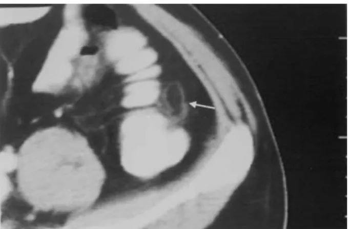
Fig. 1. Helical CT of the epiploic appendigitis. Pericolonic fat density oval shaped lesion anterolateral to the descending colon with a rim of soft tissue density is visulized (arrow). The adjacent colonic wall and nearby seg-ments were unremarkable.
Fig. 2. MRI of the same patient with epiploic appendigitis. T1-weighted breath-hold spoiled gradient echo (SGE) images (TR/TE/FA, 190/4,2/80) with 6 mm section thickness in axial plane (a), reveals high signal lesion with a thin hypointense rim (arrow) and hypointense central dot, which has similar appearance on axial respiratory gated T2-weighted fast spin echo image (TR/TE/ETL, 4800/90/8) (section thickness 6 mm) (arrow) (b). T1-weighted fat suppressed breathhold spoiled gradient echo images (TR/ TE/FA, 195/4,2/80) (section thickness 6 mm) after gadolinium application reveals enhancement of the rim (arrow) (c).
and increased mobility are predisposing factors for torsion and infarction of epiploic appendages [3–5]. PEA is the inflammatory response of an appendage to infarction caused by torsion or spontaneous venous thrombosis. Secondary epiploic appendagitis is the inflammation of the epiploic appendages caused by nearby pathologic processes, such as diverticulitis, appendicitis or cholecystitis [5].
Patients with PEA are typically obese people in the second to fifth decades of life, complaining of motion-aggravated abdominal pain. Depending on the location of the inflammatory process, the location of the pain varies but is mostly in the left or right lower-quadrant. Left lower quadrant pain is the more commonly encountered localiza-tion. White blood-cell count is normal or slightly elevated in most cases. Physical examination shows localized tender-ness and some guarding but no rigidity [1,3–5]. Symptoms usually subside within less than one week from onset. This entity is a self-limiting process with spontaneous resolution and conservative treatment with analgesics is sufficient [1,3, 4].
The CT findings of PEA have been described in several series and case reports [3,5,8]. On contrast enhanced CT images, PEA is a pericolonic, oval shaped lesion with fat density center and a soft tissue density rim representing inflammation of the serosa and associated with periappenda-geal fat stranding. In some cases, a hyperdense dot or linear high density is seen in the center representing a thrombosed vessel or hemorrhagic necrosis. Other associated CT find-ings of epiploic appendagitis are nearby parietal peritoneal thickening and adjacent colonic wall thickening [2–5].
The MRI findings of a single case of PEA has been described in a series of PEA imaged with other cross sec-tional imaging modalities [4]. According to that report, epiploic appendagitis appeared as an oval-shaped lesion with high signal intensity center and low signal intensity rim on T1-weighted images. T2-weighted images without fat suppression revealed a high intensity center and low signal intensity rim. On postgadolinium T1-weighted fat sup-pressed images, the rim revealed increased enhancement due to inflammatory response [4]. In our case a hypointense central dot was visible on both precontrast T1-and T2 -weighted images. Hypointensity of the central dot on both sequences is in agreement with the belief that central dot/ line corresponds to the histopathologic finding of fibrous septa which was detected in a few surgically confirmed cases in two previous reports [3,9]. PEA was best recog-nized on postcontrast fat suppressed images. Postcontrast T1-weighted fat suppressed MR imaging of the bowel is the commonly accepted method to visualize inflammatory bowel disease [7]. Therefore, the rim, representing the in-flammatory response of the visceral peritoneum was best seen on postcontrast T1-fat saturated images in our case. It has been reported that the imaging findings resolve in one to
two months of duration after the initial onset of symptoms, although the symptoms usually subside earlier [10]. The imaging findings also disappeared about one month after the initial symptoms in our case.
Only a few pathologic processes are considered in the differential diagnosis of PEA based on the CT findings. These are omental infarction and secondary epiploic ap-pendagitis. Infarction of the greater omentum may present with identical clinical signs of PEA of the transverse colon. However, considerably larger size (3–15 cm in diameter) of omental infarction and lack of peripheral thickened visceral peritoneum aid in the differential diagnosis. In addition, infarction of the omentum is localized medial to the colon unlike PEA which is located anterolaterally [11]. Secondary epiploic appendagitis (e.g., due to diverticulitis) presents with findings of luminal narrowing, diverticula, pericolonic fluid, air bubbles, phlegmon and/or abscess formation which are diagnostic for the original disease [3,4].
In conclusion, we have described the MRI findings of PEA in one case. MRI findings correlated well with the CT findings in our case. With the wider use of MRI in bowel disease and in abdominal emergencies, the MRI findings of epiploic appendagitis should be kept in mind for the differ-ential diagnosis of other acute abdominal conditions.
References
[1] Rao PM, Rhea JT, Wittenberg J, Warshaw, AL. Misdiagnosis of Primary Epiploic Appendagitis. Am J Surg 1998;176:81–5. [2] Torres GM, Abbitt PL, Weeks M. CT Manifestations of Infarcted
Epiploic Appendages of the Colon. Abdom Imaging 1994;19:449 – 50.
[3] S¸irvancı M, Tekeliog˘lu TH, Duran C, Yardimci H, Onat L, O¨ zer AK. Primary Epiploic Appendagitis: CT Manifestations. Clin Imaging 2000;24:357– 61.
[4] Barbier C, Denny P, Pradoura PB, et al. Primary Epiploic Ap-pendagitis:US, CT and MR Findings. J Radiol 1998;79:1479 – 85. [5] Rioux M, Langis P. Primary Epiploic Appendagitis: Clinical, US, and
CT Findings in 14 Cases. Radiology 1994;191:523– 6.
[6] Lee JK, Marcos HB, Semelka RC. MR imaging of the small bowel using the HASTE sequence. AJR 1998;170:1457– 63.
[7] Chung JJ, Semelka RC, Martin DR, Marcos HB. Colon diseases: MR evaluation using combined T2-weighted single-shot echo train spin-echo and gadolinium-enhanced spoiled gradient-spin-echo sequences. J Magn Reson Imaging 2000;12:297–305.
[8] Danielson K, Chernin MM, Amberg JR, Goff S, Durham JR. Epiploic appendicitis: CT characteristics. J Comput Assist Tomogr 1986;10: 142–3.
[9] Molla E, Ripolles T, Martinez MJ, Morote V, Rosello-Sastre E. Primary epiploic appendagitis: US and CT findings. Eur Radiol 1998; 8:435– 8.
[10] Rao PM, Wittenberg J, Lawrason JN. Primary Epiploic Appendagitis: Evolutionary Changes in CT Appearance. Radiology 1997;204: 713–7.
[11] Puylaert JBCM. Right-sided Segmental Infarction of the Omentum: Clinical US, and CT Findings. Radiology 1992;185:169 –72.
139