Investigating the relationship between internal tibial torsion and medial
collateral ligament injury in patients undergoing knee arthroscopy due
to tears in the posterior one third of the medial meniscus
Olcay Guler
a,⁎
, Mehmet Isyar
a, Dilek Karata
ş
b, Tugrul Ormeci
c, Halis Cerci
d, Mahir Mahirogullar
ı
a aDepartment of Orthopedics and Traumatology, Medipol University, Medical Faculty, Istanbul, Turkeyb
Department of Radiology, Nisa Hospital, Istanbul, Turkey
c
Department of Radiology, Medipol University, Medical Faculty, Istanbul, Turkey
d
Department of Orthopedics and Traumatology, Nisa Hospital, Istanbul, Turkey
a b s t r a c t
a r t i c l e i n f o
Article history: Received 9 July 2015
Received in revised form 7 September 2015 Accepted 30 September 2015
Keywords: Knee arthroscopy Medial collateral ligament Injury
Internal tibial torsion Foot femoral angle Transmalleolar angle Medial meniscus
Purpose: To evaluate the relationship between medial collateral ligament (MCL) injury and degree of internal tibial torsion in patients who had undergone arthroscopic resection due to tears in the posterior one third of the medial meniscus.
Methods: Seventy-one patients were allocated into two groups with respect to foot femur angle (FFA) and transmalleolar angle (TMA) (Group 1 31 patients with FFAb 8° and Group 2 40 patients with FFA ≥ 8°). The groups were compared in terms of valgus instability, Lysholm score, magnetic resonance view, FFA, and TMA, both before and after the operation.
Results: Lysholm scores were higher in Group 2 at both postoperative week 1 (pb 0.001) and month 1 (p = 0.045) relative to Group 1. Preoperative cartilage injury was encountered more frequently in Group 1 (p = 0.037) than in Group 2. MCL injury was detected more frequently in Group 1 compared to Group 2 postoperatively at week 1 (p = 0.001).
Conclusion: We conclude that FFA and TFA, indicators of internal tibial torsion, may serve as markers for foreseeing clinical improvement and complications following arthroscopic surgery.
Level of Evidence: level III retrospective comparative study.
© 2015 Elsevier B.V. All rights reserved.
1. Introduction
The knee is one of the most common sites of injury in the human body. A vast majority of knee injuries are caused by non-contact events. The most common events linked to knee and medial collateral ligament (MCL) injuries are accompanied by internal torsion of the tibia relative to the femur[1].
During torsion experiments in a previous study, internal rotation of the tibia and valgus rotation of the femur were observed during and after anterior cruciate ligament (ACL) injury. In compression experi-ments, the direction of tibial rotation shifted from internal rotation be-fore failure to external rotation after failure. Moreover, ACL injuries can be created via internal tibial rotation[1].
The ACL resists internal rotation due to its orientation in the axial plane, where it attaches medially on the anterior tibial plateau and laterally in the femoral notch[2]. Owing to the posterior displacement
of the femur and the medial tibial plateau induced by internal tibial ro-tation, the effective center of rotation is located on the medial side of the ACL. In addition, these motions occur on a coupled internal tibial and valgus rotation of the femur[3]. During these movements, the axis of ro-tation is placed beside the MCL due to tension in the ligament for valgus bending moments. Therefore, in addition to the ACL, the MCL has an im-portant function in knee motion.
Tibial torsion is defined as torsion of tibia along its longitudinal axis. Deformity of the lower limb in the coronal plane has been widely inves-tigated and found to be associated with the development of osteoarthri-tis (OA). However, rotational deformities of the lower limb are still a controversial subject. Some surgeons have advocated the correction of rotational deformity during total knee replacement and during treat-ment of complex tibial fractures[4].
Arthroscopic knee surgery is performed for various pathologies with satisfactory results. The majority of these procedures consist of meniscal resection. Adequate visualization is mandatory to achieve good results. Tightness in the medial compartment is one factor restricting sufficient visualization; the posterior horn of the medial meniscus is especially difficult. Therefore, complications can occur due to limited exposure at this location[5].
The Knee 23 (2016) 655–658
⁎ Corresponding author at: Orthopedics and Traumatology Department, Medipol University, Medical Faculty, Atatürk Bulvarı No: 27 Unkapanı, 34083, Fatih, Istanbul, Turkey. Tel.: +90 212 444 8544; fax: +90 212 531 7555.
E-mail address:[email protected](O. Guler).
http://dx.doi.org/10.1016/j.knee.2015.09.020
0968-0160/© 2015 Elsevier B.V. All rights reserved.
Contents lists available atScienceDirect
Recognition of factors that facilitate the occurrence of complications is crucial to minimize these risks. It must be kept in mind that safe and effective arthroscopic intervention is implemented on an individualized basis with respect to an integrated evaluation of personal, radiological, and clinical data.
In this study, we assessed the relationship between MCL injury and the degree of internal tibial torsion together with clinical outcomes in patients that underwent arthroscopic resection due to tears in the posterior one third of the medial meniscus. We believe that our results contribute to a better understanding of factors prone to influence the success and likelihood of complications during arthroscopic surgery. 2. Patients and methods
2.1. Study design
This retrospective study was performed using data extracted from the medicalfiles of 71 patients that underwent arthroscopic partial meniscectomy due to symptomatic tears in the posterior horn of the internal meniscus between 2011 and 2014 in the Orthopaedics and Traumatology Department of our Tertiary Care Center. This study was approved by the Institutional Ethics Committee and conducted in accor-dance with the latest version of the Helsinki Declaration.
Patients included in this study did not report any complaints, and no pathologicalfindings were found upon physical examination of the con-tralateral knee preoperatively. McMurray and Apley tests were positive, and the grade of arthrosis was≤2 according to Kellgren–Lawrence clas-sification[6,7].
Preoperatively, valgus stress tests, as well as measurements of foot femur angles (FFAs) and transmalleolar angles (TMAs), were con-ducted. In accordance with report by Craft et al., magnetic resonance imaging was used for the mainstay of diagnostic imaging, with coronal sequences allowing the full assessment of the MCL complex[8]. Preop-eratively, the degree of valgus instability was measured and all patients were evaluated with respect to Lysholm classification[9–11]. Postoper-atively, magnetic resonance images (MRIs) were routinely obtained.
Exclusion criteria were as follows: internal meniscal repair, mechan-ical axis problems, ACL injury, an arthrosis score≥3 according to Kellgren–Lawrence classification, external meniscal repair, and MRI views older than three weeks.
Patients were distributed into two groups after receiver operating characteristic (ROC) curve analysis was performed with respect to FFA and TMA. Group 1 consisted of 30 patients (11 women, 19 men) with FFAb 8°; Group 2 consisted of 41 patients (14 women, 27 men) with FFA≥ 8°.
2.2. Outcome parameters
Patients were controlled on week 1, as well as postoperative months 1, 3, 6, and 12. The two groups were compared in terms of degree of valgus instability, Lysholm scores, MRIfindings, mean FFA and TMA values, and incidence of preoperative iatrogenic cartilage injury values before and after arthroscopic surgery. The degree of valgus instability and Lysholm scores were also evaluated within each group.
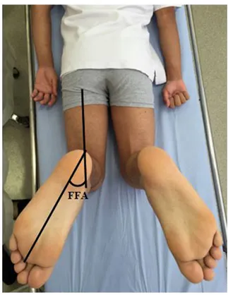
The FFA is described as the angle between the axis of the foot and the axis of the femur for a patient in supine position with legs at 90°flexion (Fig. 1). Positive values indicate external rotation, whereas negative values are consistent with internal rotation. The TMA is used for clinical evaluation of tibial torsion. In the supine position, a line connecting the tips of the medial malleolus and lateral malleolus is drawn on the heel. The TMA is located between the line perpendicular to this line and another line drawn from the middle of the femur (Fig. 2).
Valgus instability is evaluated by means of abduction. In addition, a valgus stress test can be performed with the patient in the supine posi-tion on the examining table. The contralateral normal extremity must initially be examined to gain the patient's confidence and establish
a baseline of normal ligamentous tightness. The knee to be examined is placed on the side of the table next to the examiner. The extremity is abducted off the side of the table and the knee isflexed at 30°. One of the examiner's hands is placed around the lateral aspect of the knee and the other hand supports the ankle. Gentle abduction or valgus stress is ap-plied to the knee while the hand at the ankle rotates the leg slightly[12]. 2.3. Radiological study
Conventional knee radiographs were obtained at both stance and flexion at 30°. MRI was carried out using a 1.5 T MRI device (Sigma
Fig. 1. Measurement of foot femur angle (FFA).
Fig. 2. Measurement of transmalleolar angle (TMA). 656 O. Guler et al. / The Knee 23 (2016) 655–658
HDXT, General Electric, Chicago, IL, USA). Images were taken in T1- and T2-weighted modes in the axial and sagittal planes. Fat suppression mode was selected in parallel to menisci axially in the coronal section and vertical to menisci in the sagittal section. All sections were exam-ined and evaluated by the same radiologist experienced in musculoskel-etal system radiology.
2.4. Surgical procedure
All cases were operated on by the same surgical team. Patients were in the supine position under spinal anesthesia and compres-sion (pressure, 300 mm Hg) was applied. Arthroscopy was performed while the knee wasflexed at 90° using standard anterolateral and anteromedial portals. A 30° angled scope was used. Subsequent to diag-nostic arthroscopy, partial meniscectomy was performed using a punch and shaver while the knee wasflexed at 30° and the valgus was exter-nally rotated.
Surgical procedure was performed as described by Dasic et al.[13]. Partial resection of the medial meniscus is advocated when other treat-ment modalities are not attainable. We followed guidelines described by Metcalfs et al. for arthroscopic resection that applies to most resect-able meniscal lesions[14]. Accordingly, all mobile fragments that could be pulled past the inner margin of the meniscus into the center of the joint were removed. The remaining meniscal rim was smoothed to re-move any sudden changes in contour that might lead to further tearing. The probe was used repeatedly to explore the mobility and texture of the remaining rim. Efforts were spent to protect meniscocapsular junction and the peripheral meniscal rim during resection in order to preserve meniscal stability since it is crucial for preservation of the load transmission properties of the meniscus.
Postoperatively, patients were mobilized with full load, and isomet-ric quadisomet-riceps exercises began. Lysholm scores[12], degree of valgus
instability and MRI views were evaluated at week 1 and month 1 fol-lowing the operation.
2.5. Statistical analysis
Continuous variables were expressed in descriptive statistics (mean, standard deviation, median, minimum, and maximum). Qualitative variables were analyzed using the Chi-square test, while the McNemar test was used to compare categorical and dependent variables. Two independent groups that did not display a normal distribution were compared using the Mann–Whitney U test. Student's t-test was utilized for two independent groups with normal distribution. Pairs of depen-dent variables that did not exhibit normal distribution were compared using the Wilcoxon signed-rank test, whereas the Friedman test was used to compare more than two variables. Differences determined to be statistically significant after the Friedman test were further assessed using the Wilcoxon signed-rank test. A p valueb 0.05 was considered statistically significant. Data analysis was implemented using MedCalc Statistical Software version 12.7.7 (MedCalc Software bvba, Ostend, Belgium;http://www.medcalc.org; 2013).
3. Results
The study group consisted of 25 women (35.2%) and 46 men (64.8%), with an average age of 38.3 ± 7.5 (range, 22 to 53). The symbol ± (plus sign over minus sign) defines the standard deviation of the mean value of the related parameters. Right knees were involved in 24 (33.8%) patients, while left knees were affected in 47 (66.2%) cases. A comparison of the two groups regarding the parameters under investigation is shown inTable 1. The two groups did not differ in terms of age (p = 0.66), gender distribution (p = 0.83), or involve-ment of right or left knees (p = 0.28). Preoperatively, MRI views were obtained 10.8 ± 4.0 days (range, 3 to 20) prior to the operation. Interestingly, the average FFA value for Group 2 (13.1 ± 3.0) was significantly higher than the average in Group 1 (6.2 ± 1.7) (pb 0.001). Similarly, the average TMA value in Group 2 (25.3 ± 5.7) was greater than that in Group 1 (14.8 ± 2.6) (pb 0.001).
The groups displayed similar results with respect to grade of arthrosis (p = 0.93), preoperative incidence of valgus instability (p = 0.71), preoperative Lysholm score (p = 0.63), and preoperative incidence of MCL injury (p = 0.56). However, Group 2 had a higher average Lysholm score (pb 0.001) and increased prevalence of MCL injury (p = 0.001) at week 1 postoperatively. The difference between groups for Lysholm score persisted at postoperative month 1 (p = 0.04), whereas preoperative cartilage inju-ry was more likely in Group 1 (p = 0.04). Changes in Lysholm score, physical examination findings, and frequency of MCL injury in the postoperative period are shown inTable 2.
For both groups, postoperative improvement, as reflected in Lysholm scores, was significant for postoperative week 1 and month 1 compared to the preoperative state (pb 0.001). On the other hand, the frequency of pathological findings upon physical exam-ination did not change significantly in the postoperative period.
4. Discussion
We assessed whether MCL injury is linked with internal tibial tor-sion and clinical improvement following arthroscopic surgery, which may be affected by internal tibial torsion. Our results indicate that FFA and TMA may be useful for preoperative evaluation and categoriza-tion of patients scheduled for arthroscopic surgery to weigh risks and benefits.
In clinical practice, adduction of the foot in a patient standing with the patella faced directly anterior results from torsional deformity and is termed internal tibial torsion [15]. Torsional abnormalities are
Table 1
Comparison of groups with respect to demographic, clinical, and radiological variables under investigation. Variable Group 1 (n = 30) Group 2 (n = 41) p value Age 37.8 ± 7.0 38.3 ± 7.5 0.66 Gender (male/female) 19/11 27/14 0.83 Side (right/left) 8/22 16/25 0.28 Foot femur angle 6.2 ± 1.7 13.1 ± 3.0 b0.001⁎ Transmalleolar angle 14.8 ± 2.6 25.3 ± 5.7 b0.001⁎
Grade of arthrosis (1 vs. 2) 18/12 25/16 0.93 Preoperative Lysholm score 49.1 ± 5.8 48.7 ± 6.7 0.63 Preoperative MCL injury 9/21 15/26 0.56 Preoperative valgus instability 20/10 29/12 0.71 Postoperative week 1 Lysholm score 60.3 ± 7.0 70.7 ± 4.8 b0.001⁎ Postoperative week 1 MCL injury 3/27 14/27 0.001⁎ Postoperative week 1 valgus instability 13/17 12/29 0.22 Postoperative month 1 Lysholm score 49.1 ± 5.8 48.7 ± 6.7 0.51 Postoperative 1st month MCL injury 2/28 1/40 0.84 Postoperative 1st month valgus instability 11/19 12/29 0.51 Duration of follow-up (months) 22.4 ± 9.1 20.8 ± 7.8 0.55 Abbreviations: MCL, medial collateral ligament.
⁎ Statistically significant.
Table 2
Preoperative and postoperative Lysholm scores, incidences of MCL injury and pathological physical examinationfindings.
Variable Preoperative Postoperative week 1 Postoperative month 1 p value
Lysholm score Group 1 49.1 ± 5.8 60.3 ± 7.0 89.1 ± 6.2 b0.001⁎
Group 2 48.7 ± 6.7 70.7 ± 4.8 92.0 ± 4.8 b0.001⁎ Postoperative physical examination (N/P) Group 1 20/10 17/13 19/11 N0.05‡
Group 2 29/12 29/12 29/12 N0.05ǂ
Abbreviations: MCL, medial collateral ligament; N, normal; P, pathological. ⁎ Statistically significant.
‡ Preoperative vs. postoperative week 1, p = 0.25; preoperative vs. postoperative month 1, p = 1.00; postoperative week 1 vs. postoperative month 1, p = 0.62. ǂ p = 1.00 for comparison of values at preoperative, postoperative week 1 and postoperative month 1 periods.
657 O. Guler et al. / The Knee 23 (2016) 655–658
considered both a possible cause and effect of OA. Turner[16]suggested that patients with panarticular OA of the knee had reduced external tor-sion or true internal tortor-sion. In addition, Yagi et al.[17]reported that tib-ial torsional deformities occurred in association with medtib-ial-type osteoarthritic knees.
Access to the medial part of the knee is of paramount importance for providing adequate vision during arthroscopic surgery. Therefore, selection and preoperative evaluation of patients likely to benefit from arthroscopy is crucial. Interestingly, intraoperative cartilage injury is more common in patients with smaller FFA and TMA. In contrast, patients with larger FFA and TMA are more likely to benefit from arthro-scopic surgery for meniscal tears. Thesefindings may be attributed to structural difficulties that hinder instrumentation at addressing meniscal and cartilaginous pathology during surgery[18]. These struc-tural challenges result not only in iatrogenic cartilage injury, but also some pathology that may be missed[19]. Documentation of the rela-tionship between FFA, TMA, and instrumentation during arthroscopy warrants further trials on larger cohorts.
To the best of our knowledge, no reports have addressed the rela-tionship between MCL injury and internal tibial torsion. The decline of MCL injury rate seems to indicate spontaneous healing of preoperative MCL injury, and it is slower in patients with FFA≥ 8°. Moreover, an increase in Lysholm score after surgery is more prominent in this popu-lation. Our results suggest that FFA and TMA may be important determi-nants for the classification and evaluation of patients with tears in the posterior one third of the medial menisci. Grouping patients based on parameters related with internal tibial torsion can provide valuable data on the likelihood of MCL injury, improvement in Lysholm score, and preoperative cartilage injury.
Our results on compatibility of postoperative“residual” MCL injury with good clinical results may be confusing atfirst glance. However, presumably, patients with smaller FFA/TMA may have tighter medial structures, which cause iatrogenic cartilage damage, inadequate menisectomy and insufficient clinical results. On the other hand, pa-tients with larger FFA/TMA have relative loose medial structures with less frequent cartilage injury, adequate menisectomy and improved clinical results.
The main limitations of this study include its small sample size and retrospective design. Because our series reflects the experience of a sin-gle institution, our results must be carefully interpreted. In addition, influences of social, environmental, and ethnic factors should not be ignored. Lack of comparison of the percentage of iatrogenic cartilage in-jury between groups constitutes another restriction of the present study. In conclusion, our results demonstrate that FFA and TMA, indicators of internal tibial torsion, may serve as markers for foreseeing clinical im-provement and likelihood of MCL injury in patients that underwent
arthroscopic surgery due to tears in the posterior one third of the medial meniscus.
Acknowledgments
The authors declare no competing interests. Nofinancial support was received for this paper. References
[1]Amis A, Dawkins G. Functional anatomy of the anterior cruciate ligament:fibre bundle actions related to ligament replacements and injuries. J Bone Joint Surg 1991;73-B:260–7.
[2]Aronson PA, Gieck JH, Hertel J, et al. Tibiofemoral joint positioning for the valgus stress test. J Athl Train 2010;45:357–63.
[3]Briggs KK, Lysholm J, Tegner Y, et al. The reliability, validity, and responsiveness of the Lysholm score and Tegner activity scale for anterior cruciate ligament injuries of the knee: 25 years later. Am J Sports Med 2009;37:890–7.
[4]Mullaji AB, Sharma AK, Marawar SV, et al. Tibial torsion in non-arthritic Indian adults: a computer tomography study of 100 limbs. Indian J Orthop 2008;42:309–13.
[5]Celik D, Coskunsu D, Kilicoglu O. Translation and cultural adaptation of the Turkish Lysholm knee scale: ease of use, validity, and reliability. Clin Orthop Relat Res 2013;471:2602–10.
[6]Emrani PS, Katz JN, Kessler CL, et al. Joint space narrowing and Kellgren–Lawrence progression in knee osteoarthritis: an analytic literature synthesis. Osteoarthritis Cartilage 2008;16:873–82.
[7]Fakioglu O, Ozsoy MH, Ozdemir HM, et al. Percutaneous medial collateral ligament release in arthroscopic medial meniscectomy in tight knees. Knee Surg Sports Traumatol Arthrosc 2013;21:1540–5.
[8]Craft JA, Kurzweil PR. Physical examination and imaging of medial collateral ligament and posteromedial corner of the knee. Sports Med Arthrosc 2015;23: e1–6.
[9]Javidan P, Ahmed M, Kaar SG. Arthroscopic release of the deep medial collateral ligament to assist in exposure of the medial tibiofemoral compartment. Arthrosc Tech 2014;3:e699–701.
[10]Kellgren JH, Lawrence JS. Radiological assessment of osteo-arthrosis. Ann Rheum Dis 1957;16:494–502.
[11]Lubowitz JH, MacKAy G, Gilmer B. Knee medial collateral ligament and posteromedial corner anatomic repair with internal bracing. Arthrosc Tech 2014;3:e505–8.
[12]Lysholm J, Gillquist J. Evaluation of knee ligament surgery results with special em-phasis on use of a scoring scale. Am J Sports Med 1982;10:150–4.
[13]Dasić Z, Radoicić D. Arthroscopic partial medial meniscectomy. Vojnosanit Pregl 2011;68:774–8.
[14]Metcalf RW, Burks RT, Metcalf MS, et al. Arthroscopic meniscectomy. In: McGinty JB, Caspari RB, Jackson RW, Poehling GG, editors. Operative arthroscopy. 2nd ed. Philadelphia, PA: Lippincott-Raven; 1996. p. 263–97.
[15]Meyer EG, Haut RC. Anterior cruciate ligament injury induced by internal tibial torsion or tibiofemoral compression. J Biomech 2008;41:3377–83.
[16]Turner MS. The association between tibial torsion and knee joint pathology. Clin Orthop Relat Res 1994;302:47–51.
[17]Yagi T. Tibial torsion in patients with medial-type osteoarthrotic knees. Clin Orthop Relat Res 1994;302:52–6.
[18]Matsumoto H, Suda Y, Otani T, et al. Roles of the anterior cruciate ligament in preventing valgus instability. J Orthop Sci 2001;6:28–32.
[19]Naruse K, Urabe K, Jiang S, et al. Osteoarthritic changes of the patellofemoral joint in STR/Ortcrlj mice are the earliest detectable changes and may be caused by internal tibial torsion. Connect Tissue Res 2009;50:243–55.