Ankara Univ Vet Fak Derg, 66, 379-384, 2019 DOI: 10.33988/auvfd.544263
Comparison of periapical radiography, panoramic, and cone-beam
CT in the detection of dental caries in dog teeth
Kaan ORHAN
1,a,, Mehmet Özgür ÖZEMRE
1,b, Cansu KÖSEOĞLU SEÇGİN
1,c,
Sevil ATALAY VURAL
2,d, Gürkan GÜR
3,e, Kıvanç KAMBUROĞLU
4,f1 1Başkent University, Faculty of Dentistry, Department of Dentomaxillofacial Radiology; 2Ankara University, Faculty of Veterinary Medicine, Department of Pathology; 3Ankara University, Faculty of Dentistry, Department of Restorative Dentistry; 4Ankara
University, Faculty of Dentistry, Department of Dentomaxillofacial Radiology, Ankara, Turkey aORCID: 0000-0003-1686-4746; bORCID: 0000-0001-5863-6990; cORCID: 0000-0002-7896-1165; dORCID: 0000-0003-2111-3381; eORCID: 0000-0002-4376-7848; fORCID: 0000-0002-4134-5756.
Corresponding author: [email protected]
Received date: 25.03.2019- Accepted date: 23.06.2019
Abstract: The aim of this study was to compare the effectiveness of panoramic, periapical and two different Cone Beam Computed Tomography (CBCT) devices in the detection of dental caries of dog teeth ex vivo. A total of 880 teeth were investigated, 33 of which were with caries, whereas; 33 healthy teeth were the controls. Periapical, panoramic and CBCT scans were made for the assessment of the teeth. All images were evaluated separately by two observers experienced in image interpretation. The presence or absence of occlusal caries was scored using a 5-point scale. Kappa values were calculated to assess intra and interobserver agreement. Receiver Operating Characteristic (ROC) analysis was performed to compare the effectiveness of different imaging methods in the detection of dental caries. For both observers, the order of success of the image sets in the estimation of the caries tooth was CBCT Morita, CBCT Iluma, periapical and panoramic radiograph (Area Under Curve (AUC): 0.929, 0.882, 0.861, and 0.704 for observer 1, AUC: 0.927, 0.896, 0.875, and 0.693 for observer 2, respectively). CBCT was found to be the best imaging method for the ex vivo detection of caries in dog teeth. In addition, panoramic images performed worse than all other modalities.
Keywords: CBCT, dental caries, dogs, panoramic radiography, periapical radiography.
Köpek dişlerindeki çürüklerin tespitinde periapikal, panoramik ve konik ışınlı BT görüntülerinin
karşılaştırılması
Özet: Bu çalışmanın amacı, ex vivo olarak köpeklerde diş çürüğü tespitinde panoramik, periapikal ve iki farklı Konik Işınlı Bilgisayarlı Tomografi (KIBT) cihazının etkinliğini karşılaştırmaktır. Toplam 880 diş değerlendirilmiş olup 33 çürük diş tespit edilmiştir. Sağlıklı dişlerden rastgele 33’ü seçilerek kontrol grubu oluşturulmuştur. Çürüklerin radyografik olarak değerlendirilmesi için periapikal, panoramik ve KIBT yöntemleri kullanılarak görüntüler elde edildi. Tüm görüntüler, görüntü yorumlamada deneyimli iki gözlemci tarafından ayrı ayrı değerlendirildi. Okluzal çürüğün varlığı veya yokluğu, 5 puanlık bir ölçek kullanılarak puanlandı. Kappa değerleri, gözlemci içi ve gözlemciler arası uyumu değerlendirmek için hesaplandı. Diş çürüğü tespitinde farklı görüntüleme yöntemlerinin karşılaştırılması için ROC analizi yapıldı. Her iki gözlemci için, çürük dişin tespitinde görüntüleme yöntemlerinin başarı sırası KIBT Morita, KIBT Iluma, periapikal ve panoramik radyograf olarak bulunmuştur. (1. gözlemci için Area Under Curve (AUC): 0.929, 0.882, 0.861 ve 0.704, 2. gözlemci için AUC: 0.927, 0.896, 0.875 ve 0.693). KIBT’ın köpek dişlerinde çürüklerin ex vivo tespiti için en iyi görüntüleme yöntemi olduğu belirlenmiştir. Ayrıca, panoramik görüntüler diğer tüm yöntemlerden daha kötü performans göstermiştir.
Anahtar sözcükler: Diş çürüğü, köpek, KIBT, panoramik radyografi, periapikal radyografi.
Introduction
Dental caries is plaque-induced demineralization of the teeth formed by the effect of bacteria that ferment carbohydrates. This fermentation leads to the production of acids, which demineralize enamel and dentin. As a result of this, bacteria spread into the dentin, undermining
the enamel, leading to a collapse of the enamel and cavitation of the tooth (9). Dental caries can affect any tooth. Teeth of dogs with deeper pits and fissures may be more susceptible to caries. In the dogs, dental caries most commonly occurs on the occlusal surface of the distal aspect of the mandibular first molar tooth, the remaining
mandibular molar teeth, maxillary molar teeth, and teeth with prominent developmental grooves. The maxillary first molar tooth is particularly prone to caries (2). In the literature, the incidence of dental caries is between 3.1-5.3% of dogs (2). Dental caries is less common in dogs than in humans (2). This is because dogs do not have too many fermented carbohydrates in their diet and have an increased salivary pH (9).
Imaging modalities utilized for veterinary dentistry showed an enormous development during the last decade. Consequently, veterinarians' knowledge regarding complex diagnostic imaging methods and treatments, and the importance of orofacial health for domestic animals has increased progressively (12). In consideration of several pathological conditions, radiographic imaging of domestic animal teeth plays a critical role in clinical diagnosis (6). Radiological examination in veterinary dentistry is necessary for certain conditions, such as; caries diagnosis, periodontal assessment, endodontics, restorative procedures and maxillofacial surgery etc. (10). Yet, adequacy of radiographic information seen on conventional two dimensional radiographs, such as; periapical and panoramic radiography is usually limited by the superimposition of anatomical structures. Generally, several radiographs of the suspected teeth are needed during the initial examination. A full-mouth examination is regarded as a series of radiographs describing not only existing teeth but also the toothless segment of the jaw (15). Pathologic radiographic changes are often challenging to diagnose, and for this reason, clarity and detail of radiographic images are important (6). Cone beam computed tomography (CBCT) which offered high resolution three dimensional images of dentomaxillofacial structures with relatively lower radiation doses and costs than medical computed tomography was introduced in 1999 (14). CBCT also enabled three dimensional imaging of rabbits, pigs, cats, and dogs (8).
The aim of this study was to compare the effectiveness of periapical, panoramic, and two different CBCT devices in the detection of dental caries of dog teeth ex vivo. If the diagnostic capability of conventional radiographs is found better than CBCT there would be no need to three-dimensional (3D) images.
Materials and Methods
Sample: This study was performed with local ethical
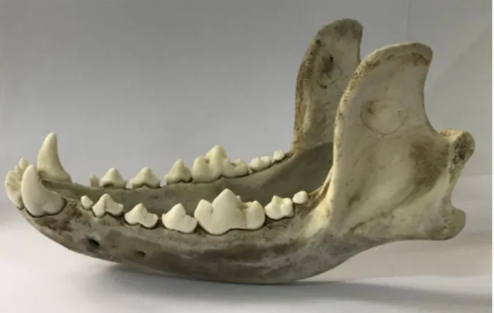
committee approval (Baskent University, D-KA 18/21). The sample consisted of randomly selected 40 fresh cadaver mandibles frozen within the post mortem 24th hour. The sample was defrosted 24-hours prior to making the scans. (Figure 1) Firstly, according to tooth size and position, three groups of teeth were formed. The first group included lower incisors, the second group consisted
of lower canines, and the third group comprised lower premolars and molars (6). However, there is no caries found on incisors, canines and premolars. All the caries lesions were on molar teeth. A total of 880 teeth were investigated, 33 of with dentine caries (on occlusal surfaces) and 33 healthy teeth were selected randomly for the control group. We selected the same type of teeth (molar) with the caries and compared them with the healthy ones. A 1.5 cm red wax material was used as a soft tissue equivalent.
Figure 1. One of the fresh cadaver mandible used in the study.
All caries lesions were diagnosed with visual inspection and the use of fine dental explorer for the gold standard.
Periapical, panoramic and CBCT assessments: A
full-mouth radiographic survey was made for the assessment of teeth. A standard wall-mounted dental radiography unit (Sirona Dental, Salzburg, Austria), along with photostimulable phosphor (PSP) digital intraoral imaging system (Digora, Optime, Soredex, Finland) was used. Intraoral radiographs of the mandibular premolars and molars were obtained by using paralleling technique. Bisecting angle technique was used to evaluate mandibular incisors and canine teeth because of the anatomy of the dogs’ mandible.
All digital panoramic images were acquired using the same machine (Veraviewpocs 2D, Morita, Japan), with the following exposure parameters: 64–66 kVp; 6–9 mA; and 10 s. The isolated mandibles were positioned with the occlusal plane perpendicular to the floor.
Two CBCT systems (3D Accuitomo 170, Morita, Japan and Iluma, OrthoCATTM, IMTEC Imaging, Ardmore, OK, US) were used to scan the sample. CBCT Iluma and Morita are two different systems that each of them has its own technical parameters and software
program. Technical parameters for 3D Accuitomo 170 and
Iluma were; 90 kV, 5 mA, 17.5 s, 10x5 cm FOV, 0.25 mm voxel size and 120 kV, 3.8 mA, 40 s, 18x14 cm FOV, 0.09
mm voxel size, respectively. The isolated mandibles were positioned with the occlusal plane perpendicular to the floor. Axial scans and multiplanar reconstructions were obtained from CBCT scans.
Two experienced observers assessed all the images, separately. Image evaluation was done in a dimly lit room without time constraints. (FUJITSU L20T1 20’’ 1600x900 resolution LCD monitor, Kawasaki, Japan). The presence or absence of occlusal/incisal caries was scored using the following 5-point scale: 1= caries definitely present; 2= caries probably present; 3= uncertain-unable to tell; 4= caries probably not present; 5= caries definitely not present. Each observer evaluated the images twice in four weeks to analyze the intra- and interobserver agreement.
Statistical analysis: Statistical analysis was performed with the SPSS (Version 22.0, SPSS Inc., Chicago, IL, USA) package program. Weighted kappa statistics with confidence intervals were calculated to determine the level of agreement between the imaging methods and the gold standard. Kappa values were calculated to assess intra-and inter-observer agreement. The kappa values were interpreted as follows: (< 0.20: Poor, 0.21 - 0.40: Fair, 0.41 - 0.60: Moderate, 0.61 - 0.80: Good, 0.81 - 1.00: Very good). Receiver Operating Characteristic (ROC) analysis was carried out to compare different imaging methods for caries detection. The areas under the curve (AUC), with 95% confidence intervals (CI) were determined. The categories used to classify the accuracy of the imaging methods in ROC analysis were as follows: excellent (0.9–1), good (0.8–0.9), fair (0.7–0.8), poor (0.6–0.7) and fail (0.5–0.6). Sensitivity, specificity, positive predictive value, negative predictive value, positive likelihood value, and accuracy were also calculated to compare classification success of dental caries (> 3 without caries, <3 caries).
Results
Fourteen of the 40 mandibles (35%) had bilaterally symmetrical lesions. Caries lesions were most common on mandibular first molar teeth, with 25 on mandibular first molar teeth and eight on mandibular second molar teeth. The incidence of caries lesions was found 4.1% and 35% of 40 mandibles had bilaterally symmetrical lesions in our study.
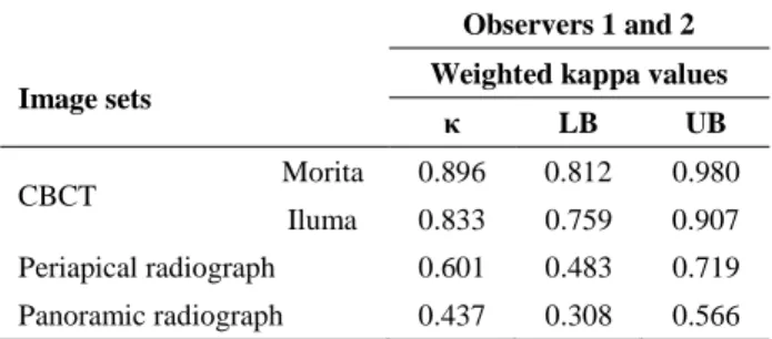
Intraobserver agreement was 0.932 (very good), 0.924 (very good), 0.835 (moderate) and 0.820 (very good) for Oberver 1, 0.940 (very good), 0.938 (very good), 0.842(very good) and 0.826 (very good) for Observer 2 for CBCT Morita, CBCT Iluma, periapical and panoramic radiography, respectively. Interobserver agreement was 0.896 (very good), 0.833 (very good), 0.601(moderate) and 0.437 (moderate) for CBCT Morita, CBCT Iluma,
periapical and panoramic radiography, respectively (Table 1).
Table 1. Interobserver weighted kappa coefficients by image sets.
Observers 1 and 2
Image sets Weighted kappa values
κ LB UB
CBCT Morita 0.896 0.812 0.980
Iluma 0.833 0.759 0.907 Periapical radiograph 0.601 0.483 0.719 Panoramic radiograph 0.437 0.308 0.566 κ: kappa value, LB: Lower Bound, UB: Upper Bound, CBCT: Cone beam computed tomography
The agreement results between the gold standard test and the image sets were calculated with weighted kappa statistics. Kappa values are presented in Table 2. According to Table 2, the highest agreement with the gold standard test for both observers was found for CBCT Morita (observer 1: κ = 0.586: moderate agreement, observer 2: κ = 0.616: high agreement). For observer 1, the agreement value of the other image sets was κ = 0.525: moderate agreement, κ = 0.515: moderate agreement, κ = 0.303: poor agreement, for periapical radiography, CBCT Iluma, and panoramic radiography, respectively. For observer 2, the agreement value of the other image sets was κ = 0.525: moderate agreement, κ = 0.485: moderate agreement, κ = 0.192: poor agreement, for CBCT Iluma, periapical and panoramic radiography, respectively. The scores obtained from panoramic radiographs for both observers showed poor agreement with the gold standard test. Images obtained from a carious tooth by using each imaging technique were shown in Figure 2 (A-C).
In order to compare prediction successes of different imaging techniques according to the gold standard the AUCs obtained by Binormal ROC analysis and post-hoc comparisons with the gold standard were presented in Table 3. ROC curves were also shown in Figure 3 (A) and (B) in consideration to observers. There was a statistically significant difference between the success of correct diagnosis of the four different devices used by the first and second observers (p = 0.001, p = 0.002, respectively; Table 3). This difference was due to the panoramic radiographic scores obtained from both observers, according to post-hoc ROC comparisons. For both observers, success order of the image sets in the estimation of the caries tooth was CBCT Morita, CBCT Iluma, periapical and panoramic radiography (AUC: 0.929, 0.882, 0.861, and 0.704 for observer 1, AUC: 0.927, 0.896, 0.875, and 0.693 for observer 2, respectively; Table 3).
Table 2. Observers’ results in image sets for area under the ROC curve, sensitivity, specificity, positive predictive value, negative predictive value, positive likelihood ratio, and accuracy (n=66).
Observer Image
set AUC (95 % CI)
P for ROC comparison Pairwise comparison Se Sp PPV NPV LR+ Ac (%) 1 A 0.929 (0.867-0.992) 0.001 1-2: 0.345 1-3: <0.001 1-4: 0.249 0.879 0.727 0.763 0.857 3.22 80.3 B 0.882 (0.797-0.967) 2-3: 0.004 2-4: 0.706 0.788 0.788 0.788 0.788 3.71 78.8 C 0.704 (0.563-0.845) 3-4: 0.014 0.697 0.636 0.657 0.677 1.92 66.7 D 0.861 (0.766-0.956) 0.818 0.727 0.750 0.800 3.00 77.3 2 A 0.927 (0.862-0.992) 0.002 1-2: 0.540 1-3: <0.001 1-4: 0.304 0.848 0.758 0.778 0.833 3.50 80.3 B 0.896 (0.818-0.974) 2-3: 0.001 2-4: 0.700 0.848 0.788 0.800 0.839 4.00 81.8 C 0.693 (0.540-0.846) 3-4: 0.015 0.879 0.455 0.617 0.789 1.61 66.7 D 0.875 (0.782-0.967) 0.879 0.667 0.725 0.846 2.64 77.3
A: CBCT Morita; B: CBCT Iluma; C: Panoramic radiograph; D: Periapical radiograph; AUC: Area under the ROC curve, CI: Confidence interval, Se: Sensitivity, Sp: Specificity, PPV: Positive predictive value, NPV: Negative predictive value, LR+: Positive likelihood ratio, Ac: Accuracy.
Figure 2. The images of left lower first molar tooth with occlusal caries were shown A: Panoramic radiograph, B: Periapical radiograph, C: CBCT Morita.
Figure 3. ROC curves were shown according to observers (A, B).
Table 3. Weighted kappa coefficients to assess agreement between image sets and gold standard.
Observer Image sets P Kappa values
κ LB UB 1 A <0.001 0.586 0.451 0.721 B <0.001 0.515 0.370 0.660 C 0.008 0.303 0.109 0.497 D <0.001 0.525 0.354 0.696 2 A <0.001 0.616 0.473 0.759 B <0.001 0.525 0.386 0.664 C 0.024 0.192 0.049 0.335 D <0.001 0.485 0.342 0.628
CI: Confidence interval, κ: kappa value, LB: Lower Bound, UB: Upper Bound A: CBCT Morita; B: CBCT Iluma; C: Panoramic radiograph; D: Periapical radiograph.
Sensitivity, specificity, positive predictive value, negative predictive value, positive likelihood and accuracy values calculated to compare correct classification success (>3 without caries, and <3 caries) were given in Table 2. For both observers, the highest correct classification was achieved with CBCT Morita (80.3-81.8% Accuracy, 84.8-87.9% sensitivity and 72.7-78.8% specificity).
Discussion and Conclusion
Veterinarians should monitor the risky or already decaying areas and recommended reasonable preventative or restorative treatments even though the incidence of dental caries is lower than humans.
To the authors’ knowledge, no previous veterinary dentistry study compared two CBCT units with periapical and panoramic radiography techniques in the detection of dental caries in dog teeth. We compared images obtained from intraoral PSP, panoramic and two CBCT units in the detection of dental caries. We found that both CBCT
systems performed similarly and better than two-dimensional (2D) systems suggesting the use of 3D imaging for better caries diagnosis. Panoramic imaging showed the worst diagnostic performance in detecting of dental caries. In recent years, to evaluate the accuracy of CBCT in detecting proximal and occlusal caries lesions, several studied reported varying results (1, 11, 16). In 2007, the study comparing a Sirona CBCT unit and conventional radiography in the detection of proximal caries (3). They did not find any differences between the two imaging methods (3). Tsuchida et al. noticed that the accuracy of the 3D Accuitomo in assessing early proximal caries was not superior to conventional radiography (13). Similarly, Haiter-Neto et al. demonstrated no differences between NewTom 3G, 3DX Accuitomo, DIGORA PSP and Kodak conventional film system in detecting occlusal caries (1).We found that the highest agreement with the gold standard test for both observers was with CBCT Morita. This result might be attributable to CBCT detector and software capabilities, observer performance or
imaging settings used. Voxel size which defines the smallest component of a three dimensional image may be detrimental in diagnostic quality and patient dose. We used voxel sizes smaller than 0.2 mm and this could positively have affected observer performance. In addition, unless the patient is anesthetized, patient motion which may be an important clinical drawback was not an issue in this ex vivo study.
On the contrary, Young et al. reported that Accuitomo 3DX CBCT images were better than Charge Coupled Device (CCD) images in detecting dentin caries lesions (16). However, the authors showed that the differences between CBCT and CCD images did not statistically significant in detecting enamel caries lesions. The reason for these differences in the studies is that both of the studies (1, 13) used a population of teeth in which the most of the proximal surface lesions were limited to the enamel, whereas Young et al. (16) evaluated both enamel and dentinal lesions equally. Another study by Rathore et al. showed no statistically significant differences between Sirona CBCT unit and conventional radiography in detecting occlusal caries (7). In our study, all caries lesions extended into the dentin and similar to Young et al. (16) we found CBCT Morita images to be superior to the periapical radiographs in detecting caries lesions. In our study, a comparison between enamel and dentin caries lesions could not be performed since all caries lesions extended into the dentin.
The concern about radiation exposure is one of the most important factors when choosing between imaging modalities. CBCT systems deliver far greater effective doses when compared to intraoral imaging in general (4, 5). Future improvements in CBCT imaging can be expected to result in innovative systems with better diagnostic abilities and lower effective doses.
Limitations of this study; all caries lesions was on occlusal surfaces of teeth, histopathologic analysis of caries lesions was not performed so that the diagnosis was made with visual inspection and dental explorer.
In conclusion, conventional 2D radiographical techniques have some advantages such as low cost, shorter irradiation time, however, we found that panoramic images performed worse than all other modalities. In our study, CBCT was found to be the best imaging method for the ex vivo detection of caries in dog teeth. So that it would be recommended to increase the use of CBCT in veterinary dentistry. In addition, further studies about comparing the different imaging modalities in the detection of occlusal and proximal caries lesions in dog teeth were encouraged.
Conflict of Interest
The authors declared that there is no conflict of interest.
References
1. Haiter-Neto F, Wenzel A, Gotfredsen E (2008): Diagnostic accuracy of cone beam computed tomography scans compared with intraoral image modalities for detection of caries lesions. Dentomaxillofac Radiol, 37, 18– 22.
2. Hale FA (1998): Dental Caries in the Dog. J Vet Dent, 15, 79–83.
3. Kalathingal SM, Mol A, Tyndall DA, et al (2007): In vitro assessment of cone beam local computed tomography for proximal caries detection. Oral Surg Oral Med Oral Pathol Oral Radiol Endod, 104, 699–704.
4. Lee G-S, Kim J-S, Seo Y-S, et al (2013): Effective dose from direct and indirect digital panoramic units. Imaging Sci Dent, 43, 77–84.
5. Ludlow JB, Timothy R, Walker C, et al (2015): Effective dose of dental CBCT—a meta analysis of published data and additional data for nine CBCT units. Dentomaxillofac Radiol, 44, 20140197.
6. Pavlica Z, Erjavec V, Erzen D, et al (2003): A full-mouth radiographic survey of periodontal bone loss in dogs. Acta Vet Brno, 72, 391–398.
7. Rathore S, Tyndall D, Wright J, et al (2012): Ex vivo comparison of Galileos cone beam CT and intraoral radiographs in detecting occlusal caries. Dentomaxillofac Radiol, 41, 489–493.
8. Riggs GG, Arzi B, Cissell DD, et al (2016): Clinical application of cone-beam computed tomography of the rabbit head: part 1 – normal dentition. Front Vet Sci, 3, 93. 9. Ritchie C (2014): Class I restoration of maxillary first
molar caries in a dog. J Vet Dent, 31, 66–9.
10. Roza MR, Silva LAF, Barriviera M, et al (2011): Cone beam computed tomography and intraoral radiography for diagnosis of dental abnormalities in dogs and cats. J Vet Sci, 12, 387–392.
11. Senel B, Kamburoglu K, Ucok O, et al (2010): Diagnostic accuracy of different imaging modalities in detection of proximal caries. Dentomaxillofacial Radiol, 39, 501–511. 12. Soukup JW, Drees R, Koenig LJ, et al (2015):
Comparison of the diagnostic image quality of the canine maxillary dentoalveolar structures obtained by cone beam computed tomography and 64-multidetector row computed tomography. J Vet Dent, 32, 80–86.
13. Tsuchida R, Araki K, Okano T. (2007): Evaluation of a limited cone-beam volumetric imaging system: comparison with film radiography in detecting incipient proximal caries. Oral Surg Oral Med Oral Pathol Oral Radiol Endod, 104, 412–416.
14. Van Thielen B, Siguenza F, Hassan B (2012): Cone beam computed tomography in veterinary dentistry. J Vet Dent, 29, 27–34.
15. Verstraete F, Kass P, Terpak C (1998): Diagnostic value of full-mouth radiography in dogs. Am J Vet Res, 59, 686– 691.
16. Young SM, Lee JT, Hodges RJ, et al (2009): A comparative study of high-resolution cone beam computed tomography and charge-coupled device sensors for detecting caries. Dentomaxillofac Radiol, 38, 445–451.